
 The Relationship Between Inflammatory Markers and Supraventricular Tachycardia in Children
Abstract
Background Supraventricular tachycardia (SVT) is a common arrhythmia in pediatric patients. Emerging evidence suggests a link between systemic inflammation and cardiovascular diseases.
Objectives This study aims to investigate the relationship between the monocyte/lymphocyte ratio (MLR) and neutrophil/lymphocyte ratio (NLR) with SVT in pediatric patients. Methods: We conducted a retrospective study at Rize Education and Research Hospital’s Pediatric Health and Diseases Department, including 33 patients diagnosed with SVT and 33 control subjects without sinus tachycardia. Hemogram (CBC) parameters, electrocardiography (ECG), and echocardiography (ECHO) were evaluated. NLR and MLR were calculated. The unpaired t-test is designed to compare the means of continuous variables between two groups, while the Mann-Whitney U test was applied for data not following a normal distribution. Correlations between values were analyzed through Pearson correlation. A significance level of p <0.05 was accepted.
Results There was no significant difference between the groups in terms of age, lymphocyte count, hemoglobin, hematocrit, platelet count, and NLR. However, leukocyte count, monocyte count, and MLR showed significant differences (p<0.05). The SVT group had higher MLR values compared to the control group.
Conclusions Our study found that MLR was significantly higher in pediatric patients with SVT compared to controls, suggesting a potential role of inflammation in SVT pathogenesis. Further large-scale, prospective, and multicenter studies are needed to confirm these findings and clarify the relationship between inflammation markers and SVT.
Supraventricular Tachycardia; Cardiovascular Diseases; Child; Inflammation; Biomarkers

Introduction
A normal heartbeat originates from an electrical impulse passing through the heart. This electrical impulse typically originates from the sinoatrial node, located mostly in the upper part of the right atrium, and travels to the ventricles. This ensures the orderly and sequential contraction of the atria and ventricles. Arrhythmia refers to an abnormal heart rhythm caused by a problem in the heart's electrical system. Supraventricular tachycardia (SVT) refers to a group of tachycardias originating from the bundle of His or structures above it. Relatively common in the pediatric population, SVT is observed in healthy children at a frequency of 0.1-0.4%.1 The resting heart rate in SVT is usually >100 beats/min, and conduction, typically originating from the AV node and His-Purkinje system, results in a narrow QRS complex (<120 ms). The most common form of SVT in children involves accessory electrical connections between the atria and ventricles.2,3Symptoms of SVT in the pediatric age group are generally mild and can be challenging to identify. Symptoms may include sweating, poor feeding, rapid pulse, pale skin, and decreased activity level and alertness, particularly in infants.4
Inflammation is a physiological response to tissue damage, injury, or destruction caused by physical, chemical, or pathogens. Infection causes (upper/lower respiratory tract, urogenital system, central nervous system infections caused by viral, bacterial, fungal, and protozoan pathogens), trauma, exposure to chemical substances, or tissue damage resulting from ischemia, autoimmune disorders (such as rheumatoid arthritis, multiple sclerosis), chronic inflammation (associated with many diseases such as Alzheimer's, Parkinson's diseases), among others.5
Inflammation-related fibrosis, ischemia, coagulation abnormalities, and intracellular changes lead to pathological alterations in the electrophysiological properties of the heart, such as ion
channel dysfunction, early and late after depolarizations, and remodeling. This condition results in the initiation of arrhythmias, reentry, and the emergence of ectopic activity.6 Hemogram (CBC) and its white blood cell component, commonly used by clinicians in the diagnosis of many diseases, are clinically useful in the treatment of infection or inflammation. The most frequent type of WBC is neutrophils, accounting for 50–70% of the total WBC in blood circulation. Neutrophils are the primary immune cells that respond to infection and are controlled under homeostatic conditions. Lymphocytes are a critical population of WBCs and play an essential role in both innate and adaptive immunity. Oxidative stress and cardiac cell damage from systemic inflammation might result in decreased electrical conduction and heightened vulnerability to reentry circuits. The risk can be partly attributed to oxidative damage caused by the release of reactive oxygen species (ROS) by circulating blood neutrophils. And this may cause endothelial dysfunction . This condition results in the initiation of arrhythmias. Several previous studies have shown that the neutrophil-to-lymphocyte ratio (NLR) is an indicator of various cardiovascular diseases because it reflects systemic inflammation and immune balance.7 Additionally, the NLR and the monocyte-to-lymphocyte ratio (MLR) are inflammatory markers used to assess disease activity, recurrence rate, surveillance, and prognosis in many diseases. Therefore, the primary objective of our study was to investigate the relationship between the MLR and the NLR in patients diagnosed with SVT. Our second objective was to review the current literature on the use of MLR and NLR as biomarkers in other diseases and compare these findings with those in pediatric SVT.
Materials and Methods
Our study was conducted retrospectively in the Department of Pediatrics at Rize Teaching and Research Hospital from September 2016 to March 2023. The minimum sample size required for evaluation in the study was calculated using the G*Power 3.1.9.7 software. The effect size was calculated for the comparison of the means between two independent groups. The means and standard deviations of the NLR and the MLR obtained from the literature review were used, resulting in an effect size of 0.7016. With an alpha error level of 0.05 and a power of 80%, it was determined that each of the case and control groups should consist of at least 33 individuals. Thirty-three patients diagnosed with SVT and 33 control subjects without sinus tachycardia were included. The inclusion criteria were SVT-diagnosed patients with little or no change in the RR interval or no change, narrow QRS complexes, and heart rate >180 beats/min, such as AVRT and AVNRT types (including those with preexcitation syndrome). In the control group, patients without sinus tachycardia who had had a CBC test within the last year, and were in the same age group as the patient group were included. Patients diagnosed with sinus tachycardia, ectopic atrial tachycardia, focal atrial tachycardia, atrial fibrillation, or atrial flutter were excluded from the study. The age parameter was matched to be the same for the patient and control groups.
The CBC parameters, ECG, and echocardiograms (ECHO) of all patients were examined. The NLR and the MLR were calculated.
Statistical analysis
Statistical analysis was performed using SPSS-29 software. Continuous variables were presented as mean ± standard deviation or as median and interquartile range (IQR), while categorical data were expressed as absolute and relative frequencies. The Shapiro-Wilk test was used to assess normality. For data with a normal distribution, the unpaired Student's t-test was used to compare means of continuous variables between two groups, while the Mann-Whitney U test was applied for data not following a normal distribution. Correlations between variables were analyzed using Pearson correlation. A two-tailed significance level of p < 0.05 was considered statistically significant.
Our study was conducted with the approval of the Recep Tayyip Erdoğan University Non-Interventional Clinical Research Ethics Committee, decision number 2023/158.
Results
The study included 2 groups, SVT patients and controls. An independent t-test revealed that the leukocyte count was significantly higher in the patient group compared to the control group. Similarly, the monocyte count and neutrophil count were significantly different between the groups. The MLR was a significant difference was found in the MLR between the control group and the patient group . There was no statistically significant difference between the groups in terms of age, lymphocyte count, hemoglobin, hematocrit, platelet count, and NLR. Significant differences were observed in white blood cell count, monocyte count, and MLR (p <0.05).
The number of individuals in each group, age, CBC values, mean (± standard deviation) of NLR and MLR values, and the statistical results of the independent t-test are provided in Table 1. The Pearson correlation coefficients between the NLR and MLR values for the two groups (control and SVT group) are 0.150 for NLR and 0.053 for MLR. These values suggest a weak positive correlation between the NLR values and a very weak correlation between the MLR values across the two groups.
In the control group, the non-normally distributed were lymphocytes, monocytes, platelets, NLO, and MLO; the normally distributed were neutrophils, hemoglobin, and hematocrit. In the SVT group, the non-normally distributed were lymphocytes, monocytes, neutrophils, NLO, and MLO; the normally distributed were hemoglobin, platelets, and hematocrit.
At the end of our study, while no significant difference was found between the patient and control groups in terms of age and NLR, a significant difference was found between the groups in terms of MLR. The MLR was found to be higher in the SVT group compared to the control group (see Central Figure and Figure 1).

Additionally, echocardiograms of 33 patients diagnosed with SVT were reviewed in our study. Mitral insufficiency was reported in 4 patients, patent foramen ovale in 2 patients, atrial septal defect in 1 patient, and interatrial septal aneurysm in 1 patient. Echocardiography (ECHO) was normal in 10 patients (see Table 1). However, echocardiographic findings could not be obtained in 15 patients.
Discussion
The studies conducted in recent years indicate an association between systemic inflammation and cardiovascular disease.8 There are many ways in which systemic inflammation might raise the risk of arrhythmias. First, oxidative stress and cardiac cell damage from systemic inflammation might result in decreased electrical conduction and heightened vulnerability to reentry circuits. The risk can be partly attributed to oxidative damage caused by the release of ROS by circulating blood neutrophils. By downregulating the eNOS gene as well as reacting with NO to form peroxynitrite, ROS reduces the amount of available NO, thus causing endothelial dysfunction. Second, the autonomic nervous system (ANS) might have its equilibrium upset by systemic inflammation like in autoimmune disorders. Third, since potassium, calcium, and magnesium are necessary for a proper ventricular action potential, systemic inflammation may have an impact on these electrolyte levels. Fourth, systemic inflammation has the potential to trigger the coagulation system and raise the risk of thromboembolism, both of which can result in myocardial infarction and ischemia.9 The reasons mentioned, inflammation-related fibrosis, ischemia, coagulation disorders, and intracellular changes, lead to pathological changes in the electrophysiological properties of the heart. This condition results in the initiation of arrhythmias, reentry, and the emergence of ectopic activity. In our study investigating the relationship between inflammation markers and SVT, we found a weak correlation between MLR and SVT.
Inflammatory markers such as IL-1, IL-6, TNF-α, and C-reactive protein are commonly used markers in clinical practice.10 It has been proven that these markers can affect membrane ion
channels and, therefore, cause arrhythmias. NLR and MLR are parameters that can be easily examined from a complete blood count and can be used by clinicians due to their cost-effectiveness.11 Additionally, research in the literature has examined the association of these markers with many diseases beyond cardiovascular diseases; they have been reported to be indicators of poor prognosis in malignancies, to predict clinical severity in acute ischemic stroke, to monitor the disease and treatment response in COVID-19 patients, to be associated with inflammatory processes in psychiatric disorders, to assess disease severity and monitoring in psoriasis, to predict mortality in sepsis, to demonstrate activation in ulcerative colitis, and to be applicable in neurological diseases such as Guillain-Barre syndrome and multiple sclerosis.12-18 Additionally, it has been reported that in patients with COPD , when the NLR is 3.3 or higher, the possibility of acute exacerbation of asthma increases by approximately 32 times; and when the NLR is > 4.02, the risk of subarachnoid hemorrhage appears to be 33 times higher.19-21 There are numerous studies conducted on these ratios in adult patients with arrhythmias. Aydın et al. found the NLR in adult patients diagnosed with SVT to be higher compared to the control group.22-23 Küçük et al. could not find a significant difference between the groups in terms of the NLR and the MLR in the assessment of white blood cell subtypes in patients diagnosed with SVT.24In our study, unlike other studies in the literature, we aimed to investigate the relationship between MLR and NLR levels in pediatric patients diagnosed with SVT, and at the end of our study, we observed a significant difference between the groups in terms of MLR (p = 0.04). Our study found significant differences between the control group and the patient group in several hematological parameters, while some measures did not show significant variation. Notably, the leukocyte count was significantly higher in the patient group, consistent with the findings of previous studies that suggest elevated leukocyte levels are associated with inflammatory conditions, including those observed in SVT patients. Similarly, monocyte counts and the MLR were also significantly elevated in the patient group, further supporting the hypothesis that monocyte levels may play a role in the disease pathology of SVT. Interestingly, we found no significant differences in lymphocyte count, hemoglobin, hematocrit, or platelet count between the control and patient groups. The lack of variation in these measures suggests that while inflammation is a key factor, it may not directly affect all hematological parameters uniformly across patient populations. The neutrophil count was significantly higher in the patient group, as was the NLR, although the latter was not statistically significant. Elevated neutrophil levels in patients could indicate an acute phase response, which has been commonly reported in various cardiovascular and thrombotic conditions. Our findings, especially regarding the leukocyte, monocyte, and neutrophil levels, are in agreement with prior research linking systemic inflammation with SVT. However, the insignificant differences in other parameters such as hemoglobin and platelet counts suggest that further research is needed to understand the full hematological profile of these patients. Additionally, in our study, we also examined the ECHO reports of SVT patients. In the literature, Harris et al. found no abnormalities in ECHO exams in 78% of the patients diagnosed with SVT in the adult age group.25
Premkumar et al. found normal echocardiographic results in 41.67% of SVT patients in their study on arrhythmias in the pediatric age group. However, they still detected abnormalities in 55% of the reports.26In a study by L'Italien et al. involving 224 SVT patients in the pediatric age group, they found a normal echocardiographic result in 96.4% of the cases. Only 2% of the cases resulted in abnormal echocardiograms. Among the patients with abnormal echocardiograms, four had minor cardiac structural abnormalities, while only one had a moderate primum atrial septal defect requiring surgery.27 In our study, we also found that in the echocardiographic results of SVT patients, we could not identify abnormalities in 30.3% of the patients. Among the remaining patients, we detected mitral insufficiency in 12.1%, patent foramen ovale in 6%, atrial septal defect in 3%, and interatrial septal aneurysm in 3%.
Thus, we determined that 36% of our patients had a normal echocardiogram result, while 18% had minor cardiac pathologies. However, it was not possible to obtain echocardiographic findings in 15 patients. Therefore, our comparisons on ECHO are not valid. We still believe that our findings are consistent with the literature. Many diseases triggering the inflammatory process could be expected to alter laboratory parameters. This provides researchers with a broad spectrum to investigate. The inadequacy of the existing literature, the absence of studies finding negative results regarding the NLR and the MLR, the lack of a common value range, our study being a single-center and a retrospective study, the relatively small number of patients, and the inability to measure other inflammation markers such as TNF-α, IL-1, and IL-6, C-reactive protein demand more comprehensive research. In our study, we could not collect detailed clinical data on patients admitted with SVT like criteria for sepsis, and respiratory or gastrointestinal symptoms in this cohort . We recommend considering this aspect in future studies for a comprehensive understanding of patient presentations.
Confounders such as age, sex, and underlying medical conditions were controlled for by including them as covariates in our regression models. Future studies should consider a larger sample size to further evaluate the impact of these confounders. The strengths of our article are that this study offers significant pediatric contributions to the understanding of SVT in patients by investigating the relationship between inflammatory markers, specifically the MLR and the NLR. By focusing on a population often underrepresented in cardiology research, this article addresses a critical gap in the literature. The findings highlight the clinical relevance of using MLR as a potential biomarker for diagnosing and managing SVT, offering insights that may improve patient outcomes.
Conclusion
We found a significant difference in MLR values between the SVT group and the control group. Additionally, we observed higher MLR values in patients with SVT. However, larger-scale, prospective, and multicenter studies are needed to confirm the relationship between NLR, MLR, and SVT.
References
-
1 Abbasi E, Vijayashankar SS, Goldman RD. Management of Acute Supraventricular Tachycardia in Children. Can Fam Physician. 2023;69(12):839-41. doi: 10.46747/cfp.6912839.
» https://doi.org/10.46747/cfp.6912839 -
2 Brugada J, Katritsis DG, Arbelo E, Arribas F, Bax JJ, Blomström-Lundqvist C, et al. 2019 ESC Guidelines for the Management of Patients with Supraventricular Tachycardia The Task Force for the Management of Patients with Supraventricular Tachycardia of the European Society of Cardiology (ESC). Eur Heart J. 2020;41(5):655-720. doi: 10.1093/eurheartj/ehz467.
» https://doi.org/10.1093/eurheartj/ehz467 -
3 University of Michigan. C.S. Mott Children's Hospital. Supraventricular Tachycardia. Ann Arbor: University of Michigan; 2024. [cited 2025 Jan 7]. Available from: https://www.mottchildren.org/conditions-treatments/ped-heart/conditions/supraventricular-tachycardia
» https://www.mottchildren.org/conditions-treatments/ped-heart/conditions/supraventricular-tachycardia -
4 Childrens Hospital of Philadelphia [Internet]. Supraventricular Tachycardia (SVT) Causes, Symptoms and Treatment. Philadelphia: Children's Hospital of Philadelphia, 2023. [cited 2025 Jan 7]. Available from: https://www.chop.edu/conditions-diseases/supraventricular-tachycardia-svt
» https://www.chop.edu/conditions-diseases/supraventricular-tachycardia-svt -
5 Kell DB, Pretorius E. No Effects without Causes: The Iron Dysregulation and Dormant Microbes Hypothesis for Chronic, Inflammatory Diseases. Biol Rev Camb Philos Soc. 2018;93(3):1518-57. doi: 10.1111/brv.12407.
» https://doi.org/10.1111/brv.12407 -
6 Vonderlin N, Siebermair J, Kaya E, Köhler M, Rassaf T, Wakili R. Critical Inflammatory Mechanisms Underlying Arrhythmias. Herz. 2019;44(2):121-9. doi: 10.1007/s00059-019-4788-5.
» https://doi.org/10.1007/s00059-019-4788-5 -
7 Seo IH, Lee YJ. Usefulness of Complete Blood Count (CBC) to Assess Cardiovascular and Metabolic Diseases in Clinical Settings: A Comprehensive Literature Review. Biomedicines. 2022;10(11):2697. doi: 10.3390/biomedicines10112697.
» https://doi.org/10.3390/biomedicines10112697 -
8 Gupta L, Thomas J, Ravichandran R, Singh M, Nag A, Panjiyar BK. Inflammation in Cardiovascular Disease: A Comprehensive Review of Biomarkers and Therapeutic Targets. Cureus. 2023;15(9):e45483. doi: 10.7759/cureus.45483.
» https://doi.org/10.7759/cureus.45483 - 9 Taghdiri A. Infammation and Arrhythmogenesis: A Narrative Review of the Complex Relationship. Int J Arrhythm. 2024;25(4):1-12. doi:10.1186/s42444-024-00110-z.
-
10 Özen C. The Neutrophil-lymphocyte Ratio and its Applications in The Emergency Department [Internet]. Borzova: Ümraniye Education and Research Hospital; 2017 [cited in 2025 Jan 7]. Available from: https://acilci.net/notrofil-lenfosit-orani-ve-acil-serviste-kullanim-alanlari/
» https://acilci.net/notrofil-lenfosit-orani-ve-acil-serviste-kullanim-alanlari/ -
11 Açikgöz M, Atasoy HT. The Relationship of Lymphocyte/Monocyte Ratio with Clinical and Radiological Parameters and Its Effect of Short-Term Results in Stroke Patients. Med J West Black Sea. 2023;7(2):148-55. doi:10.29058/mjwbs.1310680.
» https://doi.org/10.29058/mjwbs.1310680 -
12 Korkmaz C, Demirbas S, Vatansev H, Yosunkaya S, Teke T, Zamani A, et al. The Association of Neutrophil/Lymphocyte Ratio, Lymphocyte/Monocyte Ratio, Platelet/Lymphocyte Ratio and Hematological Parameters with Severity and Prognosis in Hospitalized Patients with COVID-19. Turk J Med Sci. 2021;6(2):251-61. doi:10.26453/otjhs.876015.
» https://doi.org/10.26453/otjhs.876015 -
13 Örüm MH. Neutrophil to Lymphocyte Ratio in Generalized Anxiety Disorder. Med Records. 2020 Oct;2(3):93-7. DOI: 10.37990/medr.774613.
» https://doi.org/10.37990/medr.774613 -
14 Çerman AA, Karabay EA, Altunay IK. Evaluation of Neutrophil-Lymphocyte Ratio and Mean Platelet Volume in Patients with Psoriasis. Med Bull Sisli Etfal Hosp. 2016;50(2):137-41. doi: 10.5350/SEMB.20160216020543.
» https://doi.org/10.5350/SEMB.20160216020543 -
15 Çakir E, Turan IÖ. Which Hemogram-Derived Indices Might be Useful in Predicting the Clinical Outcomes of Sepsis Patients in the Intensive Care Unit? Cukurova Med J. 2021;46(2):532-9. doi: 10.17826/cumj.856741
» https://doi.org/10.17826/cumj.856741 -
16 Tenlik I, Coskun O, Çapraz M, Gökbulut V, Öztürk Ö. Evaluation of Ulcerative Colitis Activity and Severity with Non-Invasive Biomarkers. Turk. J. Gastroenterol. 2023;22(2):59-66. doi: 10.17941/agd.1348965.
» https://doi.org/10.17941/agd.1348965 -
17 Kissling M, Fritschi N, Baumann P, Buettcher M, Bonhoeffer J, Naranbhai V, et al. Monocyte, Lymphocyte and Neutrophil Ratios - Easy-to-Use Biomarkers for the Diagnosis of Pediatric Tuberculosis. Pediatr Infect Dis J. 2023;42(6):520-7. doi: 10.1097/INF.0000000000003901.
» https://doi.org/10.1097/INF.0000000000003901 -
18 Bedel C, Korkut M. The Clinical Significance of Neutrophil-Lymphocyte Ratio, Monocyte-Lymphocyte Ratio, and Platelet-Lymphocyte Ratio in Patients with Guillain-Barré Syndrome. Haydarpasa Numune Med J, 61(3), 341-345. https://doi.org/10.14744/hnhj.2019.38233
» https://doi.org/10.14744/hnhj.2019.38233 - 19 Kurtipek E, Bekci TT, Kesli R, Sami SS, Terzi Y. The Role of Neutrophil-Lymphocyte Ratio and Platelet-Lymphocyte Ratio in Exacerbation of Chronic Obstructive Pulmonary Disease. J Pak Med Assoc. 2015;65(12):1283-7.
-
20 Eryigit U, Cakmak VA, Sahin A, Tatli O, Pasli S, Gazioglu G, et al. The Diagnostic Value of the Neutrophil-Lymphocyte Ratio in Distinguishing between Subarachnoid Hemorrhage and Migraine. Am J Emerg Med. 2017;35(9):1276-80. doi: 10.1016/j.ajem.2017.03.063.
» https://doi.org/10.1016/j.ajem.2017.03.063 -
21 Jung SK, Rhee DY, Lee WJ, Woo SH, Seol SH, Kim DH, et al. Neutrophil-to-Lymphocyte Count Ratio is Associated with Perforated Appendicitis in Elderly Patients of Emergency Department. Aging Clin Exp Res. 2017;29(3):529-36. doi: 10.1007/s40520-016-0584-8.
» https://doi.org/10.1007/s40520-016-0584-8 -
22 Kalra A, Wedd JP, Bambha KM, Gralla J, Golden-Mason L, Collins C, et al. Neutrophil-to-Lymphocyte Ratio Correlates with Proinflammatory Neutrophils and Predicts Death in Low Model for End-Stage Liver Disease Patients with Cirrhosis. Liver Transpl. 2017;23(2):155-65. doi: 10.1002/lt.24702.
» https://doi.org/10.1002/lt.24702 -
23 Aydin M, Yildiz A, Yüksel M, Polat N, Aktan A, Islamoglu Y. Assessment of the Neutrophil/Lymphocyte Ratio in Patients with Supraventricular Tachycardia. Anatol J Cardiol. 2016;16(1):29-33. doi: 10.5152/akd.2015.5927.
» https://doi.org/10.5152/akd.2015.5927 -
24 Küçük U, Arslan M. Assessment of the White Blood Cell Subtypes Ratio in Patients with Supraventricular Tachycardia: Retrospective Cohort Study. Journal of Surgery and Medicine. 2019;3(4):00-00. doi: 10.28982/josam.510973.
» https://doi.org/10.28982/josam.510973 -
25 Harris R, Sahay RN. Clinical Profile of Patients Diagnosed with Supraventricular Tachycardia: A Descriptive Study at a Tertiary Care Hospital. Int. J. Med. 2022;9(3):317-21. doi:10.18203/2349-3933.ijam20220436.
» https://doi.org/10.18203/2349-3933.ijam20220436 -
26 Premkumar S, Sundararajan P, Sangaralingam T. Clinical Profile of Cardiac Arrhythmias in Children Attending the Out Patient Department of a Tertiary Paediatric Care Centre in Chennai. J Clin Diagn Res. 2016;10(12):SC06-SC08. doi: 10.7860/JCDR/2016/21751.8992.
» https://doi.org/10.7860/JCDR/2016/21751.8992 -
27 L'Italien K, Conlon S, Kertesz N, Bezold L, Kamp A. Usefulness of Echocardiography in Children with New-Onset Supraventricular Tachycardia. J Am Soc Echocardiogr. 2018;31(10):1146-50. doi: 10.1016/j.echo.2018.05.001.
» https://doi.org/10.1016/j.echo.2018.05.001
-
Study Association
This study is not associated with any thesis or dissertation work.
-
Ethics Approval and Consent to Participate
This study was approved by the Ethics Committee on Animal Experiments of Recep Tayyip Erdogan University Non-interventional Clinical under the protocol number 2023/198.
-
Sources of Funding
There were no external funding sources for this study.
Edited by
-
Editor responsible for the review:
Ricardo Alkmim Teixeira

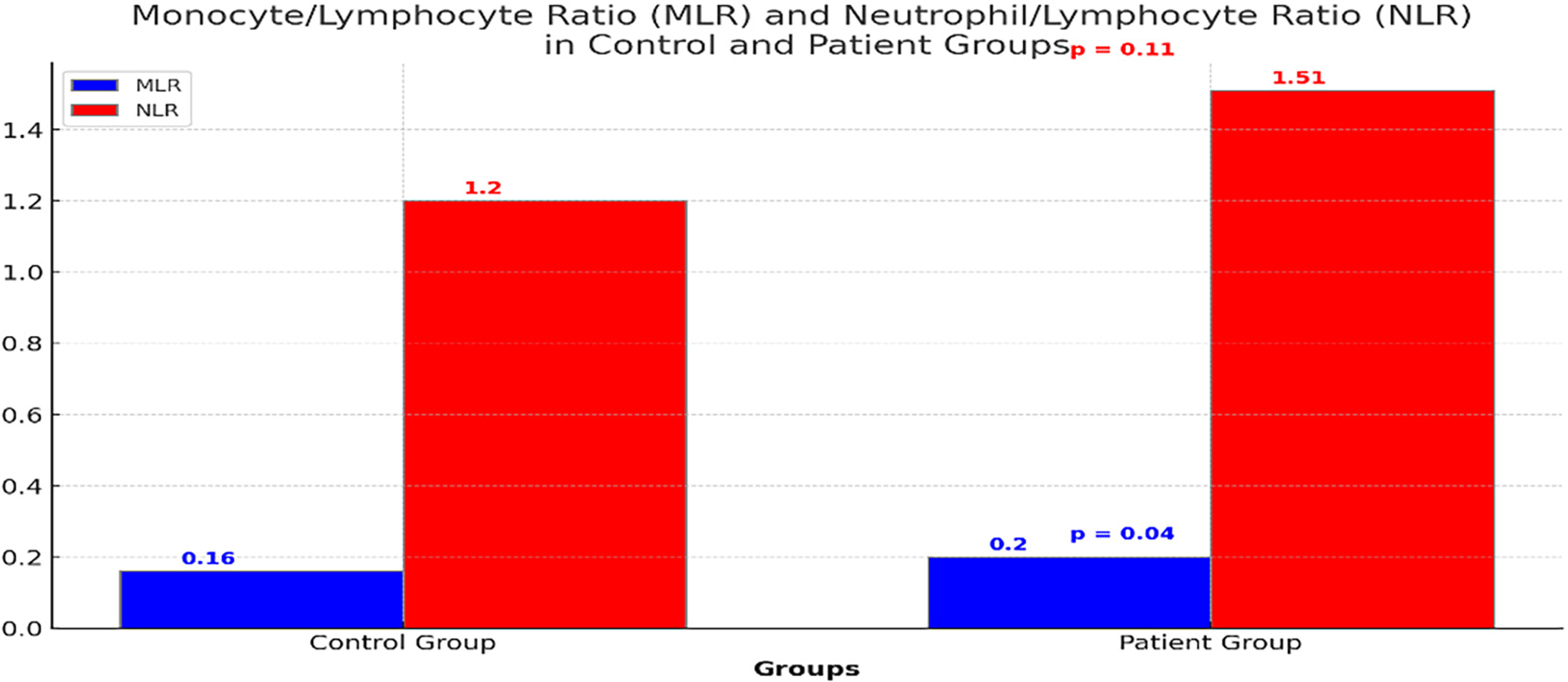 Comparison of MLR and NLR between the control and SVT patient groups. The p-value for MLR is 0.04, indicating a significant difference, whereas the p-value for NLR is 0.11, indicating no significant difference.
