 Allergy and Ischemia: A Case Report of Type II Kounis Syndrome
Keywords
Kounis Syndrome; Acute Coronary Syndrome; Anaphylaxis
Keywords
Kounis Syndrome; Acute Coronary Syndrome; Anaphylaxis
Introduction
Kounis Syndrome (KS), also known as "allergic angina," is a rare condition in which an allergic reaction triggers acute coronary events.1 Its mechanism involves mast cell degranulation, releasing inflammatory mediators such as histamine, cytokines, and proteases. These substances can induce coronary artery spasm, plaque rupture, or thrombus formation.2 KS is classified into three types: Type I (occurring in patients without coronary artery disease, CAD), Type II (in patients with pre-existing CAD), and Type III (associated with stent thrombosis). In this report, we present a case of Type II KS.3
Case report
A 56-year-old male presented to the Emergency Department with epigastric pain radiating to the back, associated with nausea and vomiting for two days. His medical history included hypertension, diabetes mellitus, dyslipidemia, and active smoking.
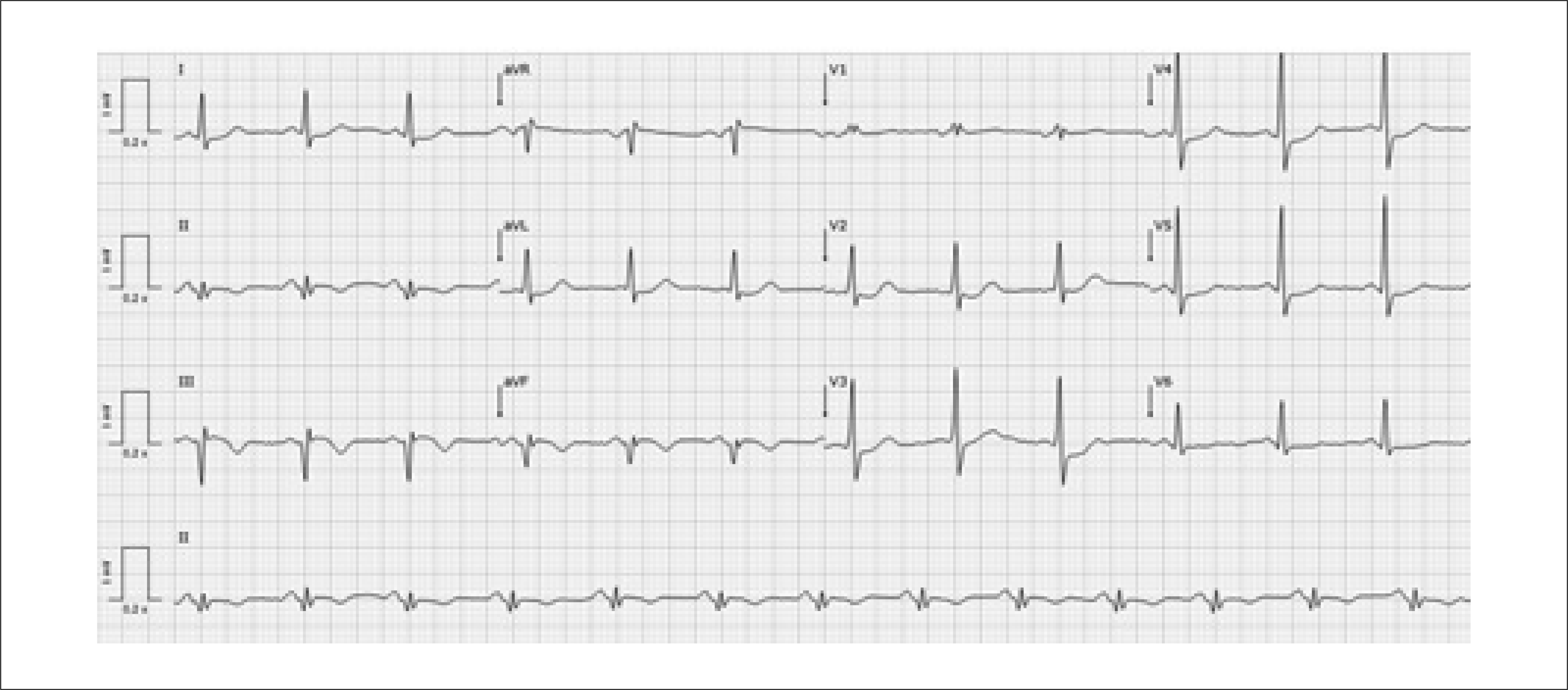
On arrival, the patient was asymptomatic, and physical examination was unremarkable. The admission electrocardiogram (ECG) demonstrated sinus rhythm with Q waves in the inferior leads (Figure 1). Transthoracic echocardiography (TTE) revealed hypokinesia of the mid-basal segments of the inferior and posterior walls, with a mildly reduced ejection fraction (estimated at 45–50%). A subacute inferior myocardial infarction was suspected, and the patient was admitted for coronary angiography during hospitalization.
On the first day of hospitalization, the patient developed fever without associated symptoms, prompting a diagnosis workup. Empirical treatment with amoxicillin/clavulanic acid was initiated. During antibiotic administration, the patient experienced clinical features consistent with anaphylactic shock, including hypotension, skin rash, and altered consciousness, which progressed to cardiorespiratory arrest. Circulation was spontaneously restored after approximately 10 minutes of advanced life support, together with administration of clemastine and corticosteroids.
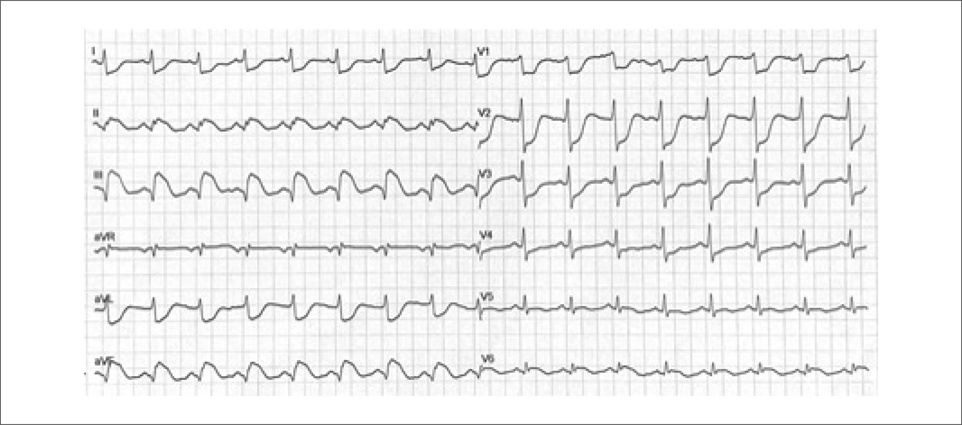
In this context, the ECG showed ST-segment elevation of approximately 5 mm in the inferior leads, with marked depression in leads I, aVL, and V1-V4 (Figure 2). A diagnosis of Type II Kounis Syndrome was considered, and emergent coronary angiography revealed a sub-occlusive lesion in the mid-right coronary artery with TIMI 3 flow. Primary angioplasty was performed with the placement of a drug-eluting stent.
Laboratory tests showed a peak high-sensitivity troponin level of 136,000 ng/mL. The complete blood count revealed leukocytosis (25,000/μL) with neutrophilia (85% of leukocytes, corresponding to 21,000/μL); eosinophils accounted for 0.1%. C-reactive protein (CRP) was elevated at 15 mg/dL, and procalcitonin was 3.43 ng/mL Tryptase, histamine, total IgE, and complement levels were not assessed.
A subsequent thoracic computed tomography revealed subsegmental ground-glass opacities in the lung parenchyma, with a "crazy-paving" pattern and interlobular septal thickening, suggesting of infectious process. Infectious screening identified Haemophilus influenzae in bronchial secretions, and targeted antibiotic therapy with levofloxacin was initiated.
A follow-up echocardiogram demonstrated preserved systolic function with hypokinesia of the mid-basal segments of the inferior wall, without pericardial effusion or significant valvular disease.
The patient showed favorable clinical progress and was discharged after sixteen days of hospitalization, with follow-up appointments scheduled in Cardiology and Pulmonology.
Discussion
KS, also referred to as "allergic angina" or "allergic myocardial infarction," was first described in 1991. This rare but increasingly recognized condition involves acute coronary events precipitated by allergic reactions.1 Epidemiological data on KS remain limited. However, according to Desai et al.4 in a cohort of 235,420 patients primarily hospitalized for allergy, hypersensitivity, or anaphylactic reactions, 2,616 patients experienced acute coronary syndrome (ACS) and were identified as having KS, representing approximately 1.1% of the studied population.
According to Cepeda et al.,5 a thorough clinical history is essential to establish a cause-effect relationship between the allergic trigger and the cardiac event, including investigation of any prior allergic conditions the patient may have.
The exact mechanism underlying KS remains unclear. However, it is thought to involve mast cell degranulation, which releases inflammatory mediators. Mast cells can activate macrophages and enhance T-cell activity, thereby promoting a cascade of inflammatory responses. During hypersensitivity reactions, mast cell degranulation releases mediators both locally and systemically, including histamine, chemokines, neutral proteases (such as chymase and tryptase), cytokines, and arachidonic acid derivatives (e.g., leukotrienes, thromboxane, prostacyclin, and tumor necrosis factor). Many of these compounds exert significant cardiovascular effects; for example, histamine can induce coronary vasoconstriction, platelet activation, and tissue factor expression. Other mediators may weaken arterial plaques by degrading the collagen cap, potentially leading to plaque rupture.2
Most of the available information on KS comes from case reports, which describe a wide range of triggers, including environmental exposures, medical conditions and medications.3 KS is classified into three subtypes:
Type I: Occurs in patients without CAD. Acute chest pain develops during an allergic reaction in individuals without coronary risk factors or lesions, likely due to coronary artery spasm triggered by mast cell mediator release. Typically, the allergic response results in coronary artery spasms without elevation of troponin or other cardiac biomarkers.
Type II: Occurs in patients with pre-existing CAD. In this subtype, allergic mediator release can destabilize an existing plaque, potentially leading to myocardial infarction.3 As Constantinides notes, even minor allergic reactions may disrupt arterial plaques.6
Type III: Associated with delayed stent thrombosis and characterized by the presence of mast cells and eosinophils within the thrombus.3
A review study by Abdelghany et al.7 summarized 175 published cases of KS and reported type I as the most common subtype followed by types II and III. Similarly, Seung et al.8 demonstrated that Type I, Type II, and Type III occur in approximately 72.6%, 22.3%, and 5.1% of cases, respectively.
In the present case, Type II KS was diagnosed, given the occurrence of an allergic reaction in association with acute coronary syndrome and documented coronary artery disease (sub-occlusive lesion in the mid-right coronary artery).
Clinical manifestations include chest pain resembling acute coronary syndrome, accompanied by allergic reactions ranging from mild to severe. Although no single laboratory test can definitively diagnose KS, investigations such as complete blood count (including eosinophil levels), cardiac biomarkers, troponin, C-reactive protein (CRP), histamine, and immunoglobulin E may help support the diagnosis.8
It is crucial to differentiate KS from other conditions, including obstructive coronary disease, unstable angina, hypersensitivity myocarditis, acute pericarditis, eosinophilic coronary periarteritis, and esophageal spasm.8
In this patient, the diagnosis was based on a confirmed allergic reaction to a specific drug, accompanied by elevated cardiac biomarkers, ECG and echocardiographic changes, and visible coronary artery spasm on angiography. Unfortunately, tryptase, histamine, total IgE, and complement levels were not assessed.
Currently, there are no established guidelines for the management of KS.3 The primary goal in treating type II KS is to control the allergic reaction while ensuring adequate myocardial revascularization.8 Management becomes particularly challenging in severe allergic reactions, as medications used for ACS and allergy may have contraindications when administered together. This is especially relevant for adrenaline, the first-line treatment for anaphylaxis, which may exacerbate ischemia, prolong the QT interval, and induce vasospasm or arrhythmias in the context of ACS. Although corticosteroids may delay myocardial healing after infarction, they are generally considered relatively safe, though further research is warranted.3–9
In this case, the in-hospital presentation enabled rapid assessment by a cardiologist and activation of the hemodynamic team, contributing to a favorable outcome through prompt diagnosis and treatment. Identifying KS cases is crucial, as recognizing and managing the allergic component – whether by desensitization or trigger avoidance – can prevent future events.
Conclusion
This case illustrates a Type II presentation of KS, a rare medical condition observed in clinical practice, occurring in a middle-aged male. The number of reported KS cases has increased since the syndrome was first described, although determining its true incidence remains challenging. Maintaining a high index of suspicion is essential in managing this fragile and potentially life-threatening disorder. The rising prevalence of cases underscores the need for heightened awareness of KS to prevent complications, particularly given the significant prognostic implications of acute coronary syndrome. While the exact mechanisms of KS are not yet fully understood, treatment should focus on both the hypersensitivity reaction and the coronary event, guided by the findings of coronary angiography.
-
Sources of Funding
There were no external funding sources for this study.
-
Study Association
This study is not associated with any thesis or dissertation work.
-
Ethics Approval and Consent to Participate
This study was approved by the Ethics Committee of the Unidade Local de Saúde do Alentejo Central under the protocol number 056/26. All the procedures in this study were in accordance with the 1975 Helsinki Declaration, updated in 2013. Informed consent was obtained from all participants included in the study.
-
Use of Artificial Intelligence
During the preparation of this work, the author(s) used ChatGPT to support textual revision, contributing to the improvement of the work's clarity, coherence, and linguistic accuracy. After using this tool/service, the author(s) reviewed and edited the content as needed and take full responsibility for the content of the published article.
Availability of Research Data
The underlying content of the research text is contained within the manuscript.
References
- 1 Kounis NG, Zavras GM. Histamine-Induced Coronary Artery Spasm: The Concept of Allergic Angina. Br J Clin Pract. 1991;45(2):121-8.
-
2 Kounis NG. Coronary Hypersensitivity Disorder: The Kounis Syndrome. Clin Ther. 2013;35(5):563-71. doi: 10.1016/j.clinthera.2013.02.022.
» https://doi.org/10.1016/j.clinthera.2013.02.022 -
3 Marinheiro R, Amador P, Semedo F, Sá C, Duarte T, Gonçalves S, et al. In-Hospital Acute Myocardial Infarction: A Case of Type II Kounis Syndrome. Rev Port Cardiol. 2017;36(5):391.e1-391.e5. doi: 10.1016/j.repc.2016.07.012.
» https://doi.org/10.1016/j.repc.2016.07.012 -
4 Desai R, Parekh T, Patel U, Fong HK, Samani S, Patel C, et al. Epidemiology of Acute Coronary Syndrome Co-Existent with Allergic/Hypersensitivity/Anaphylactic Reactions (Kounis Syndrome) in the United States: A Nationwide Inpatient Analysis. Int J Cardiol. 2019;292:35-38. doi: 10.1016/j.ijcard.2019.06.002.
» https://doi.org/10.1016/j.ijcard.2019.06.002 -
5 Cepeda PR, Herrejón EP, Aguirregabiria MMR. Kounis Syndrome. Med Intensiva. 2012;36(5):358-64. doi: 10.1016/j.medin.2011.10.008.
» https://doi.org/10.1016/j.medin.2011.10.008 -
6 Constantinides P. Infiltrates of Activated Mast Cells at the Site of Coronary Atheromatous Erosion or Rupture in Myocardial Infarction. Circulation. 1995;92(5):1083. doi: 10.1161/01.cir.92.5.1083.
» https://doi.org/10.1161/01.cir.92.5.1083 -
7 Abdelghany M, Subedi R, Shah S, Kozman H. Kounis Syndrome: A Review Article on Epidemiology, Diagnostic Findings, Management and Complications of Allergic Acute Coronary Syndrome. Int J Cardiol. 2017;232:1-4. doi: 10.1016/j.ijcard.2017.01.124.
» https://doi.org/10.1016/j.ijcard.2017.01.124 -
8 Seung J, Choo EH. Acute Coronary Syndrome Associated with Allergic Reaction: Kounis Syndrome. J Cardiovasc Interv. 2024;3(4):227-32. doi: 10.54912/jci.2024.0010.
» https://doi.org/10.54912/jci.2024.0010 -
9 Almegbel M, Alshamardl K, Aleshaiwi L, Almutairi F. Kounis Syndrome: A Case Report and a Review of Recent Literature. Cureus. 2024;16(7):e64627. doi: 10.7759/cureus.64627.
» https://doi.org/10.7759/cureus.64627
Edited by
-
Editor responsible for the review:
Glaucia Maria Moraes de Oliveira