Differential Expression of SERCA and Myosin Heavy Chain Isoforms in Rat Laryngeal Muscles
Abstract
Introduction Intrinsic laryngeal muscles (ILMs) play a fundamental role in airway protection, respiration, and phonation.
Objective Knowledge of the distribution and roles of different sarcoplasmic reticulum Ca2+ ATPase (SERCA) and myosin heavy chain (MHC) isoforms in ILMs is essential for understanding their functions and associated pathophysiological changes.
Methods We immunohistochemically characterized SERCA and MHC isoform expression and co-expression patterns in five ILMs of young, aged, and denervated rats.
Results ILMs exhibited the predominant expression of fast fibers that would be important in airway protection. ILMs showed spatially different percentages of muscle fibers and SERCA2. Slow fibers were higher in medial thyroarytenoid (TA) muscles than lateral TA. Significant increases were observed in slow and hybrid fibers with aging. SERCA2 noticeably diminished after denervation, pointing to an increased vulnerability of the denervated muscle to fatigue. A compensatory increase in SERCA2 and hybrid fibers was observed in contralateral muscles likely reflecting adaptation to increased mechanical load.
Conclusion These results indicate that ILMs, like limb muscle fibers, are subject to neural regulation. These findings may significantly advance the understanding of the Ca2+ handling system in ILMs and its role in unique laryngeal muscle functions.
Keywords
Laryngeal; Fiber; Myosin; sarcoplasmic; denervated; aging
Introduction
It is widely acknowledged that intrinsic laryngeal muscles (ILMs) play an essential role in vital functions such as respiration, airway protection, and phonation. Laryngeal muscles are commonly categorized into adductors, abductors, and tensors.1 Each ILM performs distinct roles and has unique properties that enable rapid, sustained vocal fold (VF) movements under various conditions.
ILMs have attracted significant research interest owing to their differential pathophysiological involvement in various neuromuscular diseases. ILMs are involved early in myasthenia gravis, amyotrophic lateral sclerosis, and mitochondrial myopathy.2 However, they are spared in Duchenne muscular dystrophy, with the exception of the cricothyroid muscle.3 Ferretti et al. (2015)4 observed that, compared with limb muscles, rat ILMs express higher levels of calcium-buffering proteins, which enhance their ability to handle calcium fluctuations and explain their protection from muscle dystrophy damage. Calcium (Ca2+) is the primary regulatory and signaling molecule in muscle. Alterations in the Ca2+ handling system (Ca2+ release, storage, uptake, transport, and intracellular calcium) have been implicated in many pathological processes.5
Sarcoplasmic reticulum Ca2+ ATPase (SERCA) membrane proteins play central roles in calcium regulation. SERCA proteins have a dual function: to induce and maintain muscle relaxation by pumping calcium from the sarcoplasm into the luminal spaces of the sarcoplasmic reticulum (SR), thereby lowering sarcoplasmic Ca2+ concentration; at the same time, they restore the SR luminal calcium reservoir necessary for muscle contraction and for bringing calcium levels back to baseline following release. The major proteins that participate in contraction and relaxation in skeletal muscle are the SERCA and myosin heavy chain (MHC) proteins. The former plays a key role in the muscle contraction/relaxation cycle,6 while the latter regulates the contraction force and contraction velocity of muscle fibers.7 Also, the difference between fast and slow muscle fibers in contraction and relaxation times is linked to the expression of different SERCA isoforms (SERCA1 and SERCA2) and/or MHC isoforms (MHC I and MHC II). More specifically, fast fibers, expressing MHCII, mostly co-express SERCA1, while slow fibers, expressing MHCI, mostly co-express the SERCA2 isoform.8
Despite the fundamental roles of these proteins in ILM function, studies of SERCA isoform expression and MHC composition across various ILMs are limited. A better understanding of the distribution and roles of SERCA and MHC isoforms in the larynx is necessary to elucidated the functions of the laryngeal muscle complex and associated pathophysiological changes. To address this gap, immunohistochemical immunolocalization of SERCA and MHC isoforms (SERCA1, SERCA2, MHC I, and MHC II) and their co-expression in the five ILMs were investigated in young and aged rats. Special attention was placed on the effects of denervation on ILMs.
Methods
Animals
Male adult Wistar rats aged 8–12 weeks and weighing 280–350 g (N = 10), were used as young rats. Additionally, 24-month-old rats (N = 10) were used as aged models.
Animal Tissue Preparation
Animals were anesthetized with an intraperitoneal injection of sodium pentobarbital (30–60 mg/kg) and perfused intracardially with 0.01 M phosphate-buffered saline (PBS), followed by fixation with 4% paraformaldehyde. The larynges were immediately excised and immersed in the same fixative for approximately 12 hours at 4°C. Tissues were processed in paraffin and sectioned with a microtome (8-10 μm).
The intrinsic laryngeal muscles studied were: 1) medial and lateral thyroarytenoid (MTA, LTA), 2) lateral cricoarytenoid (LCA), 3) superior cricoarytenoid (SCA), 4) posterior cricoarytenoid (PCA), and 5) cricothyroid (CT).
Previous studies of laryngeal muscles have confirmed that the TA, CT, LCA, and PCA are similarly located in humans and rats. However, the rat larynx also contains another muscle that courses posteriorly and medially from the lateral face of the arytenoid to the midline tubercle of the cricoid lamina, previously termed SCA. The SCA muscle may function to draw the arytenoids toward one another at the midline, similar to the function of the human interarytenoid (IA).9
Immunohistochemistry (IHC)
Fast fiber-type identification on frozen sections was unsuccessful, so we switched to paraffin sections. Deparaffinized sections were incubated with 3% H2O2 in PBS for 10 minutes, followed by microwave treatment (5 minutes × 3 times at 500 watts in citrate buffer, pH 6). Sections were then incubated in blocking solution (0.3 M glycine, 50 mM ammonium chloride, and 1% BSA in PBS) for 30 minutes, followed by incubation with primary antibodies. Sections were incubated with anti-SERCA1 or -SERCA2 and anti-MHC I or -MHC II (Table 1) antibodies for 1-2 days at 4 °C, then washed and incubated with Alexa Fluor® 488 donkey anti-mouse IgG and Alexa Fluor® 594 donkey anti-goat IgG secondary Abs for 1 hour at room temperature. Double IHC was performed using (anti-SERCA1 + anti-SERCA2) and (anti-SERCA2 + anti-MHCII) combinations to assess co-expression. In double IHC, the same steps were followed, using a mixture of primary or secondary antibodies at the same dilutions.
Immunostained tissue sections were examined with a FV-1000 laser confocal microscope (Olympus, Tokyo, Japan). ImageJ 1.46 was used to manually count and calculate the percentage of positive cells for each antibody.
Immunohistochemistry for the Denervated Rat
Aged rats (8-12 weeks old) were anesthetized with sodium pentobarbital, and their larynx was surgically exposed via a midline incision under an operating microscope. We identified and then denervated the right side of the larynx by resecting a 1-cm segment of the recurrent laryngeal nerve (RLN) (ligating both ends) and transecting the superior laryngeal nerve (SLN). Animals were euthanized at 2-, 4-, 8-, and 12-week post-denervation. The persistence of a gap between the nerve ends confirmed that no reinnervation had occurred. The larynges were collected and processed for immunohistochemistry (IHC) as described previously (n = 5 per group).
MHC Subtypes Using Multi-Color Fiber Typing
Initially, frozen sections were attempted. After trialing several techniques to reduce autofluorescence and background noise, adequate staining could not be achieved. We then discontinued MHC subtyping and switched to paraffin-embedded blocks.
Statistical Analysis
The relative SERCA and MHC composition for each muscle was calculated. Data on the percentage of positive cells for each antibody were analyzed using SPSS Statistics, version 16 (IBM Corp., Armonk, NY, USA) . Statistical comparisons were performed using the unpaired two-tailed Student's t-test and were considered significant at p < 0.05.
One-way analysis of variance (ANOVA) with Scheffe's post hoc test was used to compare MHC and SERCA composition between denervated and contralateral sides and between young and aged rats. Values are presented as means ± SD across rats.
Results
Young Rats
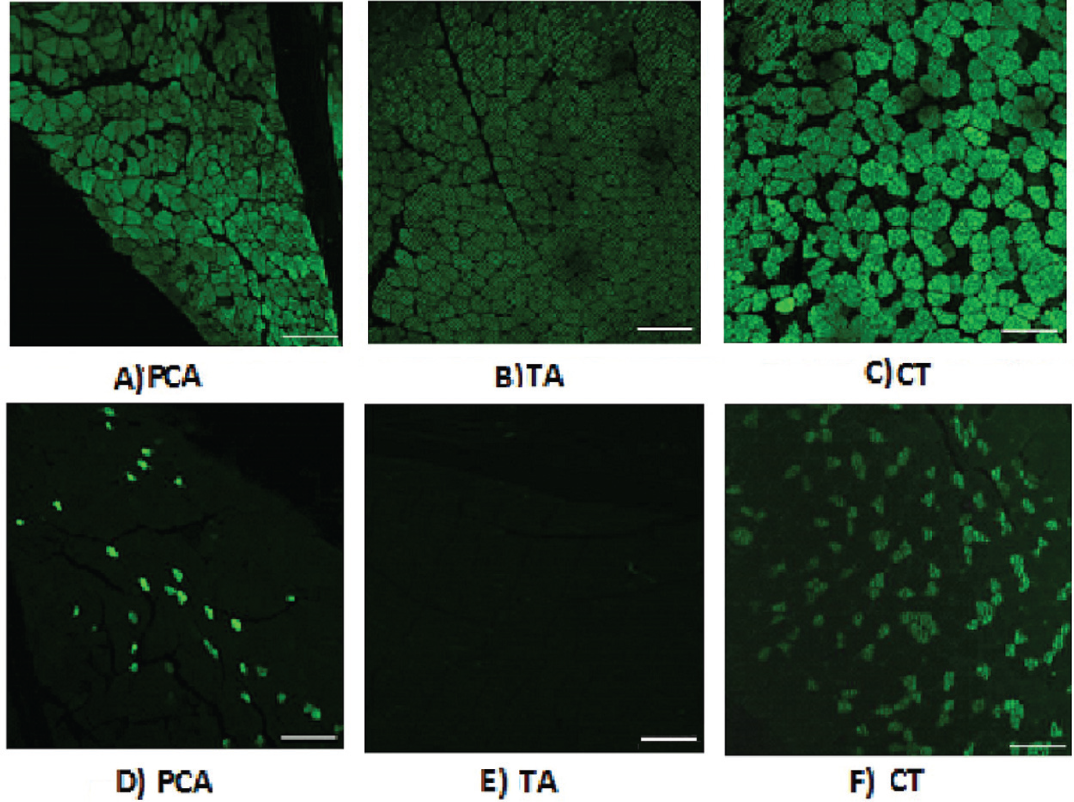
SERCA and MHC isoform expression was analyzed in individual ILMs, and the percentage of positive cells for each antibody was calculated and presented in Figure 1. Fast fibers (SERCA1- and MHCII-positive) were generally predominant in all ILMs (Figs. 1 and 2). More specifically, the highest SERCA1 expression was observed in the TA (100%), followed by the SCA (98.9 ± 1.0%), LCA (94.3 ± 1.0%), and PCA (86.6 ± 2.1%); the lowest was in the CT (75.5 ± 3.7%). The distribution and values of MHC II isoforms were similar to those of their SERCA1 counterparts. In other words, the MHCII percentages were 100, 99.4 ± 0.5, 95.1 ± 0.5, 89.3 ± 1.9, and 75.2 ± 3.3 in TA, SCA, LCA, PCA, and CT. On the other hand, significant variations in expression were observed between the slow fiber isoforms (i.e., SERCA2 and MHCI). Moreover, the SERCA2 isoform was expressed in all ILMs, whereas the MHCI isoform was not expressed in MTA and LTA (Fig. 2). SERCA2 isoforms showed higher percentages than MHCI isoforms. Additionally, substantial mismatches between SERCA2 and MHCI expression were observed: PCA (32.6 ± 1.6% vs. 10.7 ± 1.5%), LCA (28.7 ± 3.1% vs. 5.4 ± 1.0%), SCA (32.6 ± 3.8% vs. 0.7 ± 0.6%), and CT (29.5 ± 3.3% vs. 25.4 ± 3.4%), respectively. Interestingly, a spatial difference in SERCA2 expression was observed within the TA, with the MTA showing higher expression (18.9 ± 4.1%) than the LTA (7.8 ± 1.8%), suggesting that the MTA has a slower SERCA isoform profile than the LTA.

Effect of aging on SERCA and MHC isoform expression in intrinsic laryngeal muscles. C Charts show the percentages of SERCA1-, SERCA2-, and co-expression-positive fibers in young and aged rats in the posterior cricoarytenoid (PCA), lateral cricoarytenoid (LCA), superior cricoarytenoid (SCA), and cricothyroid (CT).

Immunohistochemical detection of MHCI and MHCII expression in intrinsic laryngeal muscle fibers reveals spatially distinct patterns. (A-C) MHCII (fast fiber) expression in young rats was immunohistochemically evaluated in (A) the Posterior Cricoarytenoid (PCA), (B) the thyroarytenoid (TA), and (C) the cricothyroid (CT) muscles. MHCII fast fibers were predominantly expressed by TA, but not by CT. Scale bars = 100 μm (×200). (D–F) MHCI (slow fiber) expression in young rats was immunohistochemically evaluated in (D) the posterior cricoarytenoid (PCA), (E) the thyroarytenoid (TA), and (F) the cricothyroid (CT). MHCI slow fibers were highly expressed in the CT, whereas the PCA and TA showed lower expression. Scale bars = 100 μm (x 200).
Hybrid intermediate fibers, especially SERCA1 + SERCA2, were detected in all ILM muscles. Figure 1 shows the expression percentages of the co-expression fiber (MHCII+ SERCA2) as 26.2 ± 4.8, 24.8 ± 2.2, and 7.8 ± 1.9 in LCA, PCA, and CT, respectively.
Aging-Related Changes in ILMs
Results from aged rats were used to examine the effects of age on SERCA and MHC isoform expression in the laryngeal muscles. A significant increase in slow fibers (SERCA2), particularly in MTA, PCA, and SCA, was observed with aging (Fig. 1). SERCA2 expression increased from 18.9 ± 4.1%, 32.6 ± 1.6%, and 32.6 ± 3.8% in the MTA, PCA, and SCA of young rats to 30.0 ± 4.1%, 40.8 ± 4.6%, and 41.8 ± 3.1% in aged rats, respectively. There was also a noticeable increase in hybrid fibers with aging in the PCA, MTA, and CT. Generally, there was an increase in fast fiber isoforms (SERCA1 and MHCII) in ILMs, especially in CT, where their expression was significantly increased.
Denervation Effects
Immunolocalization of SERCA and MHC isoforms, as well as their co-expression in the five ILMs on the denervated side, was examined and compared with the contralateral side in each group at 2, 4, 8, and 12 weeks.
Denervation is confirmed by the presence of obvious histological changes, including muscle atrophy and decreased fiber diameter on the affected side. On the PCA-denervated side, SERCA1 and MHCII increased, while SERCA2 and hybrid fiber decreased at weeks 2 and 4. By contrast, the PCA of the normal contralateral side showed compensatory increases in SERCA1, MHCII, SERCA2, and hybrid fiber (Figs. 3, 4, and 5). Sacrificed rats at weeks 8 and 12 maintained increases in SERCA1 and MHCII, with a subsequent rise in SERCA2 and hybrid fibers, whereas MHCI decreased on the denervated side. However, in the contralateral muscle, SERCA1 decreased toward normal; MHCI remained decreased, while MHCII, SERCA2, and hybrid fiber were increased.

Effect of denervation on SERCA and MHC isoform expression in affected and non-affected contralateral PCA muscle.

Effect of denervation on SERCA and MHC isoform expression in affected and non-affected contralateral PCA muscle after 2 weeks. Expression of MHCII (green) and SERCA2 (red) in PCA was evaluated in 2weeks after denervation. Micrographs depict the non-treated contralateral side (A-C) and the denervated side (D-F). Scale bars = 100 μm (×200).

Effect of denervation on muscle fiber expression in PCA. Expression of SERCA1 (green) and SERCA2 (red) in PCA was evaluated 12 weeks after denervation. Micrographs depict the non-treated contralateral side (A-C) and the denervated side (D-F). Scale bars = 100 μm (×200).
Denervation-related changes in CT at weeks 2 and 4 increased SERCA1, MHCII, and hybrid fiber, while decreasing SERCA2. In contrast, the normal muscle side showed compensatory increases in SERCA1, MHCII, and hybrid fiber. At 8 and 12 weeks, SERCA1, MHCII, and hybrid fiber increased on the denervated side, and SERCA1, MHCII, and hybrid fiber returned to normal values on the contralateral side (Figs. 6 and 7).


Effect of denervation on SERCA and MHC isoform expression in CT muscle, demonstrated by immunohistochemistry. Expression of SERCA1 (green) and SERCA2 (red) in CT was evaluated 2 weeks after denervation. Micrographs depict the non-treated contralateral side (A-C) and the denervated side (D-F). Scale bars = 100 μm (×100).
Regarding the medial and lateral TA, SERCA2 expression decreased at 2 and 4 weeks of denervation, then increased at 8 to 12 weeks. The contralateral side showed a compensatory increase at 2 to 12 weeks of denervation. In the denervated LCA, SERCA1 and MHCII increased, and on the contralateral side, SERCA2 and hybrid fiber increased.
Discussion
Althogh laryngeal muscles are known to express a mix of MHC isoforms, the present study provides the first detailed characterization of their corresponding SERCA isoform profiles. The fundamental finding of this study is that ILMs can be classified into three fiber types based on SERCA and MHC isoform expression: (1) fast fibers (SERCA1 and MHCII) optimized for high-speed, high-power bursts but lack endurance; (2) slow fibers (MHCI and SERCA2) that are fatigue-resistant but generate lower power; and (3) hybrid intermediate fibers (MHCII + SERCA1 + SERCA2) enabling functional flexibility. All ILMs predominantly express fast fibers, consistent with their functional demands for high twitch velocities during phonation, respiration, and swallowing, while maintaining fatigue resistance. These findings align with previous MHC-based fiber typing studies.8,11–15
Notably, slow fiber expression patterns differ between isoforms: SERCA2 is present in all ILMs, whereas MHCI is absent in the transverse and lateral thyroarytenoid (MTA and LTA), corroborating earlier observations.1,4 Moreover, SERCA2 expression ratios substantially exceeded those of MHCI. The presence of hybrid fibers expressing both fast and slow isoforms within single muscles likely enables precise fine-tuning of contraction and relaxation while preserving fatigue resistance-a functional requirement for both adductor and abductor muscles.1,4
Principal component analysis revealed that the posterior cricoarytenoid (PCA) exhibits a slow, fatigue-resistant profile (MHCI 10%, SERCA2 32.6 ± 1.6%, hybrid 25.5 ± 2.1%), reflecting its continuous activity modulating glottal opening during respiration. Conversely, TA displayed the fastest MHC profile, consistent with its very short contraction times.8 The absence of MHCI in rodent TA corroborates previous findings in small mammals13–16 and contrasts with human TA, where MHCI expression supports sustained phonatory contraction.1,2 In fact, some researchers have linked the nearly universal expression of fast isoforms in these animals to the muscle's role as a glottal adductor.17
Regional differences within the TA revealed that the medial portion (MTA) has a slower SERCA isoform profile than the lateral portion (LTA), consistent with studies across species.1,8 This organization is functionally advantageous: MTA sustains contractions for voice production while LTA participates in rapid reflex laryngeal closure for airway protection.8
Some authors have suggested that the SCA muscle may function as a posterior adductor, similar to the IA in humans, which is involved in fine-tuning phonation. Studies of the contractile properties of the IA have been limited. However, a previous study of the human IA showed that its MHC composition was similar to that of the LCA partner VF adductor.19 Similarly, the current study affirms that the SCA and LCA showed similar SERCA and MHC expression.
The CT displayed the slowest myosin profile among the laryngeal muscles, consistent with its "limb-like" fiber-type composition observed in previous studies.1,9–20 This slower, more fatigue-resistant phenotype aligns with its physiological role in tonic activation during deep breathing, which widens the laryngeal airway. The unique contractile properties of the CT, which distinguish it from the faster adductor muscles, likely stem from differences in its embryonic development or innervation pattern, leading to distinct responses to neuromuscular disease.3
Collectively, these findings confirm that laryngeal muscles represent a distinct skeletal muscle allotype, combining among the fastest contraction speeds with exceptional fatigue resistance -properties critical for airway protection .8
Aging-Related Changes
Previous studies have reported age-related decreases in total muscle mass and fiber number in laryngeal muscles, as well as alterations in MHC isoforms, changes in neuromuscular junction morphology, and reduced electromyographic activity.21 Some authors pointed to a loss of Type I and II fibers, while others suggested a loss of Type I fibers.22
Aging produced muscle-specific alterations in isoform expression. The present results show maintenance or an increase in SERCA1 expression in the LCA and CT, while the PCA, SCA, and MTA showed increased SERCA2 expression, with the PCA becoming slower (increased SERCA2 and hybrid fibers)-consistent with reported prolongation of adduction-abduction periods in aging.21−23 The PCA's unique continuous activity throughout life may explain its earlier susceptibility to aging-related changes compared to other muscles.23 Suzuki et al. (2002) reported that adductor muscle function is more critically impaired than abductor muscle function in older people.17
Potential mechanisms for these age-related changes include denervation-reinnervation cycles, supported by morphological evidence of distal axonal degeneration and end-plate alterations,24,25 as well as fiber-type grouping. The adoption of denervated fibers by neighboring motor neurons would cause reinnervated fibers to assume the donor neuron's profile, explaining the shift toward slower fiber types with aging. This neural influence on fiber-type determination was demonstrated by Buller et al.26 and subsequently confirmed by cross-innervation studies.27
Denervation Effects
In 1960, Buller et al.26 proposed that the innervation pattern is the primary determinant of speed-related properties-including time-to-peak tension, maximum shortening velocity, and MHC isoform profile-in skeletal muscle. Previous studies have shown that denervation produces similar effects in limb and laryngeal rodent muscles.28–30
In the present study, denervation produced three distinct patterns of response among the intrinsic laryngeal muscles (ILMs), reflecting their underlying allotypic differences:
The PCA (Highly Neural-Dependent) exhibited the most dramatic shift, with increased SERCA1 and MHCII (2-12 weeks) and a concurrent decrease in SERCA2, hybrid fibers (2-4 weeks), and MHCI (8-12 weeks). This confirms that the PCA's slow, fatigue-resistant phenotype is highly dependent on sustained neural activity. However, the fact that SERCA2 expression, while diminished, was not entirely abolished suggests that it is modulated by, but not absolutely dependent upon, neural input.
In contrast, TA and LCA showed relative insensitivity to denervation. This resilience may reflect their primary role in rapid, reflexive glottic closure for airway protection-a function that may be less dependent on sustained trophic neural influence in rodents, given their limited vocalization demands.28
CT (Limb-Like) responded with increased MHCII and SERCA1, decreased SERCA2 and hybrid fibers, and no change in MHCI. This profile is consistent with its classification as a "limb-like," slower muscle and aligns with previous reports of its myosin composition.31
Interestingly, the effects of aging and denervation on the PCA were not identical. Aging led to a slower profile (increased SERCA2 and hybrid fibers), while denervation pushed it toward a faster profile. This divergence is critical because it suggests that the slow, fatigue-resistant phenotype seen in the aged PCA is not a direct consequence of simple denervation (i.e., a loss of neural input). Rather, it likely results from the more complex process of repeated denervation and incomplete reinnervation cycles over a lifetime, leading to characteristic fiber-type grouping. This interpretation is consistent with the 'wear-and-tear' theory of aging, which posits that the PCA's sustained metabolic demand 30 makes it uniquely vulnerable to such neural remodeling.23
The functional consequences of these molecular shifts are significant. Denervation-induced decreases in SERCA2, for example, would elevate resting myoplasmic calcium concentration ([Ca2+]), slowing relaxation, while a reduced sarcoplasmic reticulum (SR) calcium loading capacity would diminish the calcium available for release, increasing susceptibility to fatigue.32
These muscle-specific responses to denervation have direct clinical correlations with distinct patterns of nerve injury: RLN Section disrupts the intricate balance between the adductors and the PCA, triggering a complex, muscle-specific cascade of fiber-type transformations and atrophy. This cascade underlies glottic insufficiency and places the PCA-given its high neural dependence-at significant risk for synkinetic reinnervation. In contrast, SLN section selectively targets the CT. The shift of its limb-like, slow phenotype toward faster isoforms directly correlates with the clinical deficits in pitch control and vocal fatigue observed after SLN injury.33,34
The fiber-type grouping observed in denervated ILMs, particularly for MHCI and SERCA2, provides further support for the neural regulation of both fast and slow fibers within the laryngeal allotype.17 Interestingly, the increased expression of hybrid SERCA1/SERCA2 fibers in contralateral, uninjured muscles likely reflects compensatory overwork. As the uninjured side compensates to restore vocal fold approximation and normalize function,35 the resulting mechanical overload alone may be sufficient to induce a fast-to-slow fiber-type transformation, consistent with previous studies.36
Collectively, these findings demonstrate that laryngeal muscles are not a homogeneous group but rather a collection of distinct allotypes whose unique molecular profiles are differentially regulated by neural input, mechanical load, and the aging process. Further experimental research is needed to assess the molecular changes induced by different reinnervation techniques to better understand and predict functional recovery after nerve injury.37
Conclusion
This study provides novel data on SERCA and MHC isoform expression and co-expression in young, aged, and denervated intrinsic laryngeal muscles. These data may help predict the ability of ILMs to handle calcium fluctuations in response to functional demands.
Limitations of this Study
Firstly, the findings are derived from a rodent model, and significant species differences in laryngeal muscle composition and function, particularly compared to humans, preclude direct translation. Secondly, the reliance on immunohistochemistry provides a detailed spatial map of protein presence but lacks functional correlates; observed isoform shifts are not linked to direct measurements of contraction speed, force, or calcium kinetics. Consequently, the physiological impact of aging or denervation on actual muscle performance remains inferred. Finally, the absence of complementary techniques, such as electrophysiology or isolated-muscle physiology, means that the functional consequences of the reported molecular changes are not directly validated.
-
Funding
Ministry of Education, Culture, Sports, Science and Technology >Japan Society for the Promotion of Science 15K1082, 25293350, 17H07016, and 19K18814
-
Compliance with Ethical Standards
All procedures involving animals were performed in accordance with the ethical standards of the local Committee for Animal Research No.17H07016.
Data Availability Statement
Data will be available upon request to the corresponding author.
-
Abbreviations
- IL MsIntrinsic laryngeal muscles
- IHC Immunohistochemistry
- VF Vocal fold
- Ca2+ Calcium
- SERCA Sarcoplasmic Reticulum Ca2+ ATPase
- SR Sarcoplasmic Reticulum
- MHC Myosin heavy chain
- MTA Medial thyroarytenoid
- LTA Lateral thyroarytenoid
- LCA Lateral cricoarytenoid
- SCA Superior cricoarytenoid
- PCA Posterior cricoarytenoid
- CT Cricothyroid
- IA Interarytenoid
- RLN Recurrent laryngeal nerve
- SLN Superior laryngeal nerve
Acknowledgments
This work was supported by a Grant-in-Aid for Scientific Research (15K1082, 25293350, 17H07016, and 19K18814).
References
- 1 Simpson CB, Rosen CA. Operative techniques in laryngology. Springer Nature; 2008
-
2 Mao VH, Abaza M, Spiegel JR, et al. Laryngeal myasthenia gravis: report of 40 cases. J Voice 2001;15(01):122–130. Doi: 10.1016/ S0892-1997(01)00012-1
» https://doi.org/10.1016/S0892-1997(01)00012-1 -
3 Marques MJ, Ferretti R, Vomero VU, Minatel E, Neto HS. Intrinsic laryngeal muscles are spared from myonecrosis in the mdx mouse model of Duchenne muscular dystrophy. Muscle Nerve 2007;35 (03):349–353. Doi: 10.1002/mus.20697
» https://doi.org/10.1002/mus.20697 -
4 Ferretti R, Marques MJ, Khurana TS, Santo Neto H. Expression of calcium-buffering proteins in rat intrinsic laryngeal muscles. Physiol Rep 2015;3(06):e12409. Doi: 10.14814/phy2.12409
» https://doi.org/10.14814/phy2.12409 -
5 Chemaly ER, Bobe R, Adnot S, Hajjar RJ, Lipskaia L. Sarco (endo) plasmic reticulum calcium ATPases (SERCA) isoforms in the normal and diseased cardiac, vascular and skeletal muscle. J Cardiovasc Dis Diagn 2013;1(03):1–6. Doi: 10.4172/23299517.1000113
» https://doi.org/10.4172/2329-9517.1000113 -
6 Periasamy M, Kalyanasundaram A. SERCA pump isoforms: their role in calcium transport and disease. Muscle Nerve 2007;35(04): 430–442. Doi: 10.1002/mus.20745
» https://doi.org/10.1002/mus.20745 -
7 Periasamy M, Kalyanasundaram A. SERCA pump isoforms: their role in calcium transport and disease. Muscle Nerve 2007;35(04): 430–442. Doi: 10.1007/s004240100700
» https://doi.org/10.1007/s004240100700 -
8 Hoh JF. Laryngeal muscle fibre types. Acta Physiol Scand 2005;183 (02):133–149. Doi: 10.1111/j.1365-201X.2004.01402.x
» https://doi.org/10.1111/j.1365-201X.2004.01402.x - 9 Fry LT, et al. "Effect of dystrophin deficiency on selected intrinsic laryngeal muscles of the mdx mouse.". 2010 PubMed
- 10 Bloemberg D, Quadrilatero J. Rapid determination of myosin heavy chain expression in rat, mouse, and human skeletal muscle using multicolor immunofluorescence analysis. PLoS One 2012;7 (04):e35273
-
11 Inagi K, Schultz E, Ford CN. An anatomic study of the rat larynx: establishing the rat model for neuromuscular function. Otolaryngol Head Neck Surg 1998;118(01):74–81. Doi: 10.1016/S01945998(98)70378-X
» https://doi.org/10.1016/S0194-5998(98)70378-X - 12 Toniolo L, Macchi V, Porzionato A, et al. Myosin heavy chain isoforms in human laryngeal muscles: an expression study based on gel electrophoresis. Int J Mol Med 2008;22(03):375–379
-
13 Wu YZ, Baker MJ, Crumley RL, Caiozzo VJ. Single-fiber myosin heavy-chain isoform composition of rodent laryngeal muscle: modulation by thyroid hormone. Arch Otolaryngol Head Neck Surg 2000;126(07):874–880. Doi: 10.1001/archotol.126.7.874
» https://doi.org/10.1001/archotol.126.7.874 -
14 Yokoyama T, Nonaka S, Mori S. Histochemical properties of intrinsic laryngeal muscles in cats. J Auton Nerv Syst 1995;56 (1-2):50–60. Doi: 10.1016/0165-1838(95)00064-6
» https://doi.org/10.1016/0165-1838(95)00064-6 -
15 Li ZB, Lehar M, Nakagawa H, Hoh JF, Flint PW. Differential expression of myosin heavy chain isoforms between abductor and adductor muscles in the human larynx. Otolaryngol Head Neck Surg 2004; 130(02):217–222. Doi: 10.1016/j.otohns.2003.09.009
» https://doi.org/10.1016/j.otohns.2003.09.009 -
16 McMullen CA, Andrade FH. Contractile dysfunction and altered metabolic profile of the aging rat thyroarytenoid muscle. J Appl Physiol 2006;100(02):602–608. Doi: 10.1152/japplphysiol.01066.2005
» https://doi.org/10.1152/japplphysiol.01066.2005 -
17 Suzuki T, Connor NP, Lee K, Bless DM, Ford CN, Inagi K. Age-related alterations in myosin heavy chain isoforms in rat intrinsic laryngeal muscles. Ann Otol Rhinol Laryngol 2002;111(11):962–967. Doi: 10.1177/000348940211101102
» https://doi.org/10.1177/000348940211101102 -
18 Hoh JF. Laryngeal muscles are highly specialized organs in airway protection, respiration, and phonation. InHandbook of behavioral neuroscience. 2010 Jan 1 (Vol. 19, pp. 13-21). Elsevier. https://doi.org/10.1016/B978-0-12-374593-4.00002-4
» https://doi.org/10.1016/B978-0-12-374593-4.00002-4 -
19 Shiotani A, Westra WH, Flint PW. Myosin heavy chain composition in human laryngeal muscles. Laryngoscope 1999;109(09): 1521–1524. Doi: 10.1097/00005537-199909000-00030
» https://doi.org/10.1097/00005537-199909000-00030 -
20 Rhee HS, Lucas CA, Hoh JF. Fiber types in rat laryngeal muscles and their transformations after denervation and reinnervation. J Histochem Cytochem 2004;52(05):581–590. Doi: 10.1177/002215540405200503
» https://doi.org/10.1177/002215540405200503 -
21 Lee K, Kletzien H, Connor NP, Schultz E, Chamberlain CS, Bless DM. Effects of aging on thyroarytenoid muscle regeneration. Laryngoscope 2012;122(12):2800–2807. Doi: 10.1002/lary.23589
» https://doi.org/10.1002/lary.23589 -
22 Malmgren LT, Fisher PJ, Bookman LM, Uno T. Age-related changes in muscle fiber types in the human thyroarytenoid muscle: an immunohistochemical and stereological study using confocal laser scanning microscopy. Otolaryngol Head Neck Surg 1999; 121(04):441–451. Doi: 10.1016/S0194-5998(99)70235-4
» https://doi.org/10.1016/S0194-5998(99)70235-4 -
23 Gambino DR, Malmgren LT, Gacek RR. Age-related changes in the neuromuscular junctions in the human posterior cricoarytenoid muscles: a quantitative study. Laryngoscope 1990;100(03): 262–268. Doi: 10.1288/00005537-199003000-00010
» https://doi.org/10.1288/00005537-199003000-00010 -
24 Teig E, Dahl HA, Thorkelsen H. Actomyosin ATPase activity of human laryngeal muscles. Acta Otolaryngol 1978;85(34):272–281. Doi: 10.3109/00016487809121450
» https://doi.org/10.3109/00016487809121450 -
25 McMullen CA, Andrade FH. Functional and morphological evidence of age-related denervation in rat laryngeal muscles. J Gerontol A Biol Sci Med Sci 2009;64(04):435–442. Doi: 10.1093/gerona/gln074
» https://doi.org/10.1093/gerona/gln074 -
26 Buller AJ, Eccles JC, Eccles RM. Interactions between motoneurones and muscles in respect of the characteristic speeds of their responses. J Physiol 1960;150(02):417–439. Doi: 10.1113/jphysiol.1960.sp006395
» https://doi.org/10.1113/jphysiol.1960.sp006395 -
27 Paniello RC, West SE, Lee P. Laryngeal reinnervation with the hypoglossal nerve. I. Physiology, histochemistry, electromyography, and retrograde labeling in a canine model. Ann Otol Rhinol Laryngol 2001;110(06):532–542. Doi: 10.1177/ 000348940111000607
» https://doi.org/10.1177/000348940111000607 -
28 Wu YZ, Baker MJ, Marie JP, Crumley R, Caiozzo VJ. The plasticity of denervated and reinnervated laryngeal muscle: focus on single-fiber myosin heavy-chain isoform expression. Arch Otolaryngol Head Neck Surg 2004;130(09):1070–1082. Doi: 10.1001/archotol.130.9.1070
» https://doi.org/10.1001/archotol.130.9.1070 -
29 Shiotani A, Nakagawa H, Flint PW. Modulation of myosin heavy chains in rat laryngeal muscle. Laryngoscope 2001;111(03): 472–477. Doi: 10.1097/00005537-200103000-00017
» https://doi.org/10.1097/00005537-200103000-00017 - 30 Chai S, Zhang N, Cui C, et al. Systematic review of mitochondrial dysfunction and oxidative stress in aging: A focus on neuromuscular junctions. Neural Regen Res 2026;21(05):1947–1960
-
31 Schulte L, Peters D, Taylor J, Navarro J, Kandarian S. Sarcoplasmic reticulum Ca2+ pump expression in denervated skeletal muscle. Am J Physiol 1994;267(2 Pt 1):C617–C622. Doi: 10.1152/ajpcell.1994.267.2.C617
» https://doi.org/10.1152/ajpcell.1994.267.2.C617 -
32 Liu YH, Qi J, Hou YX, Wang F. Effects of sex hormones on genioglossal muscle contractility and SR Ca2+-ATPase activity in aged rat. Arch Oral Biol 2008;53(04):353–360. Doi: 10.1016/j. archoralbio.2007.10.009
» https://doi.org/10.1016/j.archoralbio.2007.10.009 -
33 Foerster G, Mueller AH. PCA Atrophy and Synkinesis as the Main Factors for Persistent Vocal Fold Immobility in RLN Paralysis. Laryngoscope 2021;131(04):E1244–E1248. Doi: 10.1002/ lary.29195
» https://doi.org/10.1002/lary.29195 -
34 Orestes MI, Chhetri DK. Superior laryngeal nerve injury: effects, clinical findings, prognosis, and management options. Curr Opin Otolaryngol Head Neck Surg 2014;22(06):439–443. Doi: 10.1097/ MOO.0000000000000097
» https://doi.org/10.1097/MOO.0000000000000097 -
35 Shinners MJ, Goding GS, McLoon LK. Effect of recurrent laryngeal nerve section on the laryngeal muscles of adult rabbits. Otolaryngol Head Neck Surg 2006;134(03):413–418. Doi: 10.1016/j. otohns.2005.11.037
» https://doi.org/10.1016/j.otohns.2005.11.037 - 36 Vanmunster M, Rojo-Garcia AV, Pacolet A, et al. Prolonged mechanical muscle loading increases mechanosensor gene and protein levels and causes a moderate fast-to-slow fiber type switch in mice. J Appl Physiol 2023;135(04):918–931
-
37 Guarino P, RussoG, ChiariF, et al. Laryngeal ReinnervationTechniques for Unilateral Vocal Fold Paralysis-Clinical Outcomes and Surgical Approaches: A Systematic Review and Meta-Analysis. J Voice 2025; •••S0892-1997(25)00265-6. Doi: 10.1016/j.jvoice.2025.07.002
» https://doi.org/10.1016/j.jvoice.2025.07.002
Edited by
-
Editor-in-Chief:
Geraldo Pereira Jotz