Oral microbiota dysbiosis in pediatric patients undergoing treatment for acute lymphoid leukemia a preliminary study
Abstract
Acute lymphoblastic leukemia (ALL) stands out as the most prevalent neoplasm during childhood, characterized by the rapid production of abnormal lymphoid cells. Chemotherapy administered to these patients may induce a substantial imbalance in the oral microbiota. A prospective pediatric study encompassing a control group (without ALL) and ALL patients at two treatment stages (pre-induction and consolidation) was conducted. Clinical and laboratory data were meticulously collected. Moreover, DNA from saliva samples was extracted for 16S rRNA sequencing. Clinical data revealed a heightened incidence of oral mucositis during the consolidation phase. Analysis of alpha biodiversity (observed taxa) exhibited a significant reduction in bacterial richness among patients in the consolidation phase. Network analysis identified key taxa during this phase, namely Neisseria flavescens, Prevotella melaninogenica and Porphyromonas. The findings underscore the substantial impact of ALL treatment on the oral microbiota composition, indicating diminished bacterial diversity and an elevated prevalence of oral mucositis.
Keywords:
Acute lymphoblastic leukemia; human microbiome; microbiome; microbial community structure; mouth mucosa; mucositis
Introduction
Acute lymphoblastic leukemia (ALL) is a hematological neoplasm characterized by the exacerbated proliferation of blasts in the bone marrow and mainly affects children aged 2 to 15 years (Béné, 2005; Miranda et al., 2018). Patients with hematological and/or oncological diseases usually present harmful oral manifestations as a result of the intense immunosuppression obtained through chemotherapy treatment. Pathogenic bacterial growth negatively affects the oral mucosa and, thus, the associated immune system (Fattizzo et al., 2021), indicating that a healthy microbiome not only provides nutrition and a natural barrier but also influences the outcome of medical treatment (Blijlevens et al., 2000). Thus, especially in compromised individuals, such as those with hematological malignancies, the oral environment can be assisted throughout the treatment period to promote not only oral health but also general health (Santos and Soares, 2012).
In health, there is an ecological balance between the human host and the microorganisms that colonize mucosal surfaces. Pediatric patients undergo chemotherapy as part of their treatment, which alters the resident microbiota (Oldenburg et al., 2021). The cytotoxic effects of these treatments lead to greater immunosuppression, causing complications (febrile neutropenia and infections), thus bringing changes in the ecosystem that can disrupt the ecological balance of the oral and gastrointestinal microbiota (Rajagopala et al., 2019).
Understanding the dysbiosis pattern in the oral microbiome can help identify specific microbial agents that act as biomarkers in ALL patients. Thus, the aim of this study was to describe the shifts in the oral microbiota in pediatric patients with ALL undergoing antineoplastic therapy.
Subjects and Methods
Study population and oral clinical condition
From September 2020 to December 2021 eight pediatric patients (from 4 to 11 years old) and diagnosed with ALL were admitted for treatment in the oncology sector of the Hospital Pequeno Príncipe (HPP) located in Curitiba, Paraná - Brazil and included in this project. The diagnosis of ALL followed morphological, cytochemical, immunophenotyping and cytogenetic international criteria (Pui et al., 2018), These patients were treated according to the RE-LLA-2005 protocol (Pedrosa and Lins, 2002). The control group (four patients from 4 to 11 years old) attended routine dental physical examination at the dentistry service of HPP and did not present onco-hematological disease. This study was approved by the research ethics committee of HPP (Protocol Number: 3.836.067/2020). Three patients’ groups were organized in the present study:
-
pre-induction: Eight ALL patients in the phase prior to induction (D0 to D5).
-
consolidation: Eight ALL patients in the consolidation phase (D40 to D45).
-
control: Four patients without onco-hematological disease.
Demographic, clinical (medical and dental history) and laboratory data were obtained from HPP’s electronic medical record and by direct interview (Table 1). Microbiota samples and intraoral physical examination was performed. At the same time as the microbiota collection, the results of routine laboratory tests (marker dosages) of the patients were accessed from the internal system. The intraoral physical examination of all groups was performed after acceptance and before the microbioma samples collection.
The assessment of the patient’s oral condition was performed through a unique dentistry and evaluated (i) Dental biofilm: a thin adherent layer formed by bacteria and other substances from saliva, accumulating on teeth and other oral surfaces. (ii) Gingival inflammation: known as gingivitis, results from bacterial plaque on teeth, leading to red, swollen, and sensitive gums with bleeding during brushing (Franceschini et al., 2003). (iii) Mucositis: an inflammation of the mucosa, the moist lining that covers the mouth and gastrointestinal tract. On the other hand, blood leukocyte profile was obtained by leukocytes, neutrophils, lymphocytes, monocytes, and platelets traditional counts (Sonis, 2004).
Collection of biological material
Biological samples for the analysis of the oral microbiota were collected at two different times during the ALL treatment (pre-induction and consolidation). Patients in the control group collected only one time after they accepted to participate in the research. Oral rinse with 5 ml of 0.9% saline solution for 1 minute was collected from each patient and immediately frozen in liquid nitrogen and transferred to a -80 °C freezer at the central laboratory until the time of extraction of bacterial genetic material (Lim et al., 2017).
DNA extraction, sequencing, and bioinformatic analysis
Around 150 mg of oral rinse was sampled for DNA extraction using the ZymoBIOMICS DNA® kit, according to the manufacturer’s instructions. The concentrations and quality of the extracted DNA were measured using a NanoDrop spectrophotometer (Nanodrop Technologies, Wilmington, DE, USA). The integrity of the DNA was also confirmed by electrophoresis in a 1% agarose gel with 1×TAE buffer (Klindworth et al., 2021).
PCR primers (F515/R806) were used to amplify the V4 region of the 16S rRNA gene. PCR was performed at 94 ºC for 3 min to denature the DNA, followed by 28 cycles at 94 ºC for 45 s, 50 ºC for 60 s, and 72 °C for 90 s, with a final extension of 10 min at 72 ºC to ensure complete amplification (Klindworth et al., 2021). The amplicons were quantified with Qubit using HS dsDNA kit (Invitrogen, Carlsbad, CA, USA), diluted to 500 pM, and pooled. Next, 16 pM of pooled DNA was sequenced using MiSeq reagent 500V2 (Illumina, San Diego, CA, USA). Sequencing was performed using a MiSeq® sequencer (Illumina), obtaining paired reads of 250 bp, as previously described (Caporaso et al., 2011). Four samples of (mock microbiota) of ZymoBIOMICS® Microbial Community Standard kit were used as positive control.
Raw reads quality was checked using FastQC and when necessary, reads were trimmed using Trimmomatic (Bolger et al., 2014). Sequencing reads were analyzed with the QIIME2 pipeline (Bolyen et al., 2019). Sequences were sorted according to quality and chimeras were then removed from the dataset using DADA2 algorithm. Following, good quality sequences were clustered into amplicon sequence variant (ASV) using the DADA2 algorithm inserted in the QIIME2 program (Bolger et al., 2014). Furthermore, taxonomic assignment was performed using the SILVA 138 database, release 2020 and the q2-vsearch algorithm of the QIIME2 program (Yilmaz et al., 2013; Callahan et al., 2016). The reads output was normalized to 9,000 per sample, allowing alpha and beta diversity comparisons between groups (Rognes et al., 2016; Bolyen et al., 2019). Detailed information of reads per sample can be found in Table S1 and rarefaction curves (Figure S1).
Pearson correlation and network analysis
Blood leukocyte profile of patients (leukocytes, neutrophils, lymphocytes, monocytes, and platelets) was correlated with the taxa that showed significant differences between the treatments. Pearson’s correlation was calculated and plotted using the corrplot and RColorBrewer packages included in R software, statistically significant results (P<0.05) were highlighted with asterisk in the plots. Additionally, to identify key taxa at the time of consolidation, we constructed a network using Cytoscape program with the strongest positive and negative correlations (between 0.6-1) that were also statistically significant (P<0.05).
Data accessibility
The dataset was submitted to the National Center for Biotechnology Information (NCBI) under the BioSample accession code SAMN25696387.
Statistical analysis
Bacterial taxa abundance comparisons between treatments were conducted in the STAMP software using Welch’s t-test (P<0.05) and Bonferroni’s correction (Caporaso et al., 2011). Alpha diversity analysis (number of features) was compared between collected times using Wilcoxon’s test (P<0.05). Only statistically significant results were reported (P< 0.05). Beta biodiversity analysis, represented by principal component analysis (PCA) were done using a table with bacterial taxa abundance and the STAMP software.
Results
At the time of diagnosis, patients with ALL had a minimum age of four years (4.7±6.0), while the control group’s age was seven years (7.2±10.9). Both groups had an equal distribution between male and female genders. The ALL patients showed a high prevalence of neutropenia (7/8) and notable cases of gingival inflammation (5/8) accompanied by visible biofilm accumulation (6/8). Concerning mucositis, some patients experienced grade III mucositis (2/8), but the majority had grade II mucositis (4/8), leading to a significant occurrence of dysphagia (5/8). In the control group, no clinical alterations were observed.
The analysis of the bacterial community revealed distinct patterns in the control group and the pre-induction and consolidation phases of ALL treatment. In the control group, the most abundant taxa were the genera Streptococcus, Veillonella, and the species Haemophilus parainfluenzae, Prevotella melaninogenica, and Neisseria flavescens (Figure 1A). In the pre-induction phase, the genera Streptococcus, Veillonella, Actinomyces, and the species Prevotella melaninogenica and Fusobacterium periodonticum were more prevalent (Figure 1A). During the consolidation phase, the genera Streptococcus, Veillonella, Alloprevotella, and the species Prevotella melaninogenica and Neisseria flavescens dominated the microbial composition (Figure 1A). Additional analysis of the core microbiota showed taxa common to all groups and those exclusive to each treatment (Figure 1B). Thus, ten exclusive taxa were observed in the control group: Pirellula, Kurthia, Fretibacterium, Bifidobacterium, Desulfovibrio, Caproiciproducens, Anaerofilum, Anaerovorax, Clostridium sensu stricto 11, and Pseudopropionibacterium. In the pre-induction phase, 16 unique taxa were identified, and in the consolidation phase, 26 unique taxa were identified (Table S2). Statistical comparison of relative abundance between taxa in the treatments showed a significant increase of Leptotrichia, Fusobacterium periodonticum and Capnocytophaga in the pre-induction phase when compared to the control group (Figure 2A). Significant increase of Prevotella melaninogenica, Alloprevotella and Capnocytophaga in the consolidation phase when compared to the control group (Figure 2B). Between the pre-induction and consolidation, a significant increase of Prevotella melaninogenica, Neisseria flavescens and Alloprevotella were observed in the consolidation phase (Figure 2C).

The most abundant taxa per treatment. (A) Relative frequency of the most abundant taxa per treatment. (B) Venn diagram representing the core microbiota of the communities, taxa at genera and species level were used in the construction of the diagram. 55 taxa were identified as the core microbiota, and 10 taxa were exclusive to the control group.
Statistical comparison of relative abundance between taxa in the treatments. Significant increases of Leptotrichia, Fusobacterium periodonticum, and Capnocytophaga were observed in the pre-induction phase compared to the control group (A). Significant increases of Prevotella melaninogenica, Alloprevotella, and Capnocytophaga were observed in the consolidation phase compared to the control group (B). A significant increase of Prevotella melaninogenica, Neisseria flavescens, and Alloprevotella was observed between the pre-induction and consolidation phases (C).
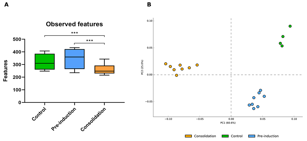
On the other hand, the analysis of alpha biodiversity (observed taxa) showed a significant decrease in bacterial richness in the patients of the consolidation group compared to the pre-induction and control group (Figure 3A). Additionally, beta biodiversity analysis, represented by principal component analysis (PCA), showed the grouping of samples, suggesting a marked differentiation of bacterial communities by treatment. Thus, microbiota changes between the control and the pre-induction group were smaller (PC2:25%) compared to microbiota changes between the control and the consolidation group (PC1:60%). This result suggests that the consolidation phase of the treatment led to more significant changes in the composition of the oral microbiota when compared to patients of the control group (Figure 3B).
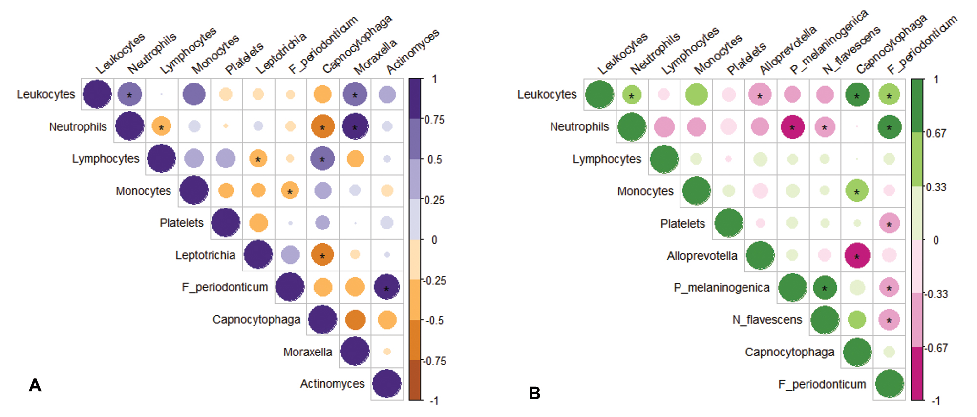
Pearson’s correlation between microbiota and leukocyte profile in the pre-induction group showed significant and negative relationships between neutrophils and Capnocytophaga; lymphocytes and Leptotrichia. On the other hand, positive relationships were identified between neutrophils and Moraxela; lymphocytes and Capnocytophaga; leukocytes and Moraxella (Figure 4A). In the consolidation, the following significant and negative correlations were identified: between neutrophils and Neisseria flavescens; neutrophils and Prevotella melaninogenica; leukocytes and Alloprevotella. On the other hand, positive relationships were identified between leukocytes and Capnocytophaga; leukocytes and Fusobacterium Periodonticum; neutrophils and Fusobacterium Periodonticum; monocytes and Capnocytophaga (Figure 4B).
Richness of communities. (A) Number of taxa identified per treatment. Bars in the plot represent the mean and standard deviation of the observed taxa in each treatment. Asterisks indicate the relationships that showed significant differences by Wilcoxon test. (p < 0.05). (B) PCA plot representing the grouping of samples by treatment. Dots of the same color indicate samples from the same treatment.
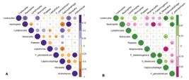
Pearson’s correlation between microbiota and leukocyte profile. (A) Pre-induction correlation matrix. (B) Consolidation correlation matrix. The figure represents the correlations between two variables on a scale from 1 to -1, where negative values indicate negative relationships. The size of the circles in the matrix represents the scale of the correlations, and asterisks indicate relationships that present significant differences.
The results of the network analysis at the time of consolidation showed a community with 18 nodes, 29 edges, and an average number of neighbors of 3.2. The presence of some abundant taxa such as Lactobacillus and Neisseria flavescens was identified in the community. However, the three taxa responsible for the highest connectivity in the network were Prevotella melaninogenica, Neisseria flavescense, and Porphyromonas, considered as the key taxa at the time of consolidation (Figure 5). Of the 29 edges in the network, 19 were negative and 10 were positive. The strongest positive correlation (0.82) was between Neisseria flavescens and Leptotrichia. On the other hand, the strongest negative correlation (-0.90) was between Leptotrichia and Porphyromonas. Additionally, Porphyromonas showed other negative correlations with Prevotella melaninogenica, Neisseria flavescens, and Lachnoanaerobaculum, and only one positive correlation with Actinomyces.

Network of consolidation. The figure represents the taxa and their relationships in the consolidation phase. Nodes represent taxa and the color of the circle around it represents its abundance in the community. The size of the nodes represents the taxon’s importance in the community given by the “Betweenness centrality” value. In yellow, the three key taxa of the community were highlighted. The edges between the nodes represent the correlations between two taxa, in red for positive correlations, and in blue for negative correlations. Only strong positive and negative correlations (between 0.6-1) and significant relationships (P < 0.05) are shown.
Discussion
The oral microbiota functions as a reservoir for planktonic-stage microorganisms that colonize/recolonize various habitats within the oral cavity. Studies by Griffen et al. (2012) and Wang et al. (2014) have highlighted Streptococcus as the most abundant genus in healthy oral microbiota, followed by Prevotella, Veillonella, Neisseria, and Haemophilus, aligning with the findings of this study. However, in immunocompromised patients, the oral microbiota can become opportunistic and pathogenic, leading to infections. This imbalance may manifest in leukemic symptoms such as pallor of the oral mucosa, gum discoloration, gingival petechiae, and ulcerative mucosal lesions, as observed in our clinical and blood profile results. Immunocompromised individuals are also more susceptible to bacterial and fungal infections (Kamasaki et al., 2005).
Our results indicate an increase in the abundance of the genus Actinomyces during the pre-induction phase and Alloprevotella during the consolidation phase among the five most abundant microorganisms. These shifts may signify a dysbiotic aspect across different chemotherapy treatment phases. Streptococcus, Veillonella, Prevotella melaninogenica, and Neisseria flavescens could be considered a central core.
Actinomyces may play a role in the bacterial community during the consolidation phase, forming a complex and diverse composition that might result from alterations in host defenses. In contrast, a less diverse commensal bacterial community during disease progression leaves individuals more susceptible to opportunistic diseases, impacting their quality of life; Actinomyces infections have been reported in cases of patients after hematopoietic stem cell transplantation (Carvalho et al., 2022). While oral infections by Actinomyces are rare in immunocompetent individuals, they become more common in immunocompromised individuals, leading to considerable morbidity with atypical and variable presentations, potentially resulting in severe outcomes if not diagnosed early (Ali and Tanwir, 2012; Bolger et al., 2014).
In our study, the abundance of Bacteroides increased, and genus/species richness decreased in children with acute leukemia. Deficient neutrophils in childhood acute leukemia may lead to reduced energy intake and storage, resulting in a state of high energy consumption due to immune disturbance. This makes the body more susceptible to bacterial invasion and infections (Van Der Meulen, 2018). In the pre-induction and consolidation phases, we observed a positive and significant relationship between neutrophils and leukocytes in the pre-induction phase with the genus Moraxella and, in the consolidation phase, with the species Fusobacterium periodonticum (Wang et al., 2020). Moraxella is associated with respiratory tract infections, while Fusobacterium periodonticum is linked to deficient oral conditions (Cauduro et al., 1999; Silva, 2016). Different Fusobacterium nucleatum strains may differentially affect neutrophil function, indicating variations in their virulence properties (Kurgan et al., 2016). In addition, data indicate that immunosuppression is also correlated with characteristic changes in the intestinal microbiota in humans, likely due to damages to gut-associated lymphoid (Lozupone et al., 2013). Thus, the intestinal microbiota seems to interact directly with the immune system of the host, stimulating local and systemic immune interactions (Sommer and Backhed, 2013). In this way, immunosuppression with cyclophosphamide led to significant decreases of several circulating leukocyte subsets in chickens and shifts in the intestinal microbiota (Mesa et al., 2020).
Fusobacterium periodonticum seems to grow less efficiently in biofilms and exhibits a more condensed localization pattern in supra and subgingival biofilm models (Thurnheer et al., 2019). However, this behavior may be induced by the new conditions resulting from antineoplastic treatment in these patients (Cocivera et al., 2022). The correlation analysis of microbiota with laboratory data reinforces previous findings regarding the presence of dysbiosis throughout the treatment process of patients with ALL.
Regarding the oral microbiota network, until the submission of this work, no articles were found that conducted a network analysis of the oral microbiota. In Wang et al. (2020) study, the authors evaluated the richness and taxonomic composition of the oral microbiota. In the study by Hou et al. (2018), the oral microbiota in patients with nasopharyngeal carcinoma undergoing radiotherapy was evaluated. Interestingly, Prevotella, Fusobacterium, Treponema, and Porphyromonas showed synchronous dynamic variations in their abundances during radiotherapy, with peaks often coinciding with the onset of severe mucositis. Our network results align with this study, highlighting the significance of taxa Prevotella melaninogenica, Neisseria flavescens, and Porphyromonas during the consolidation phase of treatment and considering them as potential biological markers of a higher degree of oral mucositis, added to the drop in biodiversity and a significant increase in this phase when compared to the control group.
Further considerations about the three main microorganisms resulting from our network analysis are essential. Porphyromonas, in the consolidation phase, appears to be a potential marker in the observed dysbiosis during chemotherapy treatment phases (Oldenburg et al., 2021). It has been found to occupy other sites, such as the upper portion of the gastrointestinal tract and the colon. In vitro studies, Porphyromonas demonstrated the ability to invade human gingival fibroblasts as a form of protection against the effects of antimicrobial use. This adaptive ability as an intracellular parasite is noteworthy and could trigger changes in essential cellular mechanisms such as the cell cycle and cell death (Irshad et al., 2012). This capacity suggests that Porphyromonas might be involved in the recurrence of other types of neoplasms in patients after initial chemotherapy treatment phases (Wang et al., 2020).
Additionally, Prevotella and Neiseria were identified as significant microbial agents in the consolidation phase. Hsiao et al. (2018) listed three periodontal pathogenic species (Prevotella tannerae, Fusobacterium nucleatum, and Prevotella intermedia) associated with an increased risk of oral squamous cell carcinoma, further emphasizing the importance of considering these microorganisms as potential biomarkers of risk (Hsiao et al., 2018).
Some studies suggest that non-pathobiont microorganisms, typically considered healthy-associated species like Neisseria flavescens, may contribute to carcinogenesis. Neisseria flavescens, along with Haemophilus parainfluenzae, have been found to induce cytotoxicity through intracellular infection. Thus, these three microorganisms could potentially be considered future targets for risk stratification in the development of secondary neoplasms (Baraniya et al., 2020).
This study has some limitations, such as a small and heterogeneous cohort size. However, it represents the largest pediatric hospital in Brazil, making it a significant clinical cohort from the country. The COVID-19 pandemic significantly constrained sampling and patient inclusion in the study, both for sick and healthy individuals. Despite these limitations, the consistent and significant results related to oral microbiota contribute to a better understanding of bacterial shifts during different moments of antineoplastic treatment.
Conclusions
The results indicate that ALL treatment can induce noteworthy alterations in the composition of the oral microbiota, characterized by decreased bacterial diversity and an elevated incidence of oral mucositis. Furthermore, specific bacterial taxa, including Prevotella melaninogenica, Neisseria flavescens, and Porphyromonas, emerged as pivotal components in the oral microbiota community during the consolidation phase of treatment, demonstrating a correlation with a heightened degree of oral mucositis. Understanding these changes is crucial for developing strategies to safeguard the oral health of patients undergoing ALL treatment.
Acknowledgements
We are grateful to Pequeno Principe Complex for their support.
References
- Ali FM and Tanwir F (2012) Oral microbial habitat: A dynamic entity. J Oral Biol Craniofac Res 2:181-187.
- Baraniya D, Jain V, Lucarelli R, Tam V, Vanderveer L, Puri S, Yang M and Al-Hebshi NN (2020) Screening of health-associated oral bacteria for anticancer properties in vitro Front Cell Infect Microbiol 10:575-656.
- Béné MC (2005) Immunophenotyping of acute leukemias. Immunol Lett 98:9-21.
- Blijleven, NM, Donnelly, JP and De Pauw, BE (2000) Mucosal barrier injury: Biology, pathology, clinical counterparts, and consequences of intensive treatment for haematological malignancy: An overview. Bone Marrow Transplant 25:1269-1278.
- Bolger AM, Lohse M, and Usadel B (2014) Trimmomatic: A flexible trimmer for Illumina sequence data. Bioinformatics 30:2114-2120.
- Bolyen E, Rideout JM, Dillon RM, Bokulich NA, Abnet CC, Al-Ghalith AG, Alexander H, Alm EJ, Arumugam M, Asnicar F et al (2019) Reproducible, interactive, scalable, and extensible microbiome data science using QIIME 2. Nat Biotechnol 37:852-857.
- Callahan BJ, McMurdie PJ, Rosen MJ, Ha W, Johnson AJA and Holmes SP (2016) DADA2: High-resolution sample inference from Illumina amplicon data. Nat Methods 13:581-583.
- Caporaso JG, Lauber CL, Walters W, Berg-Lyons D, Lozupone CA, Turnbaugh PJ, Fierer N and Knight R (2011) Global patterns of 16S rRNA diversity at a depth of millions of sequences per sample. Proc Natl Acad Sci U S A 108:4516-4522.
- Carvalho JESR, Tagliabue JF, Teixeira LDD, Menezes ACDS, Alves LDB and Antunes HS (2022) Infecção oral por actinomyces em pacientes após transplante de células tronco hematopoiéticas alogênico: Série de casos. HematolTransf Cell Therapy 4:S576-S577.
- Cauduro PF, Dias CAG and Mezzari A (1999) Moraxella catarrhalis: An old microorganism with new clothes. Cad Farm 15:15-17.
- Cocivera G, Giulian G and Panzarella V (2022) Oral microbiome pattern in hematological pediatric malignancies: Need for investigating. Qeios 1:SKGT2N.
- Fattizzo B, Cavallaro F, Folino F and Barcellini W (2021) Recent insights into the role of the microbiome in malignant and benign hematologic diseases. Crit Rev Oncol Hematol 160:103289.
- Franceschini C, Jung JE and Amante CJ (2003) Oral mucositis in post-chemotherapic patients undergoing bone marrow suppression. Rev Bras Patol Oral 2: 40-43.
- Griffen AL, Beall CJ, Campbell JH, Firestone ND, Kumar PS, Yang Z, Podar M and Ley EJ (2012) Distinct and complex bacterial profiles in human periodontitis and health revealed by 16S pyrosequencing. Isme J 6:1176-1185.
- Hou J, Zheng H, Li P, Liu H, Zhou H and Yang X (2018) Distinct shifts in the oral microbiota are associated with the progression and aggravation of mucositis during radiotherapy. Radio Onco 129:44-51.
- Hsiao JR, Chang CC and Lee WT (2018) The interplay between oral microbiome, lifestyle factors and genetic polymorphisms in the risk of oral squamous cell carcinoma. Carcinogenesis 39:778-787.
- Irshad M, Van der Reijden WA, Crielaard W and Laine ML (2012) In vitro invasion and survival of porphyromonas gingivalis in gingival fibroblasts; role of the capsule. Arch Immunol Ther Exp (Warsz) 60:469-476.
- Kamasaki Y, Satoh K, Nishiguchi M, Hoshino Y and Fujiwara T (2005) Acute oral complications in a pediatric patient with acute lymphoid leukemia. Pediatrics Int 58:484-487.
- Klindworth A, Pruesse E, Schweer T, Peplies J, Quast C and Horn M (2021) Evaluation of general 16S ribosomal RNA gene PCR primers for classical and next-generation sequencing-based diversity studies. Nucleic Acids Res 41:e1.
- Kurgan S, Kansal S, Nguyen D, Stephens D, Koroneos Y, Hasturk H, Van Dyke TE and Kantarci A (2017) Strain-specific impact of Fusobacterium nucleatum on neutrophil function. J Periodontol 88:380-389.
- Lim Y, Totsika M, Morrison M and Punyadeera C (2017) The saliva microbiome profiles are minimally affected by collection method or DNA extraction protocols. Sci Rep 7:8523.
- Lozupone CA, Li M, Campbell TB, Flores SC, Linderman D, Gebert MJ, Knight R, Fontenot AP and Palmer BE (2013) Alterations in the Gut Microbiota Associated with HIV-1 Infection. Cell Host Microbe 14:329-339.
- Mesa D, Beirão BCB, Balsanelli E, Sesti L, Caron LF, Cruz LM and Souza EM (2020) Cyclophosphamide increases lactobacillus in the intestinal microbiota in chickens. MSystems 5:e00080-20.
- Miranda FA, Piñeros M, Ferlay J, Soerjomataram L, Monnereau A and Bray F (2018) Epidemiological patterns of leukaemia in 184 countries: A population-based study. Lancet Haematol 5:e14-e24.
- Oldenburg M, Rüchel N, Janssen S, Borkhardt A and Gössling KL (2021) The microbiome in childhood acute lymphoblastic leukemia. Cancers (Basel) 13:4947.
- Pedrosa F and Lins M (2002) Acute lymphoblastic leukemia: A curable disease. Rev Bras Mater Infant 1:63-68.
- Pui CH, Robison LL and Look AT (2018) Acute lymphoblastic leukaemia. Lancet 371:1030-1043.
- Rajagopala SV, Singh H, Yu Y, Zabokrtsky KB and Nelson KE (2019) Persistent gut microbial dysbiosis in children with acute lymphoblastic leukemia (ALL) during chemotherapy. Microbial Ecology 79:1034-1043.
- Rognes T, Flouri T, Nichols B, Quince C and Mahé F (2016) VSEARCH: A versatile open-source tool for metagenomics. PeerJ 4:e2584.
- Santos PSS and Soares JAV (2012) Medicina bucal: A prática na odontologia hospitalar. Santos Editora, São Paulo.
- Silva AMCR (2016) Characterization of Haemophilus influenzae, Moraxella catarrhalis and Streptococcus pneumoniae strains isolated from nasopharyngeal colonization in children attending at a university hospital in Rio de Janeiro. M. S.c Thesis, Faculdade de Ciências Médicas, Universidade Federal do Rio de Janeiro, Brazil.
- Sommer F and Backhed F (2013) The gut microbiota - masters of host development and physiology. Nat Rev Micro 11:227-238.
- Sonis ST (2004) Oral mucositis in cancer therapy. J Support Oncol 2:3-8.
- Thurnheer T, Karygianni L, Flury M and Belibasakis GN (2019) Fusobacterium species and subspecies differentially affect the composition and architecture of supra- and subgingival biofilms models. Front Microbiol 10:1716.
- Van Der Meulen TA (2018) Reduced salivary secretion contributes more to changes in the oral microbiome of patients with primary Sjögren’s syndrome than underlying disease. Ann Rheum Dis 77 :1542-1544.
-
Wang H, Li B, Li A, Ni P, Lin L, Zhong Y and Ma L (2020) Characteristics of gut microbiome and prediction of infection in neutropenic children with acute leukemia. Res Sq. doi: 10.21203/rs.3.rs-61001/v1
» https://doi.org/10.21203/rs.3.rs-61001/v1 - Wang Y, Xue J, Xuedong Z, Meng Y and Quin D (2014) Oral microbiota distinguishes acute lymphoblastic leukemia pediatric hosts from healthy populations. PloS One 9:102-116.
- Yilmaz P, Parfrey LM, Yarza P, Gerken J, Pruesse E and Quast C (2013) The Silva and “All-species living tree project (LTP)” taxonomic frameworks. Nucleic Acids Res 42:d643-d648.
-
Ethics statement
The authors declare that the project “Analysis of environmental and genetic biomarkers in patients with acute lympoblastic leukemia” that gave rise to the article was approved by the Research Ethics Committee of Hospital Pequeno Príncipe”. CAAE: 28038619.3.0000.0097; number: 3,836,067. All those responsible for the patients signed the terms of “Assent and Free and Informed Consent” produced in accordance with the premises of the “Research Ethics Committee of Hospital Pequeno Príncipe”.
Supplementary Material
The following online material is available for this article: