 Functional capacity analysis after COVID-19 discharge
ABSTRACT
This study aimed to examine the correlation between three methods of assessing functional capacity [namely, handgrip strength (HGS), functional independence measure (FIM) and six-minute walk test (6MWT)], with clinical variables and hospitalization data in a cohort of patients recently discharged from the hospital after being treated for COVID-19. This is an observational, analytical and cross-sectional study. The study population comprised 125 patients of both sexes, aged 18 years or older, who were hospitalized due to a COVID-19 diagnosis. The patients were assessed approximately three months after hospital discharge, during which they underwent the HGS, the 6MWT, and responded to the FIM. Additionally, data from the medical records were collected and subsequently analyzed. The patients demonstrated satisfactory performance in the 6MWT (walking distance of 457m) and had handgrip strength comparable to that of healthy individuals, with a mean value of 38.9±11.7kgf in men and 27.3±2.8kgf in women. Additionally, they had an independence score on the FIM of 122.8±9.9 points, indicating a high level of autonomy. The three tests yielded positive correlations between the 6MWT and FIM (r=0.470, p<0.001), between the 6MWT and HGS (r=0.375, p<0.001), and between HGS and FIM (r=0.317, p<0.001). The analysis of the patients indicated that satisfactory functional recovery was achieved three months after discharge, regardless of the initial severity or length of hospital stay. While the three tests assess different dimensions of functional capacity, the observed correlations suggest that they can be used complementarily.
Keywords:
Functional Status; Walk Test; Muscle Strength Dynamometer; COVID-19, Rehabilitation; Oxygen Saturation
RESUMO
O objetivo deste estudo foi investigar se há associação entre três métodos de avaliação de capacidade funcional, a saber, força de preensão palmar (FPP), medida de independência funcional (MIF) e teste de caminhada de seis minutos (TC6), com variáveis clínicas e dados da internação em uma amostra de pacientes egressos de hospitalização por covid-19. Trata-se de estudo observacional, analítico e transversal. Foram incluídos 125 pacientes de ambos os sexos, com idade maior que 18 anos. Os participantes foram avaliados cerca de três meses após a alta hospitalar, quando realizaram FPP, TC6 e responderam a MIF. Os dados do prontuário foram analisados para coleta de dados clínicos e da internação. Os pacientes apresentaram distância percorrida elevada no TC6 (mediana de 457m), força de preensão palmar semelhante aos valores encontrados em indivíduos saudáveis (38,9±11,7kgf em homens, 27,3±2,8kgf em mulheres), e classificação de independência na MIF (122,8±9,9 pontos). Foram encontradas correlações positivas significativas entre o TC6 e a MIF (r=0,470, p<0,001), entre o TC6 e a FPP (r=0,375, p<0,001) e entre a FPP e a MIF (r=0,317, p<0,001). Apesar da gravidade à admissão hospitalar ou do tempo de internação hospitalar, após três meses da alta os pacientes analisados apresentaram recuperação funcional satisfatória. Os instrumentos utilizados avaliam diferentes dimensões da capacidade funcional, e as correlações encontradas entre eles sugerem que podem ser utilizados de forma complementar.
Descritores:
Estado Funcional; Teste de Caminhada; Dinamômetro de Força Muscular; Covid-19, Reabilitação; Saturação de Oxigênio
RESUMEN
El objetivo de este estudio fue analizar si existe una asociación entre tres métodos de evaluación de la capacidad funcional, la fuerza de agarre (FA), la medida de independencia funcional (MIF) y la prueba de caminata de seis minutos (6MWT), con variables clínicas y datos de hospitalización en una muestra de pacientes dados de alta de la hospitalización por covid-19. Se trata de un estudio observacional, analítico y transversal. Se incluyeron 125 pacientes de ambos sexos, mayores de 18 años de edad. Los participantes se sometieron a una evaluación aproximadamente tres meses después del alta hospitalaria cuando realizaron FA, 6MWT y respondieron a MIF. Los datos de la historia clínica se analizaron para recopilar datos clínicos y de hospitalización. Los pacientes presentaron distancia de caminata elevada en la 6MWT (mediana de 457m), fuerza de agarre similar a los valores encontrados en individuos sanos (38,9±11,7kgf en hombres, 27,3±2,8kgf en mujeres), y clasificación de independencia en MIF (122,8 ±9,9puntos). Se encontraron correlaciones positivas significativas entre 6MWT y MIF (r=0,470, p<0,001), entre 6MWT y FA (r=0,375, p<0,001) y entre FA y MIF (r=0,317, p<0,001). A pesar de la gravedad en el ingreso hospitalario o la duración de la estancia en el hospital, a los tres meses del alta los pacientes evaluados presentaron una recuperación funcional satisfactoria. Las herramientas utilizadas evalúan diferentes dimensiones de la capacidad funcional, y las correlaciones encontradas entre ellas apuntan que pueden utilizarse de manera complementaria.
Palabras clave:
Estado Funcional; Prueba de Paso; Dinamómetro de Fuerza Muscular; Covid-19, Rehabilitación; Saturación de Oxígeno
INTRODUCTION
After the acute phase of the disease caused by the coronavirus (COVID-19), it is common for respiratory or cognitive symptoms of variable duration to persist. These symptoms have been widely studied and have received the generic name of “Post-COVID-19 Syndrome”1. After the acute phase, respiratory function is affected by fatigue and dyspnea2, and tomographic and functional abnormalities are also common3. Such findings do not seem entirely related to the initial severity of the disease4 or the individual’s previous physical condition5.
In individuals with exercise intolerance after the acute phase of COVID-19, muscle changes are frequent, which seem to contribute to this state. Muscle mass loss, with consequent decrease in strength and endurance, is related to the worsening of functional capacity after hospitalization. This is mainly due to decreased muscle protein synthesis caused by the hyper-inflammatory state and hypoxemia, as well as some harmful effects of hospitalization6.
It is not yet fully understood the extent or interference of such symptoms and muscle impairment on the individual’s activities of daily living and their quality, nor what would be the personal or clinical factors associated with greater functional impairment after hospitalization. Thus, physical and functional assessment, with medium and long-term follow-up of survivors of hospitalizations, can guide the definition of the best rehabilitation strategies7.
Among the assessment tools to support rehabilitation, handgrip strength (HGS), measured by hand-held dynamometry, is a practical and widely disseminated method that provides data on overall peripheral muscle strength8. The method has been applied in intensive care unit settings (ICU), a context in which there are well-defined values to establish muscle weakness acquired during hospitalization9. However, the usefulness of this test in outpatient settings after hospital discharge has not yet been well explored or understood.
Another evaluation strategy is the use of functional scales, such as the functional independence measure (FIM)- commonly used during hospitalization and hospital discharge-to monitor the level of independence in activities of daily living and support the planning of necessary adaptations according to the functional status of the patients10.
The six-minute walk test (6MWT) analyzes the global and integrated response of the systems involved in submaximal exercise11. It is a useful test in several circumstances, such as in outpatient or inpatient evaluation of functional capacity, in the evaluation of results after clinical or surgical intervention, in the planning of a pulmonary rehabilitation program12, and even as a predictor of morbidity and mortality in patients with some lung diseases13. Recently, the 6MWT has been used to assess the functional capacity and physical fitness of patients after COVID-1914.
Our hypothesis is that the evaluation of various aspects of the individual’s functioning after hospitalization for COVID-19 can contribute to a better understanding of the functional impact of the disease and favor intervention and rehabilitation strategies. Thus, this study aimed to describe and evaluate the associations between functional capacity assessed by the HGS test, the FIM scale, and the 6MWT. All tests and the FIM completion were conducted with a sample of patients after discharge from hospitalization due to severe or moderately severe pneumonia caused by COVID-19. Associations were also sought between the tests and some clinical variables, such as pneumonia severity and use of ventilatory support during hospitalization.
METHODOLOGY
Study design and participants
This is an observational, analytical, and cross-sectional study conducted in a Brazilian university hospital. All subjects that agreed to participate in the study signed an informed consent form.
From May 1st to October 31st, 2020, patients of both sexes, aged over 18 years, who were discharged after hospitalization for pneumonia caused by COVID-19, were selected. The etiology of the disease was confirmed by RT-PCR examination. Patients who did not agree to participate in the study or who had movement limitations that hindered the performance of the tests were excluded.
Procedures
The clinical and functional evaluation of the patients was carried out between 20 and 120 days after hospital discharge, in a multidisciplinary outpatient clinic of the Hospital de Clínicas da Universidade de Campinas. Participants were served by pulmonologists and respiratory physical therapists. On the day of the consultation, the HGS test, FIM scale, and 6MWT were performed in sequence, with a 10-minute interval between them. Data related to hospitalization were collected via medical records. Two specialist pulmonologists evaluated the chest tomography scans performed at hospital admission and classified the degree of pulmonary involvement into three categories: less than 25%, between 25 and 50%, or greater than 50%, according to the criteria of the Radiological Society of North America (RSNA)15.
Handgrip strength was measured using a portable hydraulic dynamometer (Saehan®), in accordance with the guidelines of the American Society of Hand Therapists16. The evaluation was performed with the patient’s dominant hand in the grip position. The patients remained seated in a armless chair, with the spine upright and the knees flexed at 90°. The shoulders remained in adduction and neutral rotation, the elbow of the dominant arm remained flexed at 90°, and the forearm remained in half pronation, with the wrist in a neutral position, being able to move up to 30° of extension during contraction. With the dynamometer held by the evaluator, the patient performed three maximal voluntary contractions, each sustained for six seconds, with a one-minute interval between each measurement. For the analysis, the mean value of the three measurements, expressed in kgf, was used.
The functional independence measure instrument has 18 items distributed in the following domains: self-care, sphincter control, transfers, locomotion, communication, and social cognition. The scale was applied by the examiner via a structured interview. Each item was assigned a score from 1 (complete dependence for the activity) to 7 (complete independence). The total score of the FIM instrument ranges from 18 to 126 points, with higher values indicating greater functional autonomy17.
All individuals underwent the six-minute walk test under the supervision of the same evaluator, following the guidelines of the American Thoracic Society18. Blood pressure was measured at the beginning and at the end of the test using a Premium sphygmomanometer®. Heart rate (HR) and peripheral oxygen saturation (SpO2) were measured by pulse oximeter with a digital probe (3150 USB WristOX2 pulse oximeter; Nonin Medical®), at the pre-test rest and at the sixth minute of the test. The modified Borg scale, scored from 0 to 10, was shown to participants so that they could grade the sensation of dyspnea before and after the test. Patients were carefully observed throughout the procedure to ensure that their individual exercise limits were not exceeded. For the analysis, two main variables were considered: distance walked (6MWD) and the presence of exertion-induced hypoxemia, defined in this study as ΔSpO2. This value was calculated as the difference between the SpO2 initial and the SpO2 final. The variation was considered significant when ΔSpO2 ≥4%, or when SpO2 final ≤93%19. The 6MWD was shown in meters and as a percentage of the predicted value, calculated according to the Brazilian reference equation20.
By reviewing the medical records, the following variables were collected: (1) demographic variables, such as age, sex, body mass index (BMI); (2) clinical variables, such as Charlson’s comorbidity index (CCI)21, variables related to hospitalization (length of stay, days under mechanical ventilation, oxygen supplementation, need for prone position); and (3) degree of pulmonary involvement on chest CT scans.
Statistical analysis
All raw data were tabulated and organized to ensure the quality of the records.
Quantitative and qualitative variables were first subjected to a descriptive analysis, and proportions were compared using the Chi-squared test or Fisher’s exact test, when necessary. Continuous measurements between two groups were compared using the Mann-Whitney U-test, while three or more groups were compared using the Kruskal-Wallis test.
The association between continuous measurements was calculated using Spearman’s correlation test. Significance level was set at 5%. Data were analyzed using the R software22 and SAS System for Windows23.
RESULTS
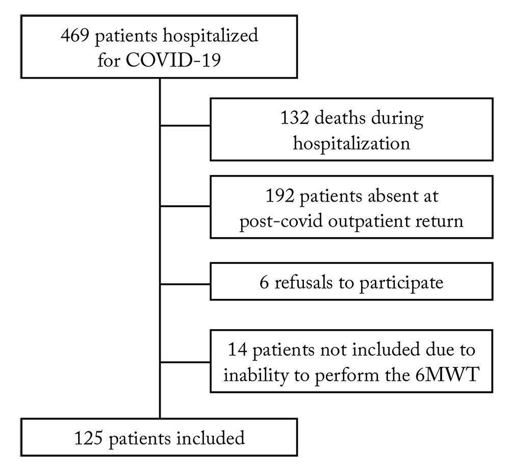
Of 469 patients hospitalized due to COVID-19 in the analyzed period, 125 were included and underwent all evaluations. Figure 1 shows the flow of recruitment and inclusion.
The mean age was 57±12.4 years, and 59.2% of the patients were men. The mean score of pre-existing comorbidities assessed via CCI was 2.1±1.6, with systemic hypertension and diabetes mellitus being the most prevalent conditions. The mean BMI was 31.1±6.2kg/m2, and 46.5% of the patients were classified as obese (BMI>30kg/m2). On admission, chest tomography showed pulmonary involvement greater than 25% in 75% of the cases.
The patients remained hospitalized for 14.5±11 days, and all of them received some type of ventilatory support. Most individuals (75.2%) were treated using noninvasive interfaces, including oxygen supplementation by nasal cannula, non-rebreather mask, high-flow nasal cannula, or noninvasive ventilation, and only 24.8% required invasive mechanical ventilation. The mean length of stay of the 31 patients who needed invasive mechanical ventilation was 28±3.5 days, with a mean duration of 15.7±9.6 days under mechanical ventilation. Table 1 summarizes demographic, clinical, and hospitalization data.
The mean 6MWD was 447.9±114.9m, and 57 (45.6%) of the patients covered less than 80% of the predicted distance according to the reference equation20. A quarter of the patients (26.4%) had desaturation during the 6MWT. The mean handgrip strength was 38.9±11.7kgf for men and 27.3±2.8kgf for women. The mean score of the FIM instrument was 122.8±9.9, classifying the participants as totally independent. Table 2 shows the functional data regarding the two tests and the FIM scale.
Participants underwent clinical and functional evaluations on average 77 days after discharge. The interval between discharge and outpatient evaluation was wide (20-120 days) due to the relative unavailability of transportation from the city of residence to the hospital and the limitation of scheduling due to the need to reduce the number of people in the same environment. Considering that the convalescence process after hospitalization could have affected the results of patients who were evaluated later (after 60 days), we compared the data from the evaluations performed before 60 days after discharge and those obtained after 60 days from discharge. No statistically significant difference was found between the groups in relation to demographic, clinical, or hospitalization-related variables. However, in the 6MWT, we have found a significant difference in the frequency of individuals who had to interrupt the test, and in the group evaluated 60 days after hospital discharge, more patients needed to interrupt the test (16.1%; p=0.046). Of the 125 patients analyzed, 11 had to interrupt the 6MWT. This subgroup of patients had a longer hospital stay and underwent longer use of mechanical ventilation, presenting lower FIM values. In total, 54.5% participants had desaturation during the 6MWT.
In the 6MWT evaluation, two main variables were considered: desaturation (decrease in SpO2 at the end of the test) and 6MWD. Considering the distance covered (447m) and the fact that more than half of the patients walked more than 80% of the predicted 6MWD, we chose to use the median for comparison between two groups (≥457.2m or <457.2m). Patients who walked shorter distances were older, had more comorbidities, were hospitalized for longer periods, and had a higher rate of use of the prone positioning (p<0.05). They also had lower handgrip strength and lower FIM scores (p<0.001) (Table 3).
In the comparison based on the desaturation level, patients who had desaturation ≥4% or SpO2 <93% at the end of the 6MWT had longer hospital stay, greater need for invasive mechanical ventilation and required prone positioning during hospitalization (p<0.005). Notably, the mean 6MWD of the 33 patients who presented desaturation was 396±138 meters, i.e., 51 meters less than the overall mean.
Spearman’s coefficient showed significant correlations between the three tests; but with a weak correlation between them (Figure 2). In the correlation analysis between demographic and clinical variables (age, BMI, CCI, length of hospital stay, days under mechanical ventilation) and each of the three tests performed, correlations were identified only between the 6MWD and age (r=0.35), CCI (r=−0.322) and days under mechanical ventilation (r=−0.366). There was no association between clinical and hospitalization variables and handgrip strength and FIM. The degree of pulmonary involvement at admission, verified by chest tomography, also showed no significant association with any of the tests (p=0.772). No serious adverse events were recorded during the evaluations and tests.
Correlation analysis between six-minute walk test, functional independence measure and handgrip strength
DISCUSSION
The patients evaluated in this study had severe COVID-19, defined by pneumonia at hospital admission. Despite the prolonged length of hospital stay and the need for mechanical ventilation in a quarter of the patients, the individuals presented good functional capacity results (6MWT and HGS) in the evaluation performed almost two months after hospital discharge. In studies conducted in a similar context of evaluation after hospitalization for severe COVID-19, similar results were found in Irish patients (average distance of 460m)14 and Thais (490m)24.
The predominance of male patients (59.2%) and the mean age (57 years) were similar to those observed in an extensive observational study of the Brazilian population hospitalized for COVID-19, carried out in parallel with our study25. Among the most prevalent comorbidities, hypertension and diabetes mellitus were the most common, as in the study by Ranzani et al.25
In 24.8% of the patients evaluated, invasive mechanical ventilation was required, and we found that the length of hospital stay was different for patients who used mechanical ventilation or not, being 28 and 14.5 days, respectively. These results are similar to those found in the Brazilian study by Ranzani et al.25, in which 72% of the hospitalized patients required some oxygen supplementation, 23% of which were invasive mechanical ventilation, with a median length of hospital stay of 8 (4-14) days.
In our study, the patients had a mean FIM score of 122.8, which classifies them as completely independent individuals26.
The mean HGS of the studied sample (38.9±11.7kgf for men and 27.3±2.8kgf for women) was similar to the reference values for healthy Brazilian adults27. HGS measurement is a non-invasive, accessible, and low-cost procedure that enables assessing muscle strength in different contexts, such as during hospitalization, at patient discharge, or after hospitalization28. Reduced HGS during hospitalization is a marker of increased ICU mortality29, and is associated with prolonged hospital stay and duration of mechanical ventilation30. In this study, the evaluation of HGS after about three months of hospital discharge indicated the absence of muscle weakness, so this result does not support its use as a possible outcome in rehabilitation programs.
Patients who walked shorter distances on the 6MWT were older, had more comorbidities, longer hospital stays, underwent the prone positioning more frequently, had lower FIM scores, and had lower HGS (p<0.05, Table 3). The correlation analysis showed a significant association between lower 6MWD values and older age, more comorbidities, and longer hospital stay. In this study, the 6MWD was similar to that found in patients evaluated after three and six months of hospitalization for acute respiratory syndrome31 and after hospitalization for COVID-1914. In a study with post-COVID-19 patients, Townsend et al.14 showed an association between 6MWD and length of stay, age, and frailty score.
In our sample, 26.1% of the patients had exertion-induced hypoxemia, i.e., a drop in SpO2 greater than or equal to 4% during the 6MWT. These patients experienced longer hospital stay, most needed mechanical ventilation, and underwent prone positioning more commonly, compared to those who did not have exertion-induced hypoxemia. Comparing the frequency of this finding with other studies is challenging, as there is scarce literature with data on exertion-induced hypoxemia after recovery from COVID-19. Paneroni et al.5 detected exertional desaturation in 24% of patients at the time of hospital discharge due to COVID-19. Morikawa et al.32 detected 6MWT-induced hypoxemia in 19.5% of patients 77 days after hospitalization for COVID-19. These authors found a correlation between this finding and age and lactate dehydrogenase levels.
Hypoxia is a primary pathophysiological event of severe COVID-19, but it can be present at all stages of the disease, being the leading cause of death. The mechanisms and factors involved differ according to the stage. In the acute phase, the onset of hypoxia in more severe cases results from multiple factors, such as an intense immune-mediated inflammatory response and thrombogenic events, which lead to impaired gas diffusion and worsening of the inadequacy between pulmonary ventilation and perfusion33. Once the acute phase is over, some patients continue to show or even develop fibrotic pulmonary interstitial involvement. In this context, hypoxemia on room air at rest or induced by exertion may be evinced.
Exertion-induced hypoxemia can occur in both sick people and healthy individuals34. It is known that the drop in oxygenation induced by exertion can be attributed to the following factors, whether isolated or in combination: relative alveolar hypoventilation; limitation in diffusion; mismatch between ventilation and perfusion; and pulmonary or right-to-left shunts34. In post-COVID patients, especially critically ill patients who survived mechanical ventilation, several mechanisms may be involved in the occurrence of exertion-induced hypoxemia. Evidence suggests that the decrease in PaO2 during light to moderate exercise, such as submaximal exertion in the 6MWT, may result from relative alveolar hypoventilation (i.e., lower PaO2)34. It is plausible to assume that alveolar hypoventilation is one of the mechanisms of desaturation observed weeks after the disease in individuals who survived COVID-19-related acute respiratory failure and were subjected to mechanical ventilation. This mechanism is associated with some reduction in diffusion through the alveolar-capillary membrane, due to inflammation and endotheliitis caused by the virus.
Despite the complete independence indicated by the mean FIM score, patients who walked shorter distances on the 6MWT also had the lowest FIM scores, a finding compatible with the moderate correlation between tests. Easy to apply, the FIM instrument can assess functional capacity in ill adults and children35, in addition to being used in outpatient settings or at patient discharge.
We found positive correlations between HGS, FIM scores, and 6MWT values. Although the HGS and the FIM instrument define our participants with good functional capacity and independence, the correlations found with the 6MWT suggest a possible complementarity between these instruments. Studies prior to the COVID-19 pandemic with critically ill ICU patients with prolonged immobilization showed persistent functional impairment after hospital discharge, evinced by changes in pulmonary function tests, reduced 6MWT, muscle weakness, and decreased ability to perform activities of daily living36-38. Among the complementary methods for assessing functional capacity, handgrip strength and the FIM instrument stand out for being easily applicable both in inpatient and outpatient settings, and even in patients with impaired mobility.
The 6MWT requires more space, staff training and patients’ ability to move around. In the results obtained here, even considering that the mean 6MWD found was satisfactory, associations were identified with clinical variables and severity of hospitalization. Exertion-induced hypoxemia was more common in people with more severe respiratory conditions and longer hospitalizations. The correlations with HGS and FIM reinforce the robustness of the 6MWT for assessing the individual’s functional capacity, and its usefulness as a significant outcome in pulmonary rehabilitation programs.
CONCLUSIONS
Despite the severe respiratory impairment at admission and the long stay in the hospital, the patients in this study showed satisfactory functional recovery three months after discharge. Both the functional tests and the FIM showed high scores. It was observed that, in the 6MWT, patients who walked shorter distances had longer hospital stays and longer duration of mechanical ventilation. Moreover, exertion-induced hypoxemia occurred more frequently in more severe patients with greater respiratory distress, who also required more invasive ventilatory support.
Although the 6MWT, FIM and HGS assess different dimensions of the individual’s functional capacity, the correlations found suggest that they can be complementarily used. FIM and handgrip strength are easily applicable and reproducible assessment test, and can be used in space-constrained environments or in patients with limited mobility.
LIMITATIONS
Our study was conducted in a single center and had some limitations. Due to the contingency in outpatient care that characterized the first wave of the COVID-19 pandemic, it was not possible to include all patients hospitalized for severe cases. In addition to the restriction of beds, there were limitations related to the transportation of patients and the functioning of primary health care. More severe patients may be underrepresented in the cohort analyzed. Furthermore, the sample size and the fact that the study was conducted in a single center limit the possibility of generalizing the findings.
ACKNOWLEDGMENTS
The authors thank the RECOVID Unicamp team (Laís B. Alves; Nayara N. Basso; Hugo Ceccato; Lucieni de O. Conterno; Henrique A. Engleitner; Ana L. Lima; Paulo R. Mendes; Mônica C. Pereira; Lígia S. Ratti; Mariangela R. Resende; Julian F. Silva; Carolina S. Tamesawa; Bruna S. Vian; Pedro M. Villar); and the coordinator of the SFTO (Physiotherapy and Occupational Therapy Service of the Unicamp Hospital de Clínicas), Luciana Castilho de Figueirêdo.
DATA AVAILABILITY
The entire dataset supporting the results of this study is available within the article.
REFERÊNCIAS
-
1 Rodriguez-Morales AJ, Cardona-Ospina JA, Gutiérrez-Ocampo E, Villamizar-Peña R, Holguin-Rivera Y, et al. Clinical, laboratory and imaging features of COVID-19: a systematic review and meta-analysis. Travel Med Infect Dis. 2020;34:101623. doi: 10.1016/j.tmaid.2020.101623
» https://doi.org/10.1016/j.tmaid.2020.101623 -
2 Carfì A, Bernabei R, Landi F, Gemelli Against COVID-19 Post-Acute Care Study Group. Persistent symptoms in patients after acute COVID-19. JAMA. 2020;324(6):603-5. doi: 10.1001/jama.2020.12603
» https://doi.org/10.1001/jama.2020.12603 -
3 Huang Y, Tan C, Wu J, Chen M, Wang Z, et al. Impact of coronavirus disease 2019 on pulmonary function in early convalescence phase. Respir Res. 2020;21(1):163. doi: 10.1186/s12931-020-01429-6
» https://doi.org/10.1186/s12931-020-01429-6 -
4 Gandhi RT, Lynch JB, del Rio C. Mild or moderate COVID-19. N Engl J Med. 2020;383(18):1757-66. doi: 10.1056/NEJMcp2009249
» https://doi.org/10.1056/NEJMcp2009249 -
5 Paneroni M, Simonelli C, Saleri M, Bertacchini L, Venturelli M, et al. Muscle strength and physical performance in patients without previous disabilities recovering from COVID-19 pneumonia. Am J Phys Med Rehabil. 2021;100(2):105-9. doi: 10.1097/PHM.0000000000001641
» https://doi.org/10.1097/PHM.0000000000001641 -
6 Silva CC, Bichara CNC, Carneiro FRO, Palacios VRDCM, van den Berg AVS, et al. Muscle dysfunction in long coronavirus disease 2019 syndrome: pathogenesis and clinical approach. Rev Med Virol. 2022;32(6):e2355. doi: 10.1002/rmv.2355
» https://doi.org/10.1002/rmv.2355 -
7 Hui DS, Wong KT, Ko FW, Tam LS, Chan DP, et al. The 1-year impact of severe acute respiratory syndrome on pulmonary function, exercise capacity, and quality of life in a cohort of survivors. Chest. 2005;128(4):2247-61. doi: 10.1378/chest.128.4.2247
» https://doi.org/10.1378/chest.128.4.2247 -
8 Kock KS, Breda Neto JA, Machado MDO. Comparação dos níveis de atividade física e força de preensão manual com o perfil bioquímico de doentes renais crônicos. Ciênc Saúde. 2017;10(1):10-17. doi: 10.15448/1983-652X.2017.1.24114
» https://doi.org/10.15448/1983-652X.2017.1.24114 -
9 Ali NA, O'Brien Jr JM, Hoffmann SP, Phillips G, Garland A, et al. Acquired weakness, handgrip strength, and mortality in critically ill patients. Am J Respir Crit Care Med. 2008;178(3):261-8. doi: 10.1164/rccm.200712-1829OC
» https://doi.org/10.1164/rccm.200712-1829OC -
10 Alves GAA, Martinez BP, Lunardi AC. Avaliação das propriedades de medida das versões brasileiras do Functional Status Score for the ICU e da Functional Independence Measure em pacientes críticos internados em unidade de terapia intensiva. Rev Bras Ter Intensiva. 2019;31(4):521-8. doi: 10.5935/0103-507X.20190065
» https://doi.org/10.5935/0103-507X.20190065 -
11 Holland AE, Spruit MA, Troosters T, Puhan MA, Pepin V, et al. An official European Respiratory Society/American Thoracic Society technical standard: field walking tests in chronic respiratory disease. Eur Respir J. 2014;44(6):1428-46. doi: 10.1183/09031936.00150314
» https://doi.org/10.1183/09031936.00150314 -
12 Dias CMCC, Pereira N, Bonfim KPP, Reis HFC, Mayer AF, et al. Desempenho no teste de caminhada de seis minutos e fatores associados em adultos jovens saudáveis. Rev Pesq Fisio. 2017;7(3):408-17. doi: 10.17267/2238-2704rpf.v7i3.1555
» https://doi.org/10.17267/2238-2704rpf.v7i3.1555 -
13 du Bois RM, Weycker D, Albera C, Bradford WZ, Costabel U, et al. Six-minute-walk test in idiopathic pulmonary fibrosis: test validation and minimal clinically important difference. Am J Respir Crit Care Med. 2011;183(9):1231-7. doi: 10.1164/rccm.201007-1179OC
» https://doi.org/10.1164/rccm.201007-1179OC -
14 Townsend L, Dowds J, O'Brien K, Sheill G, Dyer AH, et al. Persistent poor health after COVID-19 is not associated with respiratory complications or initial disease severity. Ann Am Thorac Soc. 2021;18(6):997-1003. doi: 10.1513/AnnalsATS.202009-1175OC
» https://doi.org/10.1513/AnnalsATS.202009-1175OC -
15 Simpson S, Kay FU, Abbara S, Bhalla S, Chung JH, et al. Radiological Society of North America expert consensus document on reporting chest CT findings related to COVID-19. Radiol Cardiothorac Imaging. 2020;2(2):e200152. doi: 10.1148/ryct.2020200152
» https://doi.org/10.1148/ryct.2020200152 - 16 Fess EE, Moran CA. Clinical assessment recommendations. 2nd ed. Chicago: American Society of Hand Therapists; 1992.
-
17 Riberto M, Miyazaki MH, Jucá SSH, Sakamoto H, Pinto PPN, et al. Validação da versão brasileira da Medida de Independência Funcional. Acta Fisiatr. 2004;11(2):72-6. doi: 10.5935/0104-7795.20040003
» https://doi.org/10.5935/0104-7795.20040003 -
18 American Thoracic Society Committee on Proficiency Standards for Clinical Pulmonary Function Laboratories. ATS statement: guidelines for the six-minute walk test. Am J Respir Crit Care Med. 2002;166(1):111-7. doi: 10.1164/ajrccm.166.1.at1102
» https://doi.org/10.1164/ajrccm.166.1.at1102 -
19 Lama VN, Flaherty KR, Toews GB, Colby TV, Travis WD, et al. Prognostic value of desaturation during a six-minute walk test in idiopathic interstitial pneumonia. Am J Respir Crit Care Med. 2003;168(9):1084-90. doi: 10.1164/rccm.200302-219OC
» https://doi.org/10.1164/rccm.200302-219OC -
20 Iwama AM, Andrade GN, Shima P, Tanni SE, Godoy I, et al. The six-minute walk test and the body weight-walk distance product in healthy Brazilian subjects. Braz J Med Biol Res. 2009;42(11):1080-5. doi: 10.1590/S0100-879X2009005000032
» https://doi.org/10.1590/S0100-879X2009005000032 -
21 Charlson ME, Pompei P, Ales KL, MacKenzie CR. A new method of classifying prognostic comorbidity in longitudinal studies: development and validation. J Chronic Dis. 1987;40(5):373-83. doi: 10.1016/0021-9681(87)90171-8
» https://doi.org/10.1016/0021-9681(87)90171-8 - 22 R Core Team. R: a language and environment for statistical computing. Vienna: R Foundation for Statistical Computing; 2018.
- 23 SAS Institute Inc. SAS system for Windows, version 9.4. Cary (NC): SAS Institute Inc.; 2002-2012.
-
24 Eksombatchai D, Wongsinin T, Phongnarudech T, Thammavaranucupt K, Amornputtisathaporn N, Sungkanuparph S. Pulmonary function and six-minute walk test in patients after recovery from COVID-19: a prospective cohort study. PLoS One. 2021;16(2):e0257040. doi: 10.1371/journal.pone.0257040
» https://doi.org/10.1371/journal.pone.0257040 -
25 Ranzani OT, Bastos LSL, Gelli JGM, Marchesi JF, Baião F, et al. Characterisation of the first 250000 hospital admissions for COVID-19 in Brazil: a retrospective analysis of nationwide data. Lancet Respir Med. 2021;9(4):407-18. doi: 10.1016/S2213-2600(20)30560-9
» https://doi.org/10.1016/S2213-2600(20)30560-9 -
26 Abentroth LRL, Osaku EF, Silva MMM, Jaskowiak JL, Zaponi RS, et al. Functional independence and spirometry in adult post-intensive care unit patients. Rev Bras Ter Intensiva. 2021;33(2):243-50. doi: 10.5935/0103-507X.20210031
» https://doi.org/10.5935/0103-507X.20210031 - 27 Caporrino FA, Faloppa F, Santos JBG, Ressio C, Soares FHC, et al. Estudo populacional da força de preensão manual com dinamômetro Jamar(r). Rev Bras Ortop. 1998;33(2):150-4.
-
28 Bobos P, Nazari G, Lu Z, MacDermid JC. Measurement properties of the hand grip strength assessment: a systematic review with meta-analysis. Arch Phys Med Rehabil. 2020;101(3):553-65. doi: 10.1016/j.apmr.2019.10.183
» https://doi.org/10.1016/j.apmr.2019.10.183 -
29 Chlan LL, Tracy MF, Guttormson J, Savik K. Peripheral muscle strength and correlates of muscle weakness in patients receiving mechanical ventilation. Am J Crit Care. 2015;24(6):e91-8. doi: 10.4037/ajcc2015277
» https://doi.org/10.4037/ajcc2015277 -
30 Cottereau G, Dres M, Avenel A, Fichet J, Jacobs FM, et al. Handgrip strength predicts difficult weaning but not extubation failure in mechanically ventilated patients. Respir Care. 2015;60(8):1097-104. doi: 10.4187/respcare.03604
» https://doi.org/10.4187/respcare.03604 -
31 Hui DS, Joynt GM, Wong KT, Gomersall CD, Li TS, et al. Impact of severe acute respiratory syndrome on pulmonary function, functional capacity and quality of life in a cohort of survivors. Thorax. 2005;60(5):401-9. doi: 10.1136/thx.2004.030205
» https://doi.org/10.1136/thx.2004.030205 -
32 Morikawa K, Tabira K, Takemura H, Inaba S, Suzuki Y, et al. Krebs von den Lungen-6 levels at admission predicts exercise-induced hypoxemia before and after discharge in patients with COVID-19. Respir Investig. 2024;62(3):369-74. doi: 10.1016/j.resinv.2024.02.011
» https://doi.org/10.1016/j.resinv.2024.02.011 -
33 Serebrovska ZO, Chong EY, Serebrovska TV, Tumanovska LV, Xi L. Hypoxia, HIF-1a, and COVID-19: from pathogenic factors to potential therapeutic targets. Acta Pharmacol Sin. 2020;41(12):1539-46. doi: 10.1038/s41401-020-00554-8
» https://doi.org/10.1038/s41401-020-00554-8 -
34 Dominelli PB, Sheel AW. Exercise-induced arterial hypoxemia: some answers, more questions. Appl Physiol Nutr Metab. 2019;44(6):571-9. doi: 10.1139/apnm-2018-0468
» https://doi.org/10.1139/apnm-2018-0468 -
35 Matos MA, Lopes PS, Corsini AR, Rodi J, Fong CT. Aplicação da medida de independência funcional na avaliação de pacientes com mucopolissacaridose. Colomb Med (Cali). 2020;51(3):e213996. doi: 10.25100/cm.v51i3.3996
» https://doi.org/10.25100/cm.v51i3.3996 -
36 Gruther W, Benesch T, Zorn C, Paternostro-Sluga T, Quittan M, et al. Muscle wasting in intensive care patients: ultrasound observation of the M. quadriceps femoris muscle layer. J Rehabil Med. 2008;40(3):185-9. doi: 10.2340/16501977-0139
» https://doi.org/10.2340/16501977-0139 -
37 Kiriella JB, Araujo T, Vergara M, Lopez-Hernandez L, Cameron JI, et al. Quantitative evaluation of muscle function, gait, and postural control in people experiencing critical illness after discharge from the intensive care unit. Phys Ther. 2018;98(1):8-15. doi: 10.1093/ptj/pzx102
» https://doi.org/10.1093/ptj/pzx102 -
38 Ohtake PJ, Lee AC, Scott JC, Hinman RS, Ali NA, et al. Physical impairments associated with post-intensive care syndrome: systematic review based on the WHO International Classification of Functioning, Disability and Health framework. Phys Ther. 2018;98(8):631-45. doi: 10.1093/ptj/pzy059
» https://doi.org/10.1093/ptj/pzy059