 Factors that predict poor physical functioning in women living with knee osteoarthritis: a one-year follow up
ABSTRACT
Knee osteoarthritis (OA) is ranked as the 11th highest contributor to disability globally. We must improve our understanding of its related factors with poor physical functioning to create opportunities for more efficient treatment. This study aimed to find the factors that predict poor physical functioning over one year in women with knee osteoarthritis. A total of 36 community-recruited women with knee OA underwent baseline and one-year biomechanics and physical functioning assessment. Physical function was assessed by the Western Ontario and McMaster Universities and Short Physical Performance Battery. Isometric torque and rate of torque development (RTD) of the knee extensors and gait kinematic were chosen as factors biomechanics. The paired test and area of the receiver operating characteristic curve were used in the statistical analyses. The results showed that gait speed and RTD at 30 ms and RTD at 200 ms configured the variables that better predicted poor functioning in one year. Their discriminatory thresholds equal 1.07 ms and 2.06 and 1.72 Nm.s -1Kg- 1, respectively. Factors in women with knee osteoarthritis at great risk of poor function at a one-year follow-up included rapidly force generated and gait speed. Finding these factors provides thresholds for rehabilitation work to prevent or minimize functional limitations.
Keywords:
Muscle Strength; Gait; Aged; Functional Status
RESUMO
A osteoartrite de joelho ocupa o 11º lugar no ranking mundial de disfunções que mais contribuem para incapacidades funcionais. Precisamos melhorar o entendimento sobre os fatores que estão relacionados à piora da funcionalidade a fim de oferecer estratégias de tratamento mais eficientes. O objetivo deste estudo é identificar quais fatores predizem a piora da funcionalidade em mulheres com osteoartrite (OA) de joelho em 1 ano de acompanhamento. Trinta e seis mulheres com osteoartrite de joelho foram recrutadas e submetidas a avaliação biomecânica e de função física, esta realizada por meio da aplicação do questionário Western Ontario and McMaster Universities e da escala Short Physical Performance Battery. A avaliação foi repetida após 1 ano. As variáveis biomecânicas avaliadas foram torque isométrico e taxa de desenvolvimento de torque (TDT) de extensão de joelho e cinemática da marcha. Para análise estatística, foram aplicados os testes T Pareado e área de Curva Roc. Os resultados mostraram que a velocidade de marcha e TDT 30ms e 200ms são as variáveis que melhor predizem a piora da função física. A análise discriminante determinou como limiar discriminatório dessas variáveis os valores de 1.07ms, 2.06 Nm.s-1Kg-1, 1.72 Nm.s -1Kg-1, respectivamente. Em mulheres com OA de joelho as variáveis determinantes da função em um período de 1 ano são a velocidade de marcha e a capacidade de gerar força rapidamente. A identificação desses fatores fornece limiares para serem trabalhos na reabilitação a fim de prevenir ou minimizar limitações funcionais.
Descritores:
Força Muscular; Marcha; Idoso; Índice Funcional
RESUMEN
La osteoartritis de rodilla ocupa el puesto 11 en el ranking mundial de disfunciones que más contribuyen a las discapacidades funcionales. Necesitamos mejorar la comprensión de los factores que están relacionados con el empeoramiento de la funcionalidad para ofrecer estrategias de tratamiento más eficientes. El objetivo de este estudio es identificar qué factores predicen el empeoramiento de la funcionalidad en mujeres con osteoartritis (OA) de rodilla al 1 año de seguimiento. Treinta y seis mujeres con osteoartritis de rodilla fueron reclutadas y sometidas a una evaluación biomecánica y de la función física, que se realizó mediante la aplicación del cuestionario de Western Ontario and McMaster Universities y de la escala Short Physical Performance Battery. La evaluación se repitió después de 1 año. Las variables biomecánicas evaluadas fueron el torque isométrico y la tasa de desarrollo del torque (TDT) de la extensión de la rodilla y la cinemática de la marcha. Para el análisis estadístico, se aplicaron las pruebas T pareadas y el área de la curva Roc. Los resultados mostraron que la velocidad de marcha y la TDT 30ms y 200ms fueron las variables que mejor predijeron el empeoramiento de la función física. El análisis discriminante determinó como umbral discriminatorio de estas variables los valores de 1.07ms, 2.06Nm.s -1Kg-1 y 1.72 Nm.s -1Kg-1, respectivamente. En las mujeres con OA de rodilla, las variables determinantes de la función durante un período de 1 año fueron la velocidad de la marcha y la capacidad de generar fuerza rápidamente. La identificación de estos factores proporciona umbrales en el trabajo de rehabilitación para prevenir o minimizar las limitaciones funcionales.
Palabras clave:
Fuerza Muscular; Marcha; Ancianos; Índice Funcional
INTRODUCTION
Knee osteoarthritis (KOA) is a global public health problem that imposes high costs on the healthcare system1,2. KOA is a chronic, highly prevalent condition with no known cure3. Pain and impaired physical functioning constitute characteristic symptoms, leading to disability, inactivity, and reduced quality of life3. KOA is ranked as the 11th highest contributor to disability globally4. The high rate of disability in KOA requires improving our understanding of the related factors to create opportunities for more targeted treatment.
Also, patients with KOA often demonstrate muscle weakness in lower limbs, particularly in their quadriceps. Such muscle weakness seems to be related to reduced functioning5. Extensor muscles play a role as important shock absorbers that help to stabilize joints, yet the dynamic stability of the knee joint depends on maintaining the muscle strength of these muscles. Quadriceps weakness has been identified as a potential barrier to regular practice of physical activity in this population6. A large cohort study has recently found rate of torque development (RTD) as an independent predictor of physical performance and as an even better predictor of functional limitations7. In fact, for several important activities of daily living, such as regaining balance during sudden postural perturbations, it is more important to generate force rapidly than gradually8. The neuromuscular changes resulting from knee OA negatively contribute to disability, reducing the performance of activities of daily living and simple tasks such as walking9.
Walking is the most performed daily activity. People with KOA show changes in their gait biomechanics. Patients with KOA had slower walking speed, longer stride duration, shorter stride length, and lower cadence10,11. Previous studies have reported this, identifying the decrease in the loading on the knee joint as a compensation strategy to reduce pain and increase knee joint stability12. However, treatment should address such compensatory movement patterns as it may be linked to increased vertical ground reaction force loading rates, which could lead to deleterious effects on the knee13. Kinematic changes in gait persist even after rehabilitation and have been considered important determinants of functioning in KOA.
Observational studies have found that these changes reduce the performance of activities of daily living and simple tasks such as walking and stair negotiation. The development of difficulties in performing daily activities occurs more progressively in those with OA than in those without it. Thus, identifying the risk factors for functional decline via cohort studies is important. Therefore, this study aimed to identify the factors that predict poor physical functioning over a one-year follow-up in individuals with knee OA to aid in the development of strategies to prevent such functional limitations and the ensuing disability.
METHODOLOGY
All participants were informed about this research and signed an informed consent form.
Participants
This study included 36 individuals (66.48±7.63 years) with a radiological diagnosis of tibiofemoral osteoarthritis (which was confirmed according to the criteria of the American college of rheumatology) with degree II-III based on the radiological grading scale in osteoarthritis of Kelgren-Lawrence and a Western Ontario and McMaster universities osteoarthritis index (WOMAC) score above 2113. Table 1 shows participants’ characteristics.
Sample size was determined on G*Power (effect size=0.85, power=0.95, α error=0.05, outcome variable = rate of torque development, sample size=30). The eligibility criteria for this study were women aged from 50 to 75 years who could walk without walking devices and who had no other rheumatic diseases in their lower limbs, total or partial knee and/or hip arthroplasty, lower limb injuries in the six months prior to this study, or other diseases that would make it impossible to perform the tests.
Data were collected in two days. On the first visit to a laboratory, anamnesis was carried out to characterize the sample by obtaining anthropometric data and applying the WOMAC questionnaire and the short-physical performance battery (SPPB). After these tests, individuals were familiarized with the assessment of isometric knee torque. The volunteers were reassessed one year later.
The WOMAC consists of a three-dimensional quality of life questionnaire that assesses pain, joint stiffness, and physical function in individuals with knee and hip OA. It consists of 24 questions, divided into three subscales: pain (five questions), stiffness (two questions), and physical function (17 questions). The results are obtained by summing the scores of the questions in each domain and dividing them by the number of questions in that subscale, with four being the highest possible average (or a score of 0 to 96 points when the average of the questions is ignored). The higher the score, the lower the quality of life of the evaluated individual.
The SPPB consists of three tests that sequentially assess static standing balance, usual gait speed (measured in two trials with a back-and-forth course), and an indirect measure of lower limb muscle strength via a test of standing up and sitting down from a chair five times consecutively without using the arms.
Knee muscle torque assessment
Knee extensor muscle torque was assessed on the limb affected by KOA. Before the assessment protocol, participants were familiarized with the equipment, consisting of two submaximal isometric contractions and two maximal contractions of the muscle group to be assessed. The assessment protocol consisted of three maximal voluntary isometric contractions for the extension movement of the knee joint for five seconds with a 30-second interval between each contraction14. The volunteers were positioned on an extensor chair with their knee at a 90° flexion. A load cell (Noraxon®) with a 100-Hz sampling frequency was attached to the lever of the leg extension chair to acquire joint torque data. The stem and the contralateral lower limb were stabilized by belts. The volunteers were instructed and encouraged to perform the movement as strongly and as quickly as possible in response to the light stimulus. The joint torque data was processed using routines developed on Matlab environment (Mathworks®) with a fourth-order Butterworth filter and a 3-Hz cutoff frequency. Torque data were normalized by volunteers’ body mass. Peak torque was determined by the highest torque value obtained after the onset of muscle contraction. The average of the three isometric contractions was calculated.
The rate of torque development was calculated by the torque slope versus curve time in intervals of 0-30 ms and 0-200 ms according to the following equation 15,16.
In which RTD stands for the rate of torque development; Torque n=50, for the torque value of the 50th sample; Torque n=1, for the torque value of the first sample; SF, for equipment sampling frequency; and 50, for the number of samples in the set. RTD was calculated in 0-30ms and 0-200ms windows.
Gait assessment
Gait was assessed on a 14-meter long, one-meter-wide walkway. The first 2 meters and the last 2 meters of the walkway length were disregarded in the data analysis to avoid possible influences of gait acceleration and deceleration.
After familiarization with the gait test, the volunteers were guided, via verbal stimulation, to walk on the walkway at their usual daily speed. Gait was assessed in five attempts. To obtain the kinematic data, FootSwitches pressure sensors (Noraxon®, Phoenix, USA) were bilaterally used on the calcaneus and hallux base.
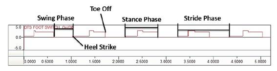
To analyze the kinematic data, 40 consecutive gait cycles were used. The temporal variables on the limb affected by knee OA were calculated: speed (distance traveled divided by time), support time (a reference time in which the limb heel was in contact with the ground), swing time (time between the heel lift and its next contact with the ground), stride time (time between two consecutive touches of the reference heel), and double support time (time during which both heels were in contact with the ground). When a heel strike occurred, the footswitch signal changed from 0 to 5 mV. When toe off occurred, the footswitch signal returned from 5 mV to baseline. Gait phases were determined based on the onset and offset of the footswitch. Figure 1 shows the way the signals from the footswitch sensor were used to determine heel strike, toe off, stance phase, and swing phase17. The average of 40 trials was calculated for each analyzed variable for data analysis.
Statistical analysis
Statistical analysis was performed on PASW statistics, 18.0® (SPSS). The paired-samples t-test was applied after data normality was assessed using the Shapiro-Wilk test. The receptor operation characteristic (ROC) curve was used to assess the prediction accuracy of the biomechanical variables of the functioning level in individuals with KOA. Subsequently, the cutoff value of these variables and their sensitivity and specificity was established by discriminant function analysis. A p<0.05 significance was adopted in all statistical tests.
RESULTS
Table 1 shows participants’ characteristics. The sample showed a 16% dropout loss.
Primary outcome
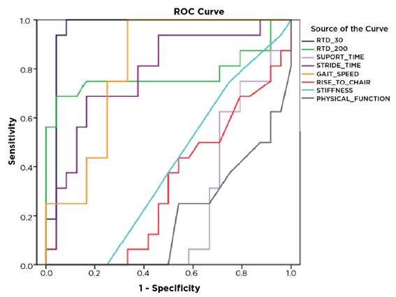
ROC curve analysis (Figure 1) showed that the variables with greater specificity and sensitivity to predict functional performance in women with KOA consisted of gait speed, RTD at 30 ms and RTD at 200 ms. Table 2 shows the area value for the variables predicting functional performance in KOA.
Secondary outcome
Regarding kinematic analysis, the test showed a difference for the variables support time (p<0.001), stride time (p<0.001, F=17.389), and gait speed (p=0.001). For neuromuscular variables, the test showed a difference between the assessments for RDT at 30 ms (p<0.001) and RDT at 200 ms (p=0.007). For objective function, test indicated a difference for stiffness (p=0.005), physical function (p<0.001), and time to rise from a chair five times (p=0.012).
The discriminant function was used to calculate the cutoff value of the predictive variables. Their discriminatory thresholds totaled 1.07 ms and 2.06 Nm.s−1Kg−1, and 1.72 Nm.s −1Kg−1, respectively.
Receiver operating characteristic curve analysis to predict functional performance in women with knee osteoarthritis
DISCUSSION
This study showed that women with KOA had poor functioning in the one-year follow-up according to the self-reported (WOMAC) and performance functioning measures (RTD, gait speed, support time, stride time, and time to rise from a chair five times). RTD and gait speed proved themselves better predictors due to their greater sensitivity and specificity in the ROC curve area.
The reduction in functional mobility constitutes the main repercussion of KOA. It refers to the difficulty of performing a variety of tasks that are important for maintaining independence, such as walking, sitting and getting up from chairs, and crossing streets18,19. The literature on physical functioning limitation in KOA includes a wealth of cross-sectional studies but few longitudinal studies. Thus, this study sought to find the biomechanical variable that could predict these individuals’ functioning level in order to propose more effective intervention strategies for this population. Among the analyzed kinematic and dynamometric variables, RTD and gait speed showed better sensitivity/specificity.
RTD comprises the ability to quickly generate strength7. According to the analyzed muscle contraction phase, several physiological factors influence RTD. Early RTD (0-30ms) is more correlated with the number of active motor units and the trigger rate of action potentials for the muscle7,16. Late RTD (>90ms) after the start of muscle contraction is more strongly associated with maximum strength7,20. The results of this study showed a reduction in RTD over its one-year follow-up. The variable predicted functional performance. A lower knee extensor RTD at the beginning of explosive contractions largely stemmed from rapid activation failure, that is, the inability to quickly activate the quadriceps21. We found a 2.06 Nm.s-1.Kg-1 for early TDT and 1.72 Nm.s-1.Kg-1 for early and late RTD cutoffs, respectively, which can serve to find individuals with KOA and limited functioning.
Unlike our initial hypothesis, knee extensor torque was unable to predict these individuals’ mobility level. This may be related to the fact that the decrease in the ability to generate strength quickly occurs more sharply than muscle strength22. Our findings corroborate with Callahan et al. (2015)23 and Ventura et al. (2019)8, who state that the ability to increase torque as quickly as possible during muscle contraction better represents performance of functional tasks than peak torque as daily activities require an appropriate combination of muscle torque and speed for its execution instead of maximum force, as for example, to cross streets. Thus, the authors of this study reinforce that RTD is fundamental to prevent physical-functional decline in women with KOA.
The WOMAC questionnaire score showed a difference in stiffness and physical functioning at the one-year follow-up. Patient-reported outcome measures prevail in clinical settings to serve these purposes despite their limited clinical value due to inherent subjectivity, potential ceiling effects, and dependence on pain rather than actual daily life activities24. This may have been occurred in the low sensitivity and specificity of the questionnaire according to the ROC curve area. As an alternative, timed performance-based tests (the speed to walk 12 meters, for example) can obtain simple measures of mobility. At the one-year follow-up, women with KOA had a reduction of 0.14m/s in gait speed. Considering that a 0.10 m/s reduction in gait speed is associated with poorer health status and a higher risk of disability, this difference between assessments has clinical importance25. The reduced gait speed aims to decrease the joint load on the limb affected by the disease and distribute body weight to both lower extremities13. Reduced gait speed is associated with quadriceps weakness since this muscle is responsible for the dynamic stability of this joint, which is greatly required in quiet stance support14. Gait speed could serve as general marker to track physical functioning over time during disease progression KOA. Clinical practice could consider a 1.07 m/s cutoff point for individuals with and without functional limitations.
The important aspect of this study refers to its finding of RTD and the gait speed as the best predictors of functional performance in women with KOA in relation to the other kinematic and dynamometric variables. Despite the greater sensitivity, specificity, and area of the ROC curve of RTD, calculating gait speed has greater applicability in clinical practice as it requires no high-cost devices or great skills for its analysis. Furthering knowledge of factors contributing to poor physical functioning will aid the development of strategies to prevent functional limitation and disability. We emphasize that the extrapolation of these data to a sample with different characteristics requires caution.
Finally, the authors recommend the inclusion of specific power exercises and resistance training for the knee extensor muscles to improve RTD. Moreover, gait training should also belong to the treatment of patients with KOA to improve their movement patterns. Further studies should be carried out to test the effectiveness of training on the functional impairment of women with KOA.
The limitations of this study are related to its approach involving only the knee extensor muscles as this muscular group is the most affected by the disease. However, muscular weakness affects the entire lower limb.
CONCLUSION
OA increases the risk of poor functioning in this one-year follow-up according to factors such as RTD and gait speed. The identification of these factors help characterize women with KOA who stand at a greater risk of disability, providing possible targets for rehabilitative and self-management strategies to prevent disability.
ACKNOWLEDGEMENTS
This study was supported by the Coordination of Superior Level Staff Improvement.
DATA AVAILABILITY
The data underlying this study are available in the published article.
REFERENCES
-
1 Bennel KL, Nelligan RK, Schwartz S, Kasza J, Kimp A. Behavior change text messages for home exercise adherence in knee osteoarthritis: randomized trial. J Med Internet Res. 2020;22(9):e21749. doi: 10.2196/21749
» https://doi.org/10.2196/21749 -
2 Gholami Z, Faezi ST, Letafatkar A, Madreseh A. Pain neuroscience education, blended exercises and booster sessions as an effective therapy for pain, functional and psychological factors in patients with knee osteoarthritis: a study protocol for a single-blind randomized controlled trial with 22 factorial design during 6-month follow-up. BMJ Open. 2023;13(5):e070336. doi: 10.1136/bmjopen-2022-070336
» https://doi.org/10.1136/bmjopen-2022-070336 -
3 Nelligan RK, Hinman RS, Atkins L, Bennel KL. A Short message service intervention to support adherence to home-based strengthening exercise for people with knee osteoarthritis: intervention design applying the behavior change wheel. JMIR Mhealth Uhealth. 2019;7(10):e14619. doi: 10.2196/14619
» https://doi.org/10.2196/14619 -
4 Nelligan RK, Hinman RS, Kasza J, Schwartz S, Kimp A, et al. Effect of a short message service (SMS) intervention on adherence to a physiotherapist-prescribed home exercise program for people with knee osteoarthritis and obesity: protocol for the ADHERE randomized controlled trial. BMC Musculoskelet Disord. 2019;20(428):428-38. doi: 10.1186/s12891-019-2801-z
» https://doi.org/10.1186/s12891-019-2801-z -
5 Baert IAC, Meeus M, Mahmoudian A, Luyten FP, Nijs J, et al. Do psychosocial factors predict muscle strength, pain, or physical performance in patients with knee osteoarthritis? J Clin Rheumatol. 2017;23(6):308-16. doi: 10.1097/rhu.0000000000000560
» https://doi.org/10.1097/rhu.0000000000000560 -
6 Aydemir B, Huang CH, Foucher KC. Strength and physical activity in osteoarthritis: the mediating role of kinesiophobia. J Orthop Res. 2022;40(5):1135-42. doi: 10.1002/jor.25151
» https://doi.org/10.1002/jor.25151 -
7 Maffiuletti NA, Aagaard P, Blazevich AJ, Folland J, Tillin N, et al. Rate of force development: physiological and methodological considerations. Eur J Appl Physiol. 2016;116(6):1091-116. doi: 10.1007/s00421-016-3346-6
» https://doi.org/10.1007/s00421-016-3346-6 -
8 Ventura A, Muendle B, Friesenbichler B, Casartelli NC, Kramers I, et al. Deficits in rate of torque development are accompanied by activation failure in patients with knee osteoarthritis. J Electromyogr Kinesiol. 2019;44:94-100. doi: 10.1016/j.jelekin.2018.11.010
» https://doi.org/10.1016/j.jelekin.2018.11.010 -
9 Machado S, Santana E, Brito V, Maciel L, Quintans Júnior LJ, et al. Knee osteoarthritis: kinesiophobia and isometric strength of quadriceps in women. Pain Res Manag. 2022;5:1466478. doi: 10.1155/2022/1466478
» https://doi.org/10.1155/2022/1466478 -
10 Ismailidis P, Hegglin L, Egloff C, Pagenstert G, Kernen R, et al. Side to side kinematic gait differences within patients and spatiotemporal and kinematic gait differences between patients with severe knee osteoarthritis and controls measured with inertial sensors. Gait Posture. 2021;84:24-30. doi: 10.1016/j.gaitpost.2020.11.015
» https://doi.org/10.1016/j.gaitpost.2020.11.015 -
11 Straaten RVD, Wesseling M, Jonkers I, Vanwanseele B, Bruijines A, et al. Functional movement assessment by means of inertial sensor technology to discriminate between movement behavior of healthy controls and persons with knee osteoarthritis. J Neuroeng Rehabil. 2020;17(1):65-76. doi: 10.1186/s12984-020-00694-2
» https://doi.org/10.1186/s12984-020-00694-2 -
12 Silva DO, Barton CH, Briani RV, Taborda B, Ferreira AS, et al. Kinesiophobia, but not strength, is associated with altered movement in women with patellofemoral pain. Gait Posture. 2019;68:1-5. doi: 10.1016/j.gaitpost.2018.10.033
» https://doi.org/10.1016/j.gaitpost.2018.10.033 -
13 Murray AM, Thomas AC, Armstrong CW, Pietrosimone BG, Tevald MA. The associations between quadriceps muscle strength, power, and knee joint mechanics in knee osteoarthritis: A cross-sectional study. Clin Biomech. 2015;30(10):1140-5. doi: 10.1016/j.clinbiomech.2015.08.012
» https://doi.org/10.1016/j.clinbiomech.2015.08.012 -
14 Spinoso DH, Bellei NC, Marques NR, Navega MT. Quadriceps muscle weakness influences the gait pattern in women with knee osteoarthritis. Adv Rheumatol. 2018;58(1):26-31. doi: 10.1186/s42358-018-0027-7
» https://doi.org/10.1186/s42358-018-0027-7 -
15 Aagaard P, Simonsen EB, Andersen JL, Magnusson P, Dyhre-Poulsen P. Increased rate of force development and neural drive of human skeletal muscle following resistance training. J Appl Physiol. 2002;93(4):1318-26. doi: 10.1152/japplphysiol.00283.2002
» https://doi.org/10.1152/japplphysiol.00283.2002 -
16 LaRoche DP, Cremin KA, Greenleaf B, Croce RV. Rapid torque development in older female fallers and nonfallers: a comparison across lower-extremity muscles. J Electromyogr Kinesiol. 2010;20(3):482-8. doi: 10.1016/j.jelekin.2009.08.004
» https://doi.org/10.1016/j.jelekin.2009.08.004 -
17 Marques NR, Hallal CZ, Spinoso DH, Morcelli MH, Crozara LF, et al. Applying different mathematical variability methods to identify older fallers and non-fallers using gait variability data. Aging Clin Exp Res. 2017;29(3):473-81. doi: 10.1007/s40520-016-0592-8
» https://doi.org/10.1007/s40520-016-0592-8 -
18 Zhang L, Wang Y, Ye T, Hu Y, Wang S, et al. Quality of clinical practice guidelines relevant to rehabilitation of knee osteoarthritis: A systematic review. Clin Rehabil. 2022; 37(7):986-1008. doi: 10.1177/02692155221144892
» https://doi.org/10.1177/02692155221144892 -
19 Sharma L, Cahue S, Song J, Hayes K, Pai YC, et al. Physical functioning over three years in knee osteoarthritis: role of psychosocial, local mechanical, and neuromuscular factors. Arthritis Rheum. 2003;48(12):3359-70. doi: 10.1002/art.11420
» https://doi.org/10.1002/art.11420 -
20 Andersen LL, Andersen JL, Zebis MK, Aagaard P. Early and late rate of force development: differential adaptive responses to resistance training? Scan J Med Sci Sports. 2010;20(1):162-9. doi: 10.1111/j.1600-0838.2009.00933.x
» https://doi.org/10.1111/j.1600-0838.2009.00933.x -
21 Park SK, Kobsar D, Ferber R. Relationship between lower limb muscle strength, self-reported pain and function, and frontal plane gait kinematics in knee osteoarthritis. Clin Biomech. 2016;38:68-74. doi: 1016/j.clinbiomech.2016.08.009
» https://doi.org/1016/j.clinbiomech.2016.08.009 -
22 Berger MJ, McKenzie CA, Chess DG, Goela A, Doherty TJ. Quadriceps neuromuscular function and self-reported functional ability in knee osteoarthritis. J Appl Physiol. 2012;113(2):255-62. doi: 10.1152/japplphysiol.00947.2011
» https://doi.org/10.1152/japplphysiol.00947.2011 -
23 Callahan DM, Tourville TW, Slauterbeck JR, Ades PA, Stevens-Lapsley JB, et al. Reduced rate of knee extensor torque development in older adults with knee osteoarthritis is associated with intrinsic muscle contractile deficits. Exp Gerontol. 2015;72:16-21. doi: 1016/j.exger.2015.08.016
» https://doi.org/1016/j.exger.2015.08.016 -
24 Boekesteijn RJ, Gerven JV, Geurts AGH, Smulders K. Objective gait assessment in individuals with knee osteoarthritis using inertial sensors: A systematic review and meta-analysis. Gait Posture. 2022;98:109-20. doi: 10.1016/j.gaitpost.2022.09.002
» https://doi.org/10.1016/j.gaitpost.2022.09.002 -
25 Purser JL, Weinberger M, Cohen H, Pieper CF, Morey MC, et al. Walking speed predicts health status and hospital costs for frail elderly male veterans. J Rehabil Res Dev. 2005;42(4):535-46. doi: 10.1682/jrrd.2004.07.0087
» https://doi.org/10.1682/jrrd.2004.07.0087