 Post-surgical rehabilitation of a rare variant of Guillain-Barré syndrome: acute motor axonal neuropathy with distal tibial fracture
Abstract
Introduction: Guillain-Barré syndrome (GBS), particularly the acute motor axonal neuropathy (AMAN) variant, presents with rapidly progressive motor weakness and often requires prolonged rehabilitation. Orthopaedic complications in such cases are rare but can increase management complexity.
Objective: To report the post-surgical rehabilitation of a patient with GBS/AMAN complicated by a distal tibial fracture and to highlight the effectiveness of a structured, interdisciplinary rehabilitation approach.
Methods: A 31-year-old male with AMAN/GBS, complicated by respiratory failure requiring ventilatory support, sustained a right distal tibial fracture during rehabilitation. The fracture was surgically managed via closed reduction and intramedullary nailing. A 12-week structured rehabilitation program was implemented, beginning with early respiratory exercises, pain management, and passive joint mobilization, followed by progressive weight-bearing, muscle strengthening, proprioceptive training, and functional gait practice. The final phase incorporated resistance exercises, dynamic balance, and task-specific functional activities. Functional outcomes were assessed using the Barthel Index, Functional Independence Measure (FIM), and Berg Balance Scale (BBS).
Results: The patient achieved substantial functional improvement. Barthel Index increased from 16 to 90/100, FIM from 59 to 106/128, and BBS from 4 to 15/56. He regained independent ambulation with minimal assistance, improved postural control, and could perform most activities of daily living safely. The outcomes demonstrate that early mobilization and progressive, individualized rehabilitation can overcome both neurological and orthopaedic limitations.
Conclusion: This case highlights that structured, multidisciplinary rehabilitation program can achieve meaningful functional recovery in GBS complicated by orthopaedic trauma, emphasizing early mobilization, strength restoration, and balance training.
Keywords:
Acute motor axonal neuropathy; Guillain-Barré syndrome; Interdisciplinary care; Rehabilitation; Tibial fracture
Resumo
Introdução: A síndrome de Guillain-Barré (SGB), particularmente a variante neuropatia axonal motora aguda (NAMA), manifestase com fraqueza motora rapidamente progressiva e frequentemente requer reabilitação prolongada. Complicações ortopédicas nesses casos são raras, mas podem aumentar a complexidade do tratamento.
Objetivo: Relatar a reabilitação póscirúrgica de um paciente com SGB/NAMA complicada por fratura distal da tíbia e destacar a eficácia de uma abordagem de reabilitação estruturada e interdisciplinar.
Métodos: Um ho-mem de 31 anos com NAMA/SGB, complicada por insuficiência respiratória com necessidade de suporte ventilatório, sofreu uma fratura distal da tíbia direita durante a reabilitação. A fratura foi tratada cirurgicamente por meio de redução fechada e fixação intramedular. Um programa de reabilitação estruturado de 12 semanas foi implementado, iniciando com exercícios respiratórios precoces, controle da dor e mobilização articular passiva, seguidos por carga progressiva, fortalecimento muscular, treinamento proprioceptivo e prática de marcha funcional. A fase final incorporou exercícios de resistência, equilíbrio dinâmico e atividades funcionais específicas. Os resultados funcionais foram avaliados utilizando o Índice de Barthel, a Medida de Independência Funcional (MIF) e a Escala de Equilíbrio de Berg (EEB).
Resultados: O paciente apresentou melhora funcional substancial. O Índice de Barthel aumentou de 16 para 90/100, a MIF de 59 para 106/128 e a EEB de 4 para 15/56. Ele recuperou a deambulação independente com assistência mínima, melhorou o controle postural e conseguiu realizar a maioria das atividades da vida diária com segurança. Os resultados demonstram que a mobilização precoce e a reabilitação progressiva e individualizada podem superar as limitações neurológicas e ortopédicas.
Conclusão: Este caso destaca que um programa de reabilitação estruturado e multidisciplinar pode alcançar uma recuperação funcional significativa na SGB complicada por trauma ortopédico, enfatizando a mobilização precoce, a recuperação da força e o treinamento do equilíbrio.
Palavras-chave:
Neuropatia axonal motora aguda; Síndrome de Guillain-Barré; Cuidado interdisciplinar; Reabilitação; Fratura da tíbia
Introduction
Guillain-Barré syndrome (GBS) is an autoimmune nerve, disorder that develops suddenly, often leading to rapidly progressing muscle weakness.1 It often necessitates both intensive medical care and rehabilitation.2 The acute motor axonal neuropathy (AMAN) variant of GBS is particularly challenging due to severe motor deficits and a prolonged, variable recovery period. Early recovery typically occurs within 1-6 months, with some patients regaining the ability to walk 5 meters with assistance in a median of 31 days. Intermediate recovery spans 6–12 months, while complete functional recovery may extend from 1 to 5 years, particularly in the presence of comorbidities or limited access to timely interventions.3,4 Epidemiologically, the AMAN variant accounts for approximately 30-50% of GBS cases in several Asian countries including India, China, Japan, and Bangladesh, and 30-47% of cases in Central and South America, whereas it represents only 3–7% of cases in Europe and North America, indicating its relative rarity in Western populations but higher prevalence in Asia.5,6
Although rarely documented, fractures from weakness in GBS patients require dual attention. A coordinated multidisciplinary approach is necessary for the rehabilitation of dual pathologies such as orthopaedic and neurological impairment, in order to guarantee functional restoration without compromising surgical outcomes.7 This case report highlights the rehabilitation course of a patient with GBS/AMAN who also experienced a right distal tibial fracture in the early stages of recovery. It highlights key clinical decisions, therapeutic challenges, and the functional gains achieved by a tailor-made neuroorthopaedic rehabilitation strategy. This report underscores the importance of integrated, practical strategies that can guide clinicians across, orthopaedics, neurology, physical medicine and rehabilitation and physiotherapy in managing multi-system conditions.
Case report
A 31-year-old male presented with acute onset weakness, initially involving the left hand. On February 26, 2022, he noticed weakness in the first and second digits of his left hand, which impaired his ability to perform fine motor tasks such as buttoning his shirt. By the following day, the weakness had progressed rapidly; he was unable to get out of bed and experienced a fall when attempting to stand. He was admitted to a tertiary care hospital, where clinical evaluation and diagnostic investigations confirmed a diagnosis of GBS/AMAN.
During the course of hospitalization, the patient developed respiratory failure, necessitating intubation and mechanical ventilation. Due to prolonged ventilator dependence and bilateral limb weakness, a tracheostomy was performed on March 3, 2022, which was maintained for approximately three months. In addition to respiratory support, the patient underwent plasmapheresis therapy to manage the underlying autoimmune process. Although his medical condition gradually stabilized, he remained functionally dependent and required structured caregiver support for basic activities of daily living due to persistent motor impairment. He received intravenous immunoglobulin therapy as part of the acute management protocol. Following the intervention, gradual neurological improvement was noted. He was successfully weaned off the ventilator, transitioned to bilevel positive airway pressure support, and subsequently decannulated as his respiratory status stabilized. He was discharged after 70 days.
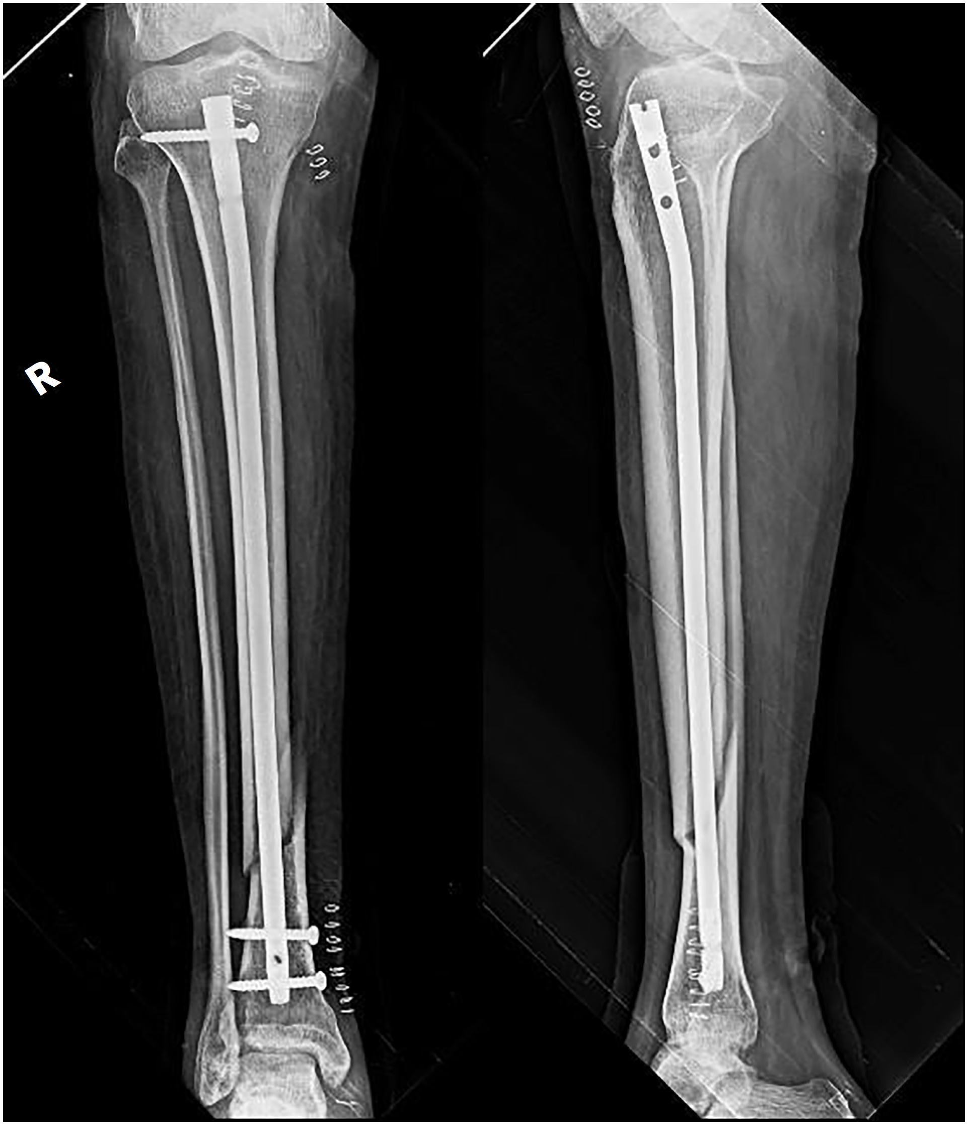
During the early phase of rehabilitation, the patient experienced a mechanical fall on February 9, 2023, resulting in a comminuted distal tibial fracture on the right side. Contributing factors included neuromuscular fatigue and an underlying osteoporotic bone structure. Radiographic evaluation (Figure 1) confirmed the fracture, which was managed with closed reduction and internal fixation using intramedullary nailing. Given the delayed callus formation and evidence of osteoporotic bone observed intraoperatively, the surgical team opted for intramedullary nailing instead of conservative management with Plaster of Paris (POP) or plate-and-screw fixation, as the thin cortex and poor bone quality increased the risk of fixation failure. A metabolic bone panel was performed to identify contributing factors, which revealed mildly low vitamin D levels (24 ng/mL) with normal calcium (9.1 mg/dL) and phosphate (3.6 mg/dL) levels. Alkaline phosphatase was within the expected range for bone healing (138 IU/L). These findings indicated bone weakening secondary to prolonged immobilization. Accordingly, calcium and vitamin D3 supplementation were optimized, and periodic monitoring continued throughout rehabilitation to support bone healing, enhance neuromuscular recovery, and reduce the risk of delayed union or refracture.

Pre-operative anteroposterior and lateral radiographs showing a comminuted distal tibial fracture with cortical thinning, indicating osteopenic bone consistent with immobilization-related weakness.
To address the residual impairments from GBS and promote fracture healing, the patient initially pursued complementary treatment, undergoing an eight-month course of Ayurvedic treatment alongside conventional medical care. Following this integrated approach, he was admitted to our neurorehabilitation unit for a comprehensive, evidence-based neurorehabilitation program, targeting both neurological and orthopaedic recovery.
On February 9, 2023, he underwent titanium tibial intramedullary nailing under general anesthesia. Under C-arm guidance, the intraoperative findings and post-operative fixation were verified as stable (Figure 2). Two distal locking screws and one distal dynamic screw were used to secure the 9 × 360 mm titanium IL nail after a patellar tendon split approach and reaming up to size 10.8 A multidisciplinary team, including a neurophysician, cardiologist, and general practitioner, evaluated the patient's surgical fitness. Preoperative assessment included cardiovascular evaluation (normal electrocardiogram - ECG - and preserved left ventricular ejection fraction > 55%), hematological screening (normal prothrombin time/international normalized ratio, activated partial thromboplastin time), and nutritional and meta-bolic optimization (albumin 3.2 g/dL, hemoglobin 11.8 g/dL, creatinine 0.9 mg/dL).

Post-operative anteroposterior and lateral radiographs demonstrating stable intramedullary fixation with proper alignment, confirming surgical stability for early mobilization.
Airway patency post-tracheostomy was confirmed, and, given the absence of infection and stable cardiopulmonary status, the patient was cleared for intramedullary nailing under general anaesthesia. Isolation and COVID-19 prevention measures were appropriately implemented.
The patient reported chest pain on the first post-operative day. The patient was moved to the intensive care unit for observation after an ECG was completed. Additional testing with USG Doppler ruled out deep vein thrombosis, though subcutaneous edema was noted around the right lower limb and ankle. An additional infectious risk was presented by the growth of Pseudomonas aeruginosa, which showed resistance to multiple antibiotics as demonstrated by microbiological culture.
Analgesics, antibiotics, and intravenous fluids were used to treat the patient. Following surgery, a comprehensive medication regimen was prescribed to aid in the patient's recovery. Pantoprazole (40 mg) and Domperidone SR (30 mg) were administered for gastrointestinal protection. Eight milligrams of Lornoxicam were used to control inflammation. Cisbind Plus, which contains Cissus quadrangularis, calcium citrate, vitamin D3, and vitamin K2-7, helped to strengthen and heal bones. The addition of Calcirol Plus, which contains calcitriol, zinc, and calcium citrate, also aided in preserving the mineral balance of the body. Furthermore, a nutritional supplement known as Vitawill 4G was added; it contains a variety of vitamins, trace minerals, omega-3 fatty acids, green tea, ginseng, spirulina, and grape seed extract among other ingredients to promote immunity and general healing.
The patient was discharged in stable condition with a comprehensive, evidence-based rehabilitation plan. To address the neurological problems caused by GBS as well as the healing process following the right distal tibial fracture, physiotherapy was initiated using a patient-centered, individualized approach. The rehabilitation process was structured into three progressive phases — early, intermediate, and advanced — each designed to address specific functional goals corresponding to the patient's medical status and individualized recovery needs. Despite the complexity of his condition, the patient remained highly motivated, cognitively intact, and actively engaged throughout the rehabilitation program. His psychological resilience and strong participation played a pivotal role in the successful progression across all phases of recovery.
Early phase rehabilitation (weeks 1-4)
The goal of treatment during the first four weeks was to support respiratory health, manage pain, prevent joint contractures, reduce edema, and facilitate early callus formation at the fracture site. The affected limb was elevated and ice therapy was applied systematically to reduce swelling and inflammation. Passive range of motion exercises for the hip and knee were performed to prevent joint stiffness. Active movement exercises were used to strengthen the upper limbs, and breathing techniques were incorporated to support lung function and prevent pulmonary complications. These targeted interventions were structured to complement the biological timeline of callus formation while promoting recovery from the neuromuscular deficits associated with GBS (Table 1).
A 12-week rehabilitation protocol outlining goals and physiotherapy interventions for a patient with acute motor axonal neuropathy variant of Guillain-Barré syndrome and a distal tibial fracture
Manual muscle testing revealed significant neuromuscular weakness at the beginning of rehabilitation. The quadriceps and ankle dorsiflexors received a score of 2 out of 5, whereas the gluteus muscles received a marginally higher score of 3 out of 5. On the right leg, a long knee immobilizer was used to provide structural support for the weakened quadriceps muscle. Bilateral ankle-foot orthoses (AFO) were fitted to compensate for ankle weakness and prevent foot drop during eventual ambulation. Standing was initially contraindicated due to the post-operative condition and risk of orthostatic intolerance associated with autonomic dysfunction in GBS. Instead, a tilt table positioned at an initial angle of 60° with appropriate head, arm, pelvis, and femur support was used to mobilize the patient gradually. This starting angle was selected based on evidence indicating that 60° provides sufficient gravitational stimulus to activate postural reflexes and cardiovascular adaptation while minimizing the risk of orthostatic hypotension, which commonly occurs at angles >70° in neurologically compromised patients during early mobilization. Blood pressure and heart rate were monitored continuously during each session; the absence of orthostatic symptoms served as the criterion for progression.9 Although cognitive functioning was unaffected, there were significant deficits in lower limb muscle power and balance. The patient's Mini-Mental State Examination score of 28/ 30 indicated that their cognitive status was preserved and that they were able to actively engage in therapy.
To develop a well-structured rehabilitation plan, the orthopaedic and neurology teams worked collaboratively. Both teams had carefully considered when and how to initiate mobilization because the patient had both physical limitations from a tibial fracture and nerve-related issues from GBS. While the orthopaedic team was cautious about protecting the healing bone, particularly with delayed callus formation, the neurology team focused on helping the patient regain function and prevent muscle deterioration.10 Through collaborative decision-making, the rehabilitation team created a plan that was safe, progressive, and aligned with both sets of recovery objectives. The patient's ability to walk short distances with bilateral elbow crutches by the end of the first phase of rehabilitation marked a significant advancement in their recovery (Table 1).
Intermediate phase rehabilitation (weeks 5-8)
The intermediate phase of rehabilitation (weeks 5-8) commenced with a focus on gradual mobilization. To activate the quadriceps and hamstring muscles, isometric exercises were introduced, and the patient began to use a walker for partial weight-bearing activities (Table 1).
To enhance coordination and balance, proprioceptive training with active-assisted range of motion exer-cises were implemented. Although post-operative X-ray taken 2-weeks after surgery confirmed that callus formation at the fracture site was still insufficient, early mobilization was carefully managed using a tilt table with a solid AFO to minimize the risk of delayed union.
During the intermediate phase, the tilt angle was systematically progressed from 70 to 90° across multiple sessions, with incremental increases of 10° every 3-4 sessions based on individual tolerance and hemodynamic stability. The 70° threshold represents a critical transition point where cardiovascular demand increases substantially, requiring careful monitoring before advancing to full vertical positioning. Reaching 90° (full upright) is essential for normalizing baroreceptor sensitivity, optimizing venous return, and preparing the neuromuscular system for weight-bearing through parallel bars. This graduated approach allowed safe adaptation to orthostatic stress while simultaneously facilitating bone loading through the fractured tibia within protected weight-bearing limits.11,12
Following his return to the unit, the patient was gradually progressed to upright standing within parallel bars, with weight distribution on each limb closely monitored using a weight scale. Given the bilateral quadriceps strength of 2/5, both lower limbs were supported with immobilizers to enhance stability during early weight-bearing. Weight loading on the right (fractured) leg was initiated cautiously, beginning at 10% of body weight and incrementally increased to 50% based on patient tolerance and serial radiographic findings. Meanwhile, full weight-bearing was permitted on the left leg, and biomechanical loading was adjusted every fourth week by progressively increasing right-side loading by 10% increments, with careful pelvic weight shift training under parallel bar support to facilitate symmetrical gait retraining.
Advanced phase rehabilitation (weeks 9-12)
Following orthopaedic clearance, full weight-bearing was authorized by the ninth week in May 2023. During this advanced phase of rehabilitation (weeks 9-12), the program focused on functional movements, such as sit-to-stand exercises, gait training, and progressive resistance training with therabands. Although the implant was stable, X-rays revealed only partial callus formation. As a result, the orthopaedic team recommended calcium supplements and continued caution during loading activities. With the assistance of a right knee immobilizer and bilateral leaf spring AFO, the patient was able to ambulate using bilateral elbow crutches for balance support. The right quadriceps and bilateral ankle dorsiflexors were graded at 2+/5, necessitating the continued use of assistive devices. Fatigue management remained a critical component of care to prevent potential exacerbation of GBS. To facilitate a safe transition back to daily activities, dynamic balance exercises were integrated into the rehabilitation program (Table 1).
The interdisciplinary approach used to treat this rare combination of orthopaedic trauma and GBS makes this complex case noteworthy. With careful surgical intervention, continuous medical supervision, and a well-structured physiotherapy program, the patient demonstrated significant progress. By the end of the 12-week period, notable improvements were observed in muscle strength, ambulatory independence, and overall quality of life, highlighting the effectiveness of a coordinated neurological and orthopaedic rehabilitation strategy.13
Outcome and follow-up
After undergoing closed reduction and internal fixation for a right distal tibia fracture in the context of GBS/AMAN, the patient demonstrated significant functional improvements during a 12-week interdisciplinary rehabilitation program. Upon admission, the patient had severe neuromuscular impairments, considerable mobility limitations, and was entirely bedridden and dependent on care-taker for all activities of daily living.
The patient progressed to independent ambulation with a rollator walker through the structured rehabilitation program. Toileting and lower body dressing required minimal assistance, representing significant progress toward functional independence. The Modified Barthel Index score increased from 16/100 to 90/100, particularly in activities like walking, toilet use, and bed mobility, indicating substantial improvements in self-care and mobility (Figure 3). Gains in self-care, transfers, mobility, and social interaction were also demonstrated by the Functional Independence Measure, which increased from 59/128 to 106/128. Similarly, the Berg Balance Scale improved from 4/56 to 15/56, indicating improved static and dynamic balance, although more complex balancing tasks still required supervision (Figure 3).
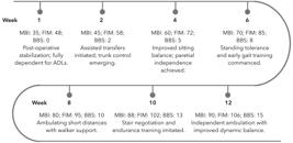
Twelve-week functional recovery profile, demonstrating progressive improvements in activities of daily living (ADLs) (Functional Independence Measure - FIM), ambulatory capacity (Modified Barthel Index - MBI), and balance (Berg Balance Scale - BBS) following a structured, multidisciplinary neuroorthopaedic rehabilitation protocol.
Lower limb strength increased from an initial grade of 2/5 to a functional 3+/5, enabling greater independence during daily tasks. Crucially, throughout the entire rehabilitation process, no secondary complications were noted, including refracture, pressure sores, or respiratory problems. This case demonstrates the efficacy and safety of a well-coordinated neurological and orthopaedic rehabilitation approach in managing complex multi-system conditions.
Discussion
GBS, particularly in its AMAN variant, is characterized by axonal damage causes rapidly progressing motor weakness.14 This frequently results in the requirement for prolonged rehabilitation assistance and critical care.15 In this case report, a rare and difficult consequence occurred where a 31-year-old male patient with AMAN suffered a right distal tibial fracture after a mechanical fall in the early stages of inpatient neurorehabilitation. Although such orthopaedic injuries are uncommon in GBS, they can occur, especially in the presence of neuromuscular fatigue and bone weakening due to prolonged corticosteroid use or immobility.16
Managing the dual challenges of GBS and orthopaedic trauma required a timely team-based strategy that addressed both neurological and orthopaedic requirements. Regaining neuromuscular control, minimizing muscle loss, and preventing fatigue-related setbacks are the usual goals of recovery from GBS.17 In contrast, orthopaedic care emphasizes safe healing, tissue protection, gradual loading, and avoiding issues such as implant failure or delayed bone healing.18 Achieving a balance between these objectives is rarely covered in the literature and poses significant clinical challenges particularly for patients with severe lower limb weakness.
Unlike standard GBS rehabilitation which permits immediate full weight-bearing mobilization, this case required modified progression limited by fracture healing, with weight-bearing restricted to 10-50% during weeks 1-8 and delayed resistance training until radiographic callus formation was confirmed. Bilateral immobilizers and AFO were essential to offload the healing tibia while compensating for severe quadriceps weakness, and tilt table progression was extended to monitor both orthostatic tolerance and mechanical bone stability.19
Patient's rehabilitation program adhered to established GBS rehabilitation guidelines, including early respiratory care, tilttable mobilization, and passive range of motion exercises at the beginning.20 In addition to preserving lung function and joint flexibility, these early interventions also helped prevent avoid orthostatic intolerance which is a common problem in GBS patients because of autonomic system involvement.21
Progressive weight-bearing was introduced cautiously and guided by radiographic findings because delayed callus formation is a common issue in post-operative patients with underlying metabolic fragility.22 Following the guidelines, the switch from tilttable mobilization to walker-assisted gait training was predicated on continuing strength assessments.23 The patient utilized AFO and a knee immobilizer to enhance walking and stability which was aligned with standard guidelines for people with drop foot caused by peripheral neuropathy or weak quadriceps.24
This patient's recovery was greatly aided by fatigue management which is often overlooked in GBS rehabilitation. In a study conducted by Gawande et al.,25 the rehabilitation plans involved plan incorporated pacing techniques, assistive technology, and planned rest intervals through workload modification and the monitoring of exertional fatigue symptoms. This case demonstrates the importance of a phased and individualized rehabilitation approach in managing combined neurological and orthopaedic conditions. Collaboration among neurology, orthopaedics, and physiotherapy enabled the development of a recovery strategy that supported both neurological stability and fracture healing. Regular assessments ensured that therapy remained aligned with the patient's evolving needs and progress.
Conclusion
This case demonstrates the effectiveness of an interdisciplinary rehabilitation strategy in managing GBS alongside a tibial fracture. A structured, phased program led to significant improvements in mobility, strength, and independence. Early mobilization, assistive support, and fatigue management were essential to the patient's recovery. Beyond physical gains, the rehabilitation emphasized restoring the patient's independence, thereby reducing the caregiver's burden. Successful reintegration into society and return to occupational roles further underline the importance of individualized rehabilitation plans in complex multi-system conditions.
These outcomes highlight the critical role of physiotherapy within an interdisciplinary team, where coordinated planning and tailored interventions are essential for optimizing recovery in complex neuroorthopaedic cases. Structured interdisciplinary rehabilitation is costeffective, reduces complications and caregiver dependency, and future studies are needed to evaluate long-term follow-up for sustained functional recovery and early detection of late complications.
Data availability statement
Research data are available from the corresponding author upon reasonable request.
References
-
1 Willison HJ, Jacobs BC, van Doorn PA. Guillain-Barré syndrome. Lancet. 2016;388(10045):717-27. https://doi.org/10.1016/s0140-6736(16)00339-1
» https://doi.org/10.1016/s0140-6736(16)00339-1 -
2 Sejvar JJ, Baughman AL, Wise M, Morgan OW. Population incidence of Guillain-Barré syndrome: a systematic review and meta-analysis. Neuroepidemiology. 2011;36(2):123-33. https://doi.org/10.1159/000324710
» https://doi.org/10.1159/000324710 -
3 Sulli S, Scala L, Berardi A, Conte A, Baione V, Belvisi D, et al. The efficacy of rehabilitation in people with Guillain-Barrè syndrome: a systematic review of randomized controlled trials. Expert Rev Neurother. 2021;21(4):455-61. https://doi.org/10.1080/14737175.2021.1890034
» https://doi.org/10.1080/14737175.2021.1890034 -
4 Vishnuram S, Abathsagayam K, Suganthirababu P. Physiotherapy management of a rare variant of Guillain Barre Syndrome, acute motor and sensory axonal neuropathy (AMSAN) along with COVID-19 in a 35-year-old male–a case report. Afr Health Sci. 2022;22(3):520-6. https://doi.org/10.4314/ahs.v22i3.56
» https://doi.org/10.4314/ahs.v22i3.56 -
5 Shrivastava M, Nehal S, Seema N. Guillain–Barre syndrome: Demographics, clinical profile & seasonal variation in a tertiary care centre of central India. Indian J Med Res. 2017;145(2):203-8. https://doi.org/10.4103/ijmr.ijmr_995_14
» https://doi.org/10.4103/ijmr.ijmr_995_14 -
6 Oliveira DRCAB, Fernandez RNM, Grippe TC, Baião FS, Duarte RL, Fernandez DJ. Epidemiological and clinical aspects of Guillain-Barré syndrome and its variants. Arq Neuropsiquiatr. 2021;79(6):497-503. https://doi.org/10.1590/0004-282X-ANP-2020-0314
» https://doi.org/10.1590/0004-282X-ANP-2020-0314 -
7 Shende K, Ratnani G, Deodhe NP, Gandhi KM. Promoting flexibility and functionality in a surgically managed tibial fracture: a case report on physiotherapeutic interventions for postoperative stiffness. Cureus. 2023;15(12):e50589. https://doi.org/10.7759/cureus.50589
» https://doi.org/10.7759/cureus.50589 -
8 Devarakonda S, Subramanian AK, Alabdulmuhsin FH, Shawky AS, Minervini G. Resorbable We43 versus titanium fixation devices-evaluation of treatment outcomes: a systematic review. Bull Stomatol Maxillofac Surg. 2025;21(1):119-31. https://tinyurl.com/25jm3tnc
» https://tinyurl.com/25jm3tnc -
9 Meythaler JM. Rehabilitation of Guillain-Barré syndrome. Arch Phys Med Rehabil. 1997;78(8):872-9. https://doi.org/10.1016/s0003-9993(97)90203-3
» https://doi.org/10.1016/s0003-9993(97)90203-3 -
10 Stephen GG, Mohanan K, Kandakurti PK, Mathukrishnan R. Comparative analysis of the effects of real-time audio-visual feedback, yoga and pressure biofeedback stabilizer in core strengthening: a randomised controlled trial. Biomed Hum Kinet. 2025;17(1):135-46. https://doi.org/10.2478/bhk-2025-0013
» https://doi.org/10.2478/bhk-2025-0013 -
11 Kiper P, Chevrot M, Godart J, Cieślik B, Kiper A, Regazzetti M, et al. Physical exercise in Guillain-Barré syndrome: a scoping review. J Clin Med. 2025;14(8):2655. https://doi.org/10.3390/jcm14082655
» https://doi.org/10.3390/jcm14082655 -
12 Arsenault NS, Vincent PO, Yu BHS, Bastien R, Sweeney A. Influence of exercise on patients with Guillain-Barré syndrome: a systematic review. Physiother Can. 2016;68(4):367-76. https://doi.org/10.3138/ptc.2015-58
» https://doi.org/10.3138/ptc.2015-58 -
13 M K, Josyula S, S JA, M N, J H, J V, et al. Comparative analysis of high-intensity interval training and traditional rehabilitation programs for accelerated recovery from musculoskeletal injuries. J Back Musculoskelet Rehabil. 2024;37(2):437-43. https://doi.org/10.3233/bmr-230146
» https://doi.org/10.3233/bmr-230146 -
14 Yuki N, Hartung HP. Guillain-Barré syndrome. N Engl J Med. 2012;366(24):2294-304. https://doi.org/10.1056/nejmra1114525
» https://doi.org/10.1056/nejmra1114525 - 15 Lennon S, Ramdharry G, Verheyden G. Physical management for neurological conditions. Amsterdam: Elsevier; 2018.
-
16 Sri Lekha M, Vishnuram S, K R, Abathsagayam K, Suganthirababu P. Efficacy of dynamic neuromuscular stabilization exercises on balance and fall risk in subjects with diabetic peripheral neuropathy among geriatrics - A pilot study. Phys Occup Ther Geriatr. 2025;43(4):285-99. https://doi.org/10.1080/02703181.2025.2467803
» https://doi.org/10.1080/02703181.2025.2467803 -
17 Harbo T, Markvardsen LK, Hellfritzsch MB, Severinsen K, Nielsen JF, Andersen H. Neuromuscular electrical stimulation in early rehabilitation of Guillain-Barré syndrome: A pilot study. Muscle Nerve. 2019;59(4):481-4. https://doi.org/10.1002/mus.26396
» https://doi.org/10.1002/mus.26396 -
18 Malasani S, Jha G, Ganesh S. A prospective study on the suprapatellar approach for tibial shaft fractures: Insights into functional and radiological outcomes. Cureus. 2024;16(11): e74862. https://doi.org/10.7759/cureus.74862
» https://doi.org/10.7759/cureus.74862 -
19 Sung EJ, Kim DY, Chang MC, Ko EJ. Prediction of functional outcome in axonal Guillain-Barre syndrome. Ann Rehabil Med. 2016;40(3):481-8. https://doi.org/10.5535/arm.2016.40.3.481
» https://doi.org/10.5535/arm.2016.40.3.481 -
20 Leonhard SE, Mandarakas MR, Gondim FAA, Bateman K, Ferreira MLB, Cornblath DR, et al. Diagnosis and management of Guillain-Barré syndrome in ten steps. Nat Rev Neurol. 2019; 15(11):671-83. https://doi.org/10.1038/s41582-019-0250-9
» https://doi.org/10.1038/s41582-019-0250-9 -
21 Higgins R, Parker J, O’Flaherty L, Perkins N, Swayne O. The neurorehabilitation of people with Guillain-Barré Syndrome. Adv Clin Neurosci Rehabil. 2024;22(4):22–5. https://doi.org/10.47795/PUEK6754
» https://doi.org/10.47795/PUEK6754 -
22 Subash Y. Evaluation of functional outcome following hybrid external fixation in the management of Schatzkers type V and VI tibial plateau fractures - A prospective study of 30 patients. Malays Orthop J. 2021;15(1):48-54. https://doi.org/10.5704/moj.2103.008
» https://doi.org/10.5704/moj.2103.008 -
23 Orsini M, Freitas MRG, Presto B, Mello MP, Reis CHM, Silveira V, et al. Guideline for neuromuscular rehabilitation in Guillain-Barré syndrome: What can we do? Rev Neurocienc. 2010;18(4):572-80. https://doi.org/10.34024/rnc.2010.v18.8443
» https://doi.org/10.34024/rnc.2010.v18.8443 -
24 Tanaka M, Wada Y, Kawate N. Effectiveness of gait training with lower limb orthosis for a patient with severe Guillain-Barré syndrome at a Kaifukuki rehabilitation ward. Jpn J Compr Rehabil Sci. 2021;12:48-52. https://doi.org/10.11336/jjcrs.12.48
» https://doi.org/10.11336/jjcrs.12.48 -
25 Gawande I, Akhuj A, Samal S. Effectiveness of physiotherapy intervention in Guillain Barre syndrome: a case report. Cureus. 2024;16(1):e52062. https://doi.org/10.7759/cureus.52062
» https://doi.org/10.7759/cureus.52062
Edited by
-
Associate editor:
Emmanuel Souza da Rocha