 Severe bleeding diathesis after dental extractions: a complex case of coagulation disturbances in a patient with multiple myeloma
ABSTRACT
This case report describes a male patient diagnosed with multiple myeloma who experienced significant bleeding from both the maxillary and mandibular dental sockets following dental extraction, lasting several days. Laboratory tests revealed prolonged prothrombin time and thrombin time, accompanied by reduced factor VII levels. Initial management with intravenous vitamin K and fresh frozen plasma failed to achieve hemostasis. Despite the risk of thrombosis, low-dose tranexamic acid was initiated, resulting in effective hemorrhage control and improved healing of the alveolar sockets. This case illustrates the complex hemostatic imbalance between pro- and anticoagulant factors in a patient with multiple myeloma. It also underscores the diagnostic and therapeutic challenges associated with managing bleeding in patients with a high risk of thrombosis.
Keywords:
Multiple myeloma; Hemorrhage; Tooth extraction; Blood coagulation
INTRODUCTION
Hematological malignancies are frequently associated with disturbances in hemostasis, resulting in an increased risk of both thrombosis and bleeding. Multiple myeloma (MM) is a clonal plasma cell neoplasm that poses the highest risk of thromboembolic events among hematologic neoplasms owing to various coagulation abnormalities, including elevated thrombin levels, impaired fibrinolysis, and elevated M-protein plasma concentration.(1) In addition, prolonged immobilization, surgical procedures, and chemotherapy further predispose patients with MM to venous thromboembolism, which can affect overall survival.(2)
Subsequent studies on patients with MM have also documented instances of bleeding with reported frequencies ranging from 1% to 28%.(1) Common clinical manifestations include epistaxis, easy bruising, and bleeding from the gums or gastrointestinal tract, often occurring without significant abnormalities on coagulation tests. This case report describes an imbalance between pro- and anti-coagulant factors in a patient with MM who developed prolonged bleeding following dental extraction. The diagnostic process was particularly challenging due to the absence of a specific detectable coagulation abnormality in laboratory tests that could account for persistent bleeding, as well as the need to carefully balance hemostatic management against the heightened risk of thrombosis associated with procoagulant therapy.
CLINICAL REPORT
In September 2023, an 82-year-old man was admitted to a private hospital with significant weight loss of approximately 20kg, sarcopenia, anemia, elevated liver enzyme levels, reduced appetite, and weakness. He had been diagnosed with consumptive syndrome a year prior to this admission. The patient also had cardiac arrhythmia, depression, diabetes, and chronic lower back pain secondary to a previous fracture. These conditions were being managed with metoprolol, dapagliflozin, venlafaxine, mirtazapine, and buprenorphine. Additionally, the patient had a history of bleeding following colonoscopy with polyp resection that required reintervention with clipping, and a fall resulting in an occipital lesion with abnormal bleeding.
Serum and imaging tests were conducted to evaluate the patient's significant body weight loss and spinal orthopedic conditions. Elevated levels of serum IgG (2.113mg/dL) and free kappa light chains (704.5mg/dL) indicated monoclonal gammopathy. Further laboratory assessment revealed an increase in free light chains (combined value 7.39mg/dL; individual kappa and lambda levels were not available in the medical records).
Computed tomography (CT) and magnetic resonance imaging (MRI) revealed rarefaction of the trabecular bone network and multiple vertebral fractures. Bone marrow biopsy revealed 25% clonal plasma cells with atypical CD56 and CD117 positivity, kappa-free chain immunoglobulin restriction, and amyloid deposits, confirming the presence of a plasma cell neoplasm. Multiple myeloma was diagnosed based on marrow biopsy findings, evidence of monoclonal gammopathy, and the presence of anemia and bone lesions (Table 1).
Values in blood count, coagulogram, liver and kidney functions, and ionized calcium were performed prior to dental extractions and repeated at 5 and 10 days post-extraction
In October 2023, prior to the oncohematological treatment, the patient experienced gingival bleeding and dental pain. Dental examination revealed a fractured crown and signs of gingival inflammation. Panoramic radiographs demonstrated advanced carious lesions involving multiple sites on the upper premolars and molars, radicular remnants in the lower right premolar, and dental implants in the lower right molar region (Figure 1A). Dental extractions were indicated, and unfractionated heparin, administered since the diagnosis of MM for thrombosis prevention, was discontinued. Following prophylactic antibiotic coverage with azithromycin due to penicillin allergy, the right maxillary molars and radicular remnants were extracted without complications.

A) Dental panoramic radiograph of a patient with multiple myeloma displaying infectious areas and the necessity for dental extractions. B) Intense dental bleeding 24 hours post-extraction. C) Occasional bleeding continued for a week following the dental procedure, requiring ongoing hemostatic interventions
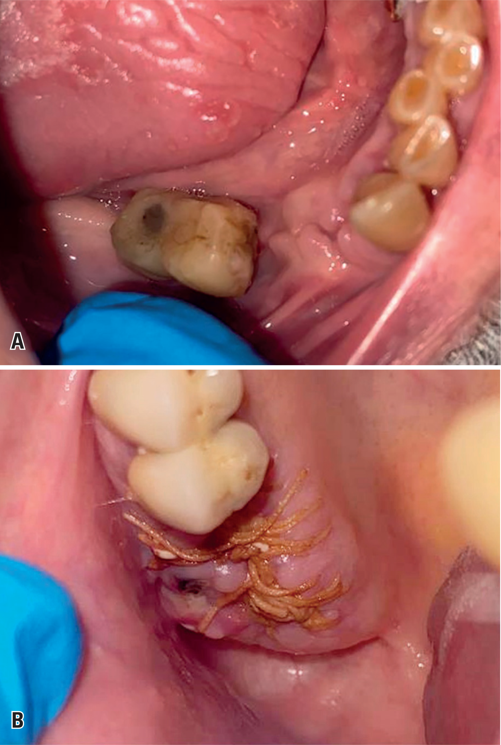
On the first postoperative day, the patient experienced significant bleeding in the upper and lower dental sockets, which was managed with local compression (Figures 1B and 1C). Daily bleeding from the extraction site was observed for eight days, mostly occurring in the early morning. The application of topical tranexamic acid and noradrenaline anesthetics helped stop the bleeding (Figures 2A and 2B). However, poor mucosal regeneration was observed in this area, consistent with delayed wound healing. Laboratory tests demonstrated prolonged prothrombin time (PT) and thrombin time (TT) (Table 1). Bleeding on postoperative day eight required emergency intervention, including systemic administration of tranexamic acid and other hemostatic measures (Figures 2A and 2B). Treatment with a high-power laser, fibrin sponge, n-butyl-2-cyanoacrylate glue, and tranexamic acid solution effectively controlled the bleeding (Figures 2C and 2D).

A and B) Severe dental bleeding occurred eight days following dental extraction, requiring immediate intervention with local and systemic hemostatic measures. C and D) Bleeding was successfully controlled using a high-power laser, fibrin sponge, n-butyl-2-cyanoacrylate glue, and tranexamic acid solution
On the ninth day following the dental extractions, the patient experienced a fall from standing height, resulting in the recurrence of dental bleeding. The previously described interventions were repeated to manage the hemorrhage. Given the persistent bleeding and delayed healing, a coagulopathy related to amyloidosis or local tumor infiltration was suspected. A gingival biopsy near the extraction site was performed without complications. Although amyloidosis in the bone marrow was previously confirmed, the gingival biopsy revealed no evidence of amyloid deposits or plasma cell infiltration.
To analyze the prolonged PT, a 50:50 test (mixed with plasma control) was conducted (Table 1). The results indicated a deficiency in coagulation factors, as PT normalized after mixing. Further analysis revealed a factor VII deficiency. Although there was no evidence of liver dysfunction or other clinical signs of vitamin K deficiency, intravenous vitamin K was administered based on the results of the PT mixture test; however, PT remained prolonged, and bleeding continued. Given this complex laboratory profile and the broader composition of fresh frozen plasma (FFP) compared to prothrombin complexes, systemic FFP therapy was initiated but also failed to achieve hemostatic control.
Episodes of bleeding occurred sporadically for days following the dental extraction. Low-dose intravenous tranexamic acid was prescribed (500mg TID) to control the dental hemorrhage. In the first week of November 2023, the patient began treatment for multiple myeloma with lenalidomide 10 mg (adjusted dose) and dexamethasone 20mg (weekly dose). Despite the thrombogenic risk of this therapy, anticoagulants were not prescribed, considering the bleeding episodes. After treatment with tranexamic acid and subsequent targeted therapy, the oral bleeding resolved completely, and wound healing progressed satisfactorily (Figure 3). The patient's nutritional status improved significantly following the resumption of normal mastication and diet. Despite the resolution of the hemorrhagic episodes, prolonged PT and TT persisted during follow-up. After hospital discharge, the patient had a better performance status and a partial response, evidenced by increased hemoglobin and decreased M protein levels. Unfortunately, the patient died in March 2024 from septic shock secondary to multilobar pneumonia associated with Influenza A infection (Table 2).

A and B) Dental socket showed efficient healing 30 days following the dental extraction. Dental bleeding stopped once the multiple myeloma treatment began
This study was approved by the Ethics Committee of the Hospital Israelita Albert Einstein (CAAE: 84127824.0.0000.0071; #7.207.169), and written informed consent was obtained from the patient.
DISCUSSION
This case highlights the complex hemostatic imbalance in a patient with MM who is simultaneously at risk of both thrombosis and bleeding. Thrombocytopenia, dysfibrinogenemia, and fibrinolysis disturbances, which are commonly associated with hemorrhage in patients with MM(3,4) were not observed in this case, as evidenced by normal thromboelastography findings. Nevertheless, other conditions such as hyperviscosity syndrome, macroglobulinemia, monoclonal gammopathy, abnormal MM proteins(1) and platelet dysfunction(4) can lead to prolonged thrombin time, affecting fibrin polymerization and increasing the risk of bleeding. Furthermore, the M protein can lead to the suppression of the von Willebrand factor, impairing primary hemostasis.(1) The identification of these alterations is seldom documented in patients with MM and often underrecognized, potentially being underestimated.(1)
In this case, laboratory abnormalities associated with bleeding episodes included prolonged PT and TT. Prolonged PT has a prevalence ranging from 25% to 48% in patients with MM.(3,5,6) The correction of INR following a prothrombin mixing test indicated a factor coagulation deficiency, which was further confirmed by the decreased level of factor VII detected in blood tests. Other cases of MM with factor VII deficiency were diagnosed when an increase in the prothrombin time was detected.(7,8) This phenomenon has been previously documented in two additional patients with MM who presented with severe epistaxis and esophageal hemorrhage.(8)
Acquired factor VII deficiency is significantly less common than the hereditary form, which represents the most prevalent of the rare congenital coagulation factor deficiencies.(8) In this case, it was unclear whether the deficiency was hereditary or induced by MM. In MM cases, research indicates that IgG1 with kappa and lambda chains may bind to the activated form of factor VII, thereby inhibiting its procoagulant function, including its interaction with tissue factor.(9) In addition, liver dysfunction, which could be associated with factor VII deficiency, was not observed. The AST, ALT, and bilirubin levels were normal, with moderately elevated GGT and alkaline phosphatase levels. There were no signs of cirrhosis, and the spleen appeared normal on ultrasonography. Upper endoscopic findings were normal, without signs of portal hypertension. Repeat factor VII testing was not performed, considering that bleeding was associated with changes in both PT and TT levels.
Skin hemorrhage, periorbital purpura, and life-threatening bleeding are common symptoms of light-chain amyloidosis and are often associated with factor X deficiency.(1,8) In this case, we considered a potential amyloid deposit in the gingiva; however, biopsy results did not confirm the presence of amyloidosis. Additionally, factor X levels were normal, indicating that the oral bleeding was not attributable to a coagulopathy linked to light-chain amyloidosis.
Other cases of MM mentioned in the literature have also reported challenges in achieving hemostasis following dental extraction(10,11) as well as spontaneous gingival bleeding and oral blood blisters.(6) In one case, the bleeding was attributed to tumor cell infiltration of the jawbones.(11) Additionally, a review of patients with MM presenting with oral hemorrhage identified deficiencies in factors I, II, VIII, and X; however, no factor VII deficiency was observed in any of these cases of dental bleeding.
CONCLUSION
We report a unique case of a patient with multiple myeloma complicated by prolonged bleeding following dental extractions, characterized by prolonged prothrombin and thrombin time and low factor VII levels, likely associated with myeloma. The challenges in diagnosing and treating this condition are compounded by the risk of thrombosis, making this case particularly complex.
DATA AVAILABILITY
The underlying content is contained within the manuscript.
REFERENCES
- 1 Coppola A, Tufano A, Di Capua M, Franchini M. Bleeding and thrombosis in multiple myeloma and related plasma cell disorders. Semin Thromb Hemost. 2011;37(8):929-45.
- 2 Jarchowsky O, Avnery O, Ellis MH. Thrombosis in multiple myeloma: mechanisms, risk assessment and management. Leuk Lymphoma. 2023; 64(12):1905-13.
- 3 Gogia A, Sikka M, Sharma S, Rusia U. Hemostatic Abnormalities in Multiple Myeloma Patients. Asian Pac J Cancer Prev. 2018;19(1):127-30.
- 4 Li L, Roest M, Sang Y, Remijn JA, Fijnheer R, Smit K, et al. Patients with multiple myeloma have a disbalanced whole blood thrombin generation profile. Front Cardiovasc Med. 2022;9:919495.
- 5 Pandey S, Post SR, Alapat DV, Smock KJ, Post GR. Prolonged prothrombin time correlates with serum monoclonal protein concentration in patients with plasma cell dyscrasia. Int J Lab Hematol. 2013;35(4):421-7.
- 6 Zhang Y, Xu F, Wen JJ, Shi L, Zhou QL. Isolated coagulopathy without classic CRAB symptoms as the initial manifestation of multiple myeloma: A case report. World J Clin Cases. 2022;10(12):3822-7.
- 7 Zaidi MH, Stanley A, Khan M. Acquired Factor VII deficiency - a rare but important consideration. Scott Med J. 2019;64(3):119-22.
- 8 Furlan A, Sartori F, Gherlinzoni F. Acquired isolated factor VII deficiency in plasma cell dyscrasias: a brief presentation of two plasma-cell-leukemia-related cases and review of literature. J Clin Med. 2023;12(18):5837.
- 9 Kamikubo Y, Miyamoto S, Iwasa A, Ishii M, Okajima K. Purification and characterization of factor VII inhibitor found in a patient with life threatening bleeding. Thromb Haemost. 2000;83(1):60-4.
- 10 Urade M, Sugi M, Nishimura K, Sugiyama M, Yakushiji N, Miyazaki T. IgA K type myeloma with severe post extraction bleeding. Int J Oral Surg. 1985;14(2):162-8.
- 11 Mozaffari E, Mupparapu M, Otis L. Undiagnosed multiple myeloma causing extensive dental bleeding: report of a case and review. Oral Surg Oral Med Oral Pathol Oral Radiol Endod. 2002;94(4):448-53.
Edited by
-
Associate Editor:
Marcos de Lima The Ohio State University – Columbus, OH, USA ORCID: https://orcid.org/0000-0002-8568-4522