 Proposing the ValvUS approach: integrating bedside tests and ultrasonography for severe valvular heart disease diagnosis
ABSTRACT
Valvular heart disease is increasingly prevalent, and bedside confirmation or exclusion of severe disease is needed to enable a rapid and cost-effective diagnostic workup. The physical examination skills of clinicians are insufficient for accurate diagnosis, making complementary tests generally necessary. Despite being commonly requested, electrocardiography and chest radiography present low positive and negative likelihood ratios. Incipient studies involving artificial intelligence have shown promising opportunities to support the diagnosis. In addition, solid current evidence demonstrates that point-of-care ultrasound enhances bedside diagnosis of several cardiovascular conditions. Echocardiographic skills can be acquired after only a few hours of training, which encourages routine bedside use with handling equipment. Despite the routine use of sonography in emergencies, large-scale simplified screening protocols for valvular disease remain lacking. Therefore, improving the accuracy of valvular heart disease diagnosis by integrating all bedside modalities needs to be better understood. We propose a simple, reproducible five-step point-of-care ultrasound protocol for diagnosing valvular heart disease (the ValvUS approach), applicable to all patients. The proposed visual assessment involves evaluating valvular movement, thickness, regurgitant flow, aliasing, and chamber dimensions. This evaluation should be interpreted in the context of traditional clinical probability to ensure the most accurate bedside diagnosis. Typical findings of severe valvular disease on electrocardiography and chest radiography, and particularly on point-of-care ultrasound, may improve the accuracy of bedside diagnosis after clinical assessment in the near future.
Heart valve diseases; Aortic valve disease; Mitral valve; Physical examination; Ultrasonography; Point-of-care testing; Point-of-care systems
INTRODUCTION
Valvular heart disease (VHD) is increasingly prevalent among outpatients, particularly in the older population, and the final diagnosis is typically established by comprehensive echocardiography following a clinical suspicion.( 1 ) Most patients present with non-severe VHD, which has no hemodynamic consequences and no specific treatment recommendations.( 2 ) However, identifying severe VHD, which represents the advanced phase of the natural history of the disease, is crucial for recognizing at-risk patients and is a cornerstone of management.( 3 ) Bedside confirmation or exclusion of severe VHD is presumably associated with time and cost savings, improved clinical reasoning, more appropriate treatment, and greater physician-patient confidence.( 4 )
Although the best cardiovascular diagnostic reasoning starts with generating hypotheses through clinical assessment and subsequent testing, current medical practices are firmly based on requesting and reviewing imaging tests.( 5 , 6 ) However, this modern approach has not been associated with better outcomes and probably decreases practitioners’ appreciation of the diagnostic value of history-taking and physical examination.( 7 ) Regarding bedside VHD diagnosis, previous evidence has demonstrated low to moderate positive and negative likelihood ratios (LR) related to murmur detection.( 8 ) Likewise, the physical examination skills of clinicians are generally limited for diagnosing VHD, and great heterogeneity exists in the experience of professionals, including clinicians and teachers.( 9 )
Solid current evidence suggests that electrocardiography, chest radiography, and point-of-care ultrasound (POCUS) enhance the bedside diagnosis of several cardiovascular conditions, thereby complementing the physical examination.( 10 ) Electrocardiography and chest radiography may suggest chamber overload related to severe VHD; however, their diagnostic accuracy is generally limited, with low LR.( 11 , 12 ) Echocardiographic skills can be acquired after only a few hours of training, which encourages routine bedside use with handling equipment.( 13 ) Large-scale, simplified screening protocols for valvular disease are lacking.( 14 ) More recently, artificial intelligence programs have been developed to assist physicians by analyzing complementary tests and highlighting red-flag situations, including VHD.( 15 )
However, despite this evidence, the overall diagnostic accuracy of VHD assessment integrating all bedside modalities remains poorly evaluated. This analytical review synthesizes previous and modern diagnostic strategies to propose a practical and effective bedside diagnostic approach for severe VHD, primarily based on POCUS.
DIAGNOSTIC FLOW
The primary objective of clinical evaluation is to generate diagnostic hypotheses. Cardiology diagnoses are usually probabilistic, and these probabilities are generally conditional.( 16 ) Following the estimation of pre-test probabilities, physicians may refine them using specific complementary tests according to each hypothesis. The likelihood ratios represent the capacity of the test to positively or negatively impact the initial hypothesis. Physicians should avoid the “shotgun approach” (requesting multiple tests without appropriate reasoning) and should be familiar with test-specific LR. A positive LR greater than 10 and a negative LR less than 0.1 are more likely to modify the diagnostic hypothesis.( 17 ) The initial post-test probability may be sufficient to establish the diagnosis; if not, the diagnostic flow resumes from this point using an additional test.
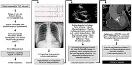
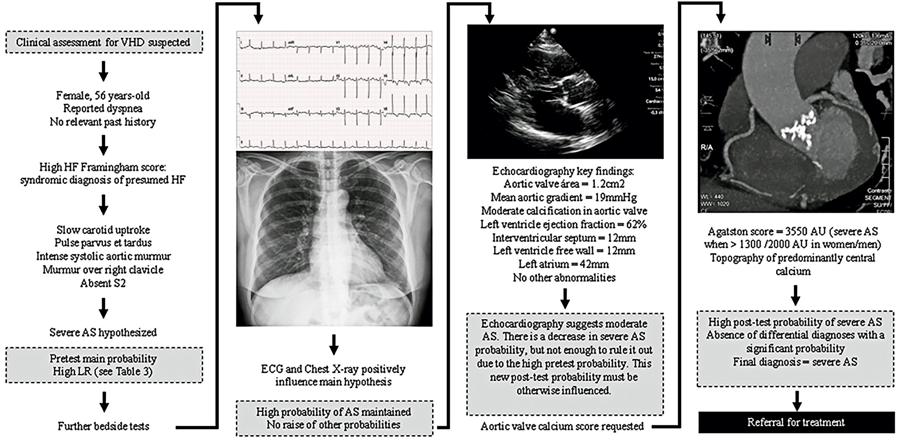
Regarding severe VHD, the pre-test probability is determined primarily through cardiac auscultation.( 18 ) As patient complexity increases, cardiac comorbidities become more common. Paradoxically, simply reviewing an echocardiogram may be insufficient for establishing a valvular diagnosis, underscoring the importance of a structured diagnostic flow. An illustration of the importance of clinical pre-test probability is presented in figure 1 .
PERIPHERAL PHYSICAL EXAMINATION
The major arterial pulses must be examined to determine their patency, rhythm, waveform, and characteristics. Because arterial pulse waves progressively decrease toward the periphery, the carotid pulse – a central pulse – provides more reliable information regarding left ventricular ejection and aortic valve function.( 19 ) A hypokinetic (or hypodynamic) arterial pulse is small and weak and is typically observed in stenosis of the aortic, mitral, or pulmonic valves. Pulsus parvus et tardus (or an anacrotic pulse) refers to a small, delayed systolic peak, usually observed in severe aortic stenosis (AS). A hyperkinetic (or hyperdynamic) arterial pulse is large and strong and is commonly observed in regurgitant valvular disease, which reflects an increased blood volume ejected from the left ventricle. This finding is more pronounced in aortic regurgitation (AR), also known as water hammer pulse , than in mitral regurgitation (MR), where it may present only as brisk.( 20 ) Other cardiac conditions may also produce this type of pulse; however, interobserver variability has not been well studied, and the LR remains poorly defined.
AUSCULTATION FLOW
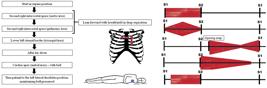
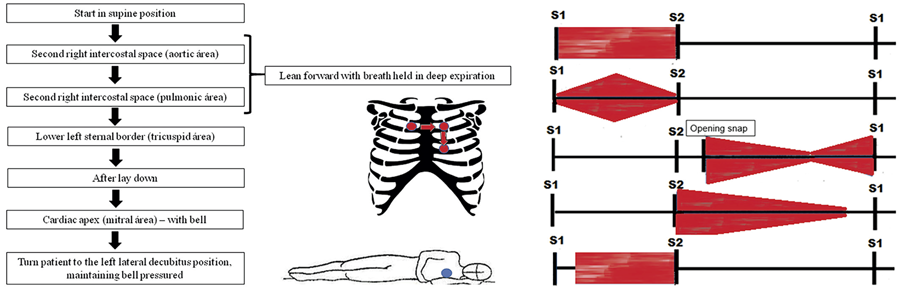
Chest palpation may help detect a valvular thrill, which is usually associated with severe VHD, and can also provide information about ventricular remodeling. Chest percussion has limited clinical value and is rarely emphasized in contemporary practice. A systematic approach to cardiac auscultation is illustrated in figure 2 .
The first split (S1) occurs just before the upstroke of the carotid pulse and is best heard at the apex and along the lower left sternal border, where it is louder than the second split (S2). Because S1 is a high-frequency sound, it is best heard with the diaphragm of the stethoscope.( 21 - 23 ) S2 occurs after the peak of the carotid pulse and coincides with the downslope of the pulse. S2 is louder than S1 and is best heard at the left second intercostal space near the sternal border. S2 is best heard in the semi-recumbent position (approximately 30° upright) during quiet inspiration using the stethoscope.( 21 , 22 , 24 ) Several abnormalities may affect S1 and S2. The causes of VHD are summarized in table 1 .
When a murmur is heard, it must be classified according to the timing within the cardiac cycle, its configuration ( Figure 2 ), and its intensity, using the Levine scale: grade I, very faint and audible only to an expert in a specific position; grade II, soft but audible in all places; grade III, moderately loud without a thrill; grade IV, loud with a thrill; grade V, very loud with a thrill and audible with the stethoscope partially off the chest; and grade VI, audible with the stethoscope completely off the chest.( 25 ) To assess the radiation of the murmur, auscultation is performed over the suprasternal notch, axilla, and epigastrium.
Murmurs may also occur in conditions other than VHD. Several bedside maneuvers have been described to aid in identifying the underlying cause of a murmur. Although these maneuvers do not determine disease severity, they may improve diagnostic accuracy when combined with appropriate investigations.( 26 ) These maneuvers are summarized in table 2 .( 22 , 27 )
Suspicion of VHD arises primarily from physical examination, particularly through the detection of the peripheral manifestations of low cardiac output or high regurgitant volume, but most importantly from the presence of a heart murmur.( 28 ) An evidence-based summary of physical examination signs, along with their positive and negative LR (LR+ and LR-) for the diagnosis of severe chronic VHD, is shown in table 3 .( 29 - 33 )
ELECTROCARDIOGRAPHY AND CHEST RADIOGRAPHY
A complete electrocardiogram can now be performed rapidly at the bedside using increasingly portable equipment with reliable image transmission.( 34 ) Although chest radiography is not strictly a bedside test, it remains an easily accessible examination.( 35 ) These tests may reveal multiple changes associated with significant VHD. However, no comprehensive compilation of LR exists for each disease. In summary, left atrial overload combined with signs of pulmonary hypertension and right-sided chamber overload may suggest mitral stenosis. Left chamber overload is typically associated with MR. Left ventricular concentric hypertrophy may indicate AS, whereas left ventricular eccentric hypertrophy may suggest AR.
ARTIFICIAL INTELLIGENCE
Artificial intelligence-driven medical technologies are developing rapidly into applicable solutions in clinical practice. Deep learning algorithms can process increasing amounts of data from various sources, yielding models that improve the diagnosis and management of different cardiac conditions.( 36 ) Regarding valvular disease, a recent study evaluated an electronic murmur detection algorithm in 603 outpatients, with a total of 3,180 heart sound recordings. The software identified pathologic cases with a sensitivity of 93% and a specificity of 81%, corresponding to a positive LR of 4.8 and a negative LR of 0.08.( 37 ) Likewise, an intelligent diagnostic system developed by Sun et al.( 38 ) discriminated AR, MR, and pulmonary stenosis with accuracies of 98.9%, 98.4%, and 98.7%, respectively. However, this emerging technology could not accurately recognize areas distant from the sternum.
In a retrospective study of more than 250,000 patients, Cohen-Shelly et al. identified moderate to severe AS in 3.7% of cases by echocardiography. Artificial intelligence analysis was applied to half of the patients using electrocardiograms, and further validation was conducted in 10% of the patients. The diagnostic performance showed a positive LR of 3 and a negative LR of 0.29 for AS.( 39 ) Another study using a similar methodology, but including external validation, reported a positive LR of 4.3 and a negative LR of 0.24 for artificial intelligence-based electrocardiogram analysis in the diagnosis of AS.( 40 )
Automatic detection of multiple diseases from chest imaging is a popular topic in radiology. However, only one study has examined the detection of VHD through analysis of the cardiovascular border on posteroanterior chest radiography. In this pilot study, the agreement between manual and software interpretation of border parameters was high, but current data are insufficient to support VHD diagnosis using this algorithm.( 41 )
Several studies have evaluated artificial intelligence in echocardiographic assessment, reporting high mean accuracy for automated software interpretation.( 42 ) Other studies have demonstrated that artificial intelligence algorithms can be applied across various modalities in POCUS.( 43 )
POCUS
POCUS has been widely used, mainly over the last decade, as an extension of physical examinations. Its use is now well established in clinical practice for scenarios such as cardiac arrest, trauma, acute undifferentiated dyspnea, and shock.( 44 , 45 ) By assessing the lungs, left ventricular function, dynamic volume status parameters, and the abdominal cavity, POCUS provides physicians with a bedside tool that yields high positive and negative LR, consistently improving the diagnostic accuracy for the etiology of shock and hemodynamic disorders, while also saving time.( 46 , 47 ) However, in the outpatient setting of VHD, the usefulness and applicability of POCUS remain underexplored, and no specific algorithms have yet been proposed for clinical practice.
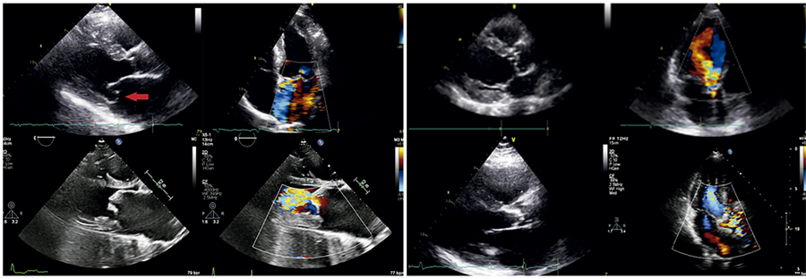
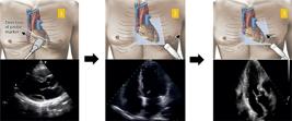
Standard echocardiographic evaluation of patients with VHD is complex. It includes several qualitative and quantitative criteria, making bedside assessment challenging and requiring simplification into a few objective points to be feasible.( 3 ) The main criteria used to assess VHD severity include: i) valvular morphology, valve area, and transvalvular pressure gradient, which determine flow turbulence visualized as color Doppler aliasing; ii) left atrial and ventricular dimensions; iii) left ventricular function; and iv) assessment of the regurgitant jet, mainly its width, in cases of valve regurgitation.( 1 , 47 - 51 ) A POCUS approach should first assess these anatomic criteria of valvular severity, followed by evaluation of the hemodynamic repercussions.( 48 ) Adapting standard echocardiographic recommendations to bedside feasibility, we propose a focused valvular ultrasound approach (ValvUS protocol) comprising five simple steps: evaluation of valve leaflet motion, thickness, regurgitant flow analysis, detection of aliasing, and visual estimation of chamber size and function. All criteria can be evaluated using only three echocardiographic views: the parasternal long-axis view and the apical four- and three-chamber views (Figures 3 and 4 ). It is important to emphasize that the ValvUS approach should be integrated with the traditional clinical pre-test probability to achieve the most accurate bedside diagnosis. The greater the number of consistent clinical and ultrasound findings, the higher the positive LR for severe VHD – and conversely, fewer findings reduce diagnostic certainty. It should be noted that the ValvUS approach has not yet been extensively validated. Future studies across various VHD scenarios are needed to further evaluate the role of POCUS and its associated LR in each valvular condition. As illustrated in the central figure, we believe that, when combined with clinical suspicion, physical examinations, and emerging artificial intelligence technologies, POCUS may become an important bedside tool for identifying severe VHD in the near future.
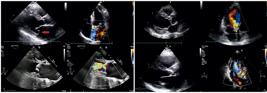
Mitral Valve: Mitral regurgitation: A) parasternal long-axis view - systolic displacement of the mitral leaflets into the left atrium (red narrow) with associated left atrial enlargement; B) parasternal long-axis view - regurgitant flow from the left ventricle to the left atrium during systole. Mitral stenosis: C) parasternal long-axis view - thickening and calcification of the mitral valve with left atrial enlargement; D) parasternal long-axis view - flow aliasing observed distal to the mitral valve.
Aortic Valve: Aortic regurgitation: A) parasternal long-axis view - thickened aortic valve with marked eccentric left ventricular hypertrophy; B) apical three-chamber view - color Doppler demonstrating aortic regurgitation from the aorta to the left ventricular apex during diastole (red narrow). Aortic stenosis: C) parasternal long-axis view - thickening and calcification of the aortic valve with concentric or eccentric left ventricular hypertrophy; D) apical three-chamber view - flow aliasing distal to the aortic valve. Laminar flow appears as a uniform blue color before the aortic valve, whereas after the stenotic region, an acceleration pattern is observed on color Doppler, with a mosaic of blue, red, and yellow signals.
COMMENTS
The physical examination skills of clinicians are insufficient for an accurate diagnosis of severe VHD; however, they usually initiate the diagnostic flow. Electrocardiography and chest radiography, which are easily obtained, may contribute to the diagnosis of severe VHD by demonstrating left-sided chamber remodeling. This process may soon be enhanced by artificial intelligence-based analyses in clinical practice. Importantly, extending the physical examination with POCUS is fundamental in many aspects of patient assessment. The proposed ValvUS approach, which evaluates five components (valve leaflet motion, thickness, regurgitant flow, aliasing, and chamber dimensions), may also assist in identifying severe VHD at the bedside. This information needs to be integrated with other clinical features, as traditional ultrasonographic findings can increase the pre-test probability and thereby prompt an urgent comprehensive echocardiographic assessment. Future studies with large patient cohorts are required to confirm the overall role of POCUS in clinical practice and to establish its diagnostic accuracy across various VHD pathologies.
DATA AVAILABILITY:The underlying content is contained within the manuscript.
REFERENCES
- 1 Otto CM, Nishimura RA, Bonow RO, Carabello BA, Erwin JP 3rd, Gentile F, Jneid H, Krieger EV, Mack M, McLeod C, O'Gara PT, Rigolin VH, Sundt TM 3rd, Thompson A, Toly C. 2020 ACC/AHA Guideline for the Management of Patients With Valvular Heart Disease: Executive Summary: A Report of the American College of Cardiology/American Heart Association Joint Committee on Clinical Practice Guidelines. Circulation. 2021;143(5):e35-e71. Erratum in: Circulation. 2021;143(5):e228. Erratum in: Circulation. 2021;143(10):e784.
- 2 Lung B, Vahanian A. Epidemiology of valvular heart disease in the adult. Nat Rev Cardiol. 2011;8(3):162-72.
- 3 Tarasoutchi F, Montera MW, Ramos AI, Sampaio RO, Rosa VE, Accorsi TA, et al. Update of the Brazilian Guidelines for Valvular Heart Disease - 2020. Arq Bras Cardiol. 2020;115(4):720-75.
- 4 Clark BW, Derakhshan A, Desai SV. Diagnostic errors and the bedside clinical examination. Med Clin North Am. 2018;102(3):453-64.
- 5 Brush JE Jr, Sherbino J, Norman GR. Diagnostic reasoning in cardiovascular medicine. BMJ. 2022;376:e064389.
- 6 Iglehart JK. Health insurers and medical-imaging policy-a work in progress. N Engl J Med. 2009;360(10):1030-7.
- 7 Abou Ziki MD, Podell DN, Schiliro DM. The value of the history and physical examination-sailing through medicine with modern tools: a teachable moment. JAMA Intern Med. 2015;175(12):1901-2.
- 8 Fosnocht KM. The rational clinical examination: detecting abnormal systolic murmurs. JAMA. 1997;277(20):1593-4.
- 9 Birrane JP, Lim ZL, Liew CH, Rosseel L, Heerey A, Coleman K, et al. A survey of general practitioners' knowledge and clinical practice in relation to valvular heart disease. Ir J Med Sci. 2022;191(2):777-84.
- 10 Kimura BJ. Point-of-care cardiac ultrasound techniques in the physical examination: better at the bedside. Heart. 2017;103(13):987-94.
- 11 Goldstein MA, Michelson EL, Dreifus LS. The electrocardiogram in valvular heart disease. Cardiovasc Clin. 1993;23:55-64.
- 12 Sultana H, Rahman MM, Begum M, Ferdousi RA, Begum K, Sangma MA, et al. Chest radiography in the evaluation of mitral valvular disease and it's correlation with echocardiography. Mymensingh Med J. 2021;30(2):292-300.
- 13 Cawthorn TR, Nickel C, O'Reilly M, Kafka H, Tam JW, Jackson LC, et al. Development and evaluation of methodologies for teaching focused cardiac ultrasound skills to medical students. J Am Soc Echocardiogr. 2014;27(3):302-9.
- 14 Hammadah M, Ponce C, Sorajja P, Cavalcante JL, Garcia S, Gössl M. Point-of-care ultrasound: closing guideline gaps in screening for valvular heart disease. Clin Cardiol. 2020;43(12):1368-75.
- 15 Long Q, Ye X, Zhao Q. Artificial intelligence and automation in valvular heart diseases. Cardiol J. 2020;27(4):404-20.
- 16 Sackett DL. The rational clinical examination. A primer on the precision and accuracy of the clinical examination. JAMA. 1992;267(19):2638-44.
- 17 Sackett DL, Rennie D. The science of the art of the clinical examination. JAMA. 1992;267(19):2650-2.
- 18 Mrsic Z, Hopkins SP, Antevil JL, Mullenix PS. Valvular heart disease. Prim Care. 2018;45(1):81-94.
- 19 Bedford DE. The ancient art of feeling the pulse. Br Heart J. 1951;13(4):423-37.
- 20 Schlant RC, Felner JM. The arterial pulse-clinical manifestations. Curr Probl Cardiol. 1977;2(5):1-50.
-
21 Meyer TE. Auscultation of heart sounds. UpToDate; 2022 [cited 2022 May 10]. Available from: https://www.uptodate.com/contents/1079
» https://www.uptodate.com/contents/1079 - 22 Meyer CR. The stethoscope. Minn Med. 2008;91(1):34.
- 23 Karnath B, Thornton W, Frye AW. Teaching and testing physical examination skills without the use of patients. Acad Med. 2002;77(7):753.
- 24 Issenberg SB, Felner JM, Brown DD. Cardiac auscultation skills of physicians in training. JAMA. 1997;278(21):1740-1.
- 25 Grewe K, Crawford MH, O'Rourke RA. Differentiation of cardiac murmurs by dynamic auscultation. Curr Probl Cardiol. 1988;13(10):669-721.
- 26 Voin V, Oskouian RJ, Loukas M, Tubbs RS. Auscultation of the heart: the basics with anatomical correlation. Clin Anat. 2017;30(1):58-60.
- 27 David L, Dumitrascu DL. The bicentennial of the stethoscope: a reappraisal. Clujul Med. 2017;90(3):361-3.
- 28 Spiteri MA, Cook DG, Clarke SW. Reliability of eliciting physical signs in examination of the chest. Lancet. 1988;1(8590):873-5.
- 29 Aronow WS, Kronzon I. Correlation of prevalence and severity of valvular aortic stenosis determined by continuous-wave Doppler echocardiography with physical signs of aortic stenosis in patients aged 62 to 100 years with aortic systolic ejection murmurs. Am J Cardiol. 1987;60(4):399-401.
- 30 Choudhry NK, Etchells EE. The rational clinical examination. Does this patient have aortic regurgitation? JAMA. 1999;281(23):2231-8.
- 31 Etchells E, Bell C, Robb K. Does this patient have an abnormal systolic murmur? JAMA. 1997;277(7):564-71.
- 32 Abbasi AS, DeCristofaro D, Anabtawi J, Irwin L. Mitral valve prolapse: comparative value of m-mode, two-dimensional and doppler echocardiography. J Am Coll Cardiol. 1983;2(6):1219-23.
- 33 Guzik P, Malik M. ECG by mobile technologies. J Electrocardiol. 2016; 49(6):894-901.
- 34 Bansal T, Beese R. Interpreting a chest X-ray. Br J Hosp Med (Lond). 2019;80(5):C75-9.
- 35 Thompson WR, Reinisch AJ, Unterberger MJ, Schriefl AJ. Artificial intelligence-assisted auscultation of heart murmurs: validation by virtual clinical trial. Pediatr Cardiol. 2019;40(3):623-9.
- 36 Seetharam K, Shrestha S, Sengupta PP. Cardiovascular imaging and intervention through the lens of artificial intelligence. Interv Cardiol. 2021;16:e31.
- 37 Cohen-Shelly M, Attia ZI, Friedman PA, Ito S, Essayagh BA, Ko WY, et al. Electrocardiogram screening for aortic valve stenosis using artificial intelligence. Eur Heart J. 2021;42(30):2885-96.
- 38 Sun S. An innovative intelligent system based on automatic diagnostic feature extraction for diagnosing heart diseases. Knowl Base Syst. 2015;75:224-38.
- 39 Kwon JM, Lee SY, Jeon KH, Lee Y, Kim KH, Park J, et al. Deep learning-based algorithm for detecting aortic stenosis using electrocardiography. J Am Heart Assoc. 2020;9(7):e014717.
- 40 Kim C, Lee G, Oh H, Jeong G, Kim SW, Chun EJ, et al. A deep learning-based automatic analysis of cardiovascular borders on chest radiographs of valvular heart disease: development/external validation. Eur Radiol. 2022;32(3):1558-69.
- 41 Alsharqi M, Woodward WJ, Mumith JA, Markham DC, Upton R, Leeson P. Artificial intelligence and echocardiography. Echo Res Pract. 2018;5(4):R115-25.
- 42 Akkus Z, Cai J, Boonrod A, Zeinoddini A, Weston AD, Philbrick KA, et al. A survey of deep-learning applications in ultrasound: artificial intelligence-powered ultrasound for improving clinical workflow. J Am Coll Radiol. 2019;16(9 9 Pt B):1318-28.
- 43 Accorsi TA, Cardoso RG, Paixão MR, De Amicis Lima K, de Souza Júnior L. Uso do ultrassom na parada cardiorrespiratória: estado da arte: ultrassom na parada cardíaca. JBMEDE. 2021;1(2):e21015.
- 44 Martindale JL, Wakai A, Collins SP, Levy PD, Diercks D, Hiestand BC, et al. Diagnosing acute heart failure in the emergency department: a systematic review and meta-analysis. Acad Emerg Med. 2016;23(3):223-42.
- 45 McKaigney CJ, Krantz MJ, La Rocque CL, Hurst ND, Buchanan MS, Kendall JL. E-point septal separation: a bedside tool for emergency physician assessment of left ventricular ejection fraction. Am J Emerg Med. 2014;32(6):493-7.
- 46 Mojoli F, Bouhemad B, Mongodi S, Lichtenstein D. Lung ultrasound for critically ill patients. Am J Respir Crit Care Med. 2019;199(6):701-714. Erratum in: Am J Respir Crit Care Med. 2020;201(8):1015. Erratum in: Am J Respir Crit Care Med. 2020;201(11):1454. Review.
- 47 Mitchell C, Rahko PS, Blauwet LA, Canaday B, Finstuen JA, Foster MC, et al. Guidelines for Performing a Comprehensive Transthoracic Echocardiographic Examination in Adults: Recommendations from the American Society of Echocardiography. J Am Soc Echocardiogr. 2019;32(1):1-64.
- 48 Baumgartner H, Hung J, Bermejo J, Chambers JB, Edvardsen T, Goldstein S, et al. Recommendations on the Echocardiographic Assessment of Aortic Valve Stenosis: A Focused Update from the European Association of Cardiovascular Imaging and the American Society of Echocardiography. J Am Soc Echocardiogr. 2017;30(4):372-92.
- 49 Zoghbi WA, Adams D, Bonow RO, Enriquez-Sarano M, Foster E, Grayburn PA, et al. Recommendations for Noninvasive Evaluation of Native Valvular Regurgitation: A Report from the American Society of Echocardiography Developed in Collaboration with the Society for Cardiovascular Magnetic Resonance. J Am Soc Echocardiogr. 2017;30(4):303-71.
- 50 Baumgartner H, Hung J, Bermejo J, Chambers JB, Evangelista A, Griffin BP, Iung B, Otto CM, Pellikka PA, Quiñones M; American Society of Echocardiography; European Association of Echocardiography. Echocardiographic assessment of valve stenosis: EAE/ASE recommendations for clinical practice. J Am Soc Echocardiogr. 2009;22(1):1-23; quiz 101-2. Erratum in: J Am Soc Echocardiogr. 2009;22(5):442. Erratum in: J Am Soc Echocardiogr. 2023;36(4):445.
- 51 Lancellotti P, Moura L, Pierard LA, Agricola E, Popescu BA, Tribouilloy C, Hagendorff A, Monin JL, Badano L, Zamorano JL; European Association of Echocardiography. European Association of Echocardiography recommendations for the assessment of valvular regurgitation. Part 2: mitral and tricuspid regurgitation (native valve disease). Eur J Echocardiogr. 2010;11(4):307-32.
- 52 Aly I, Rizvi A, Roberts W, Khalid S, Kassem MW, Salandy S, et al. Cardiac ultrasound: an anatomical and clinical review. Transl Res Anat. 2021;22:100083.
Edited by
-
Associate Editor:
Carlos Vicente Serrano Jr Instituto do Coração (InCor), Hospital das Clínica, Faculdade de Medicina, Universidade de São Paulo, São Paulo, SP, Brazil ORCID: https://orcid.org/0000-0002-9171-1224

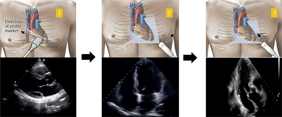 Source: Aly I, Rizvi A, Roberts W, Khalid S, Kassem MW, Salandy S, et al. Cardiac ultrasound: an anatomical and clinical review. Transl Res Anat. 2021;22:100083.(