 Clinical decisions in Orthodontics using x-ray-based images and artificial intelligence approaches: a scoping review
ABSTRACT
Introduction: Artificial intelligence (AI) in health has increased its applications over the last years. The large amount of data available due to the improvement of data storage and digital exams provided better knowledge in treatment planning and opened new possibilities of applications of AI in Orthodontics’ diagnosis and treatment planning.
Objective: The aim of this scoping review was to examine when AI models enhance the clinical decision-making process in orthodontic diagnosis and treatment planning, with a focus on the utilization of X-ray-based imaging.
Methods: Individual eletronic search strategies were developed and conducted on PubMed/Medline, Scopus, Web fo Science, Embase, Lilacs, and Cochrane Library (only English language articles published from January 2000 to October 20, 2021), aiming for relevant studies that met the eligibility criteria.
Results: 12 studies were included, categorized in 5 different groups: Orthognathic surgery, Temporomandibular Joint (TMJ) osteoarthritis, skeletal pattern, obstructive sleep apnea (OSA), and skeletal maturation/development. Most of the AI models used were Deep Learning (DL) based and the X-ray image that was most used was lateral radiographs.
Conclusion: Integrating AI into clinical practice will likely continue to evolve, enhancing treatment planning in selective cases. The best applications were over TMJ osteoarthritis, skeletal maturation, and classification, OSA, and the need for orthognathic surgery.
Keywords:
Artificial intelligence; Orthodontics; X-rays
RESUMO
Introdução: A inteligência artificial (IA) na saúde aumentou suas aplicações nos últimos anos. A grande quantidade de dados disponível, devido ao aprimoramento do armazenamento e dos exames digitais, proporcionou um melhor conhecimento no planejamento do tratamento e abriu novas possibilidades de aplicação da IA no diagnóstico e planejamento ortodôntico.
Objetivo: O objetivo desta revisão de escopo foi examinar quando os modelos de IA aprimoram o processo de tomada de decisão clínica no diagnóstico e planejamento ortodôntico, com foco na utilização de imagens radiográficas.
Métodos: Estratégias eletrônicas de busca individual foram desenvolvidas e realizadas nas bases PubMed/Medline, Scopus, Web of Science, Embase, Lilacs e Cochrane Library (somente artigos em inglês publicados de janeiro de 2000 a 20 de outubro de 2021), buscando estudos relevantes que atendessem aos critérios de elegibilidade.
Resultados: Doze estudos foram incluídos, categorizados em cinco grupos distintos: cirurgia ortognática, osteoartrite da articulação temporomandibular (ATM), padrão esquelético, apneia obstrutiva do sono (AOS) e maturação/desenvolvimento esquelético. A maioria dos modelos de IA utilizados baseava-se em aprendizado profundo (Deep Learning - DL), sendo as radiografias laterais o tipo de imagem mais utilizado.
Conclusão: A integração da IA na prática clínica provavelmente continuará a evoluir, aprimorando o planejamento do tratamento em casos seletivos. As melhores aplicações foram observadas na osteoartrite da ATM, maturação esquelética, classificação esquelética, AOS e necessidade de cirurgia ortognática.
Palavras-chave:
Inteligência artificial; Ortodontia; Raios X
INTRODUCTION
Artificial intelligence (AI) is a term used to describe the ability that computational models can learn and improve their accuracy based on experience or labeled data, imitating the way that humans can learn. Its application is over streaming services, cellphones, websites, social medial etc.1,2
The inclusion of AI systems in the health area is an emerging topic, that has increased over the last years mainly because clinicians and researchers have more access to different exams generating a large volume of data.1 However, this data may be underused due to the lack of proper decision-making support systems, lack of standardization, and there is still a need for more collaborative data pooling to fuel databases.3 Ideally any data must follow the “4vs” (volume, velocity, variety, and veracity), which could lead to more robust data analysis.1
Several studies with AI have been made in different fields of dentistry,3-5 with different purposes, from detecting teeth and caries,6-9 periapical pathosis,10-12 and tonsils hypertrophy,13,14 to predict treatments, such as the need for extractions15-18 or orthognathic surgery.19-25
One subset of AI that is commonly used in the health area is convolutional (artificial) neural networks (CNN or ANN) that can be used to identify and classify images. They are called neural networks because their structure reminds the human brain networks. It can be used for raw to high-definition specific images, detection of cephalometric landmarks on lateral cephalograms, or cone-beam computed tomography (CBCTs).26-30 AI can also be used to assist and improve the decision-making process. It may happen because these models take large amounts of data and usually are based on expert experience or gold standard patterns.5,18,20,31-34
Dentistry usually provides unstructured data such as clinical data, models, photographs, and X-ray images. This type of data is difficult to analyze than structured because it is usually composed by qualitative data. However, it is’ possible to conduce researches using only unstructured data, like X-ray-based images, by using non-relational databases or by transferring unstructured data (qualitative) to structured data (quantitative).1,35,36
Radiographic image exams, such as CBCTs and lateral radiographs, are widely used because of the standardization and capability to provide quantitative data and images that can be extracted, segmented, and enhanced so imaging deep learning (DL) models can recognize them. In addition, the X-ray images play an essential role in diagnosis and planning.26,27
Still, there is a large amount of information on this field, and the aim of this scoping review was to examine when AI models enhance the clinical decision-making process in orthodontic diagnosis and treatment planning, with a focus on the utilization of X-ray-based imaging.
METHODS
PROTOCOL AND REGISTRATION
The protocol of this scoping review was registered in PROSPERO under the number CRD42021259994 in the NHS (National Institute for Health Research) database at https://www.crd.york.ac.uk/prospero/. The present review follows the Preferred Reporting Items for Systematic Reviews and Meta-Analyses extension for Scoping Reviews (PRISMA-ScR) guidelines.37
ELIGIBILITY CRITERIA
The PICOS plus inclusion criteria was used as inclusion criteria (Table 1). All included manuscripts were experimental or observation studies focused on AI applied to radiographic imaging exams and its application to Orthodontics diagnosis and treatment planning. Also, they should provide performance metrics or predictive outcomes that could be quantified, such as accuracy, sensibility, ROC curves, and there should be a clear mention regarding the data sets used for assessing the model. The exclusion criteria were articles that were not AI related, used more than just X-ray-based images, were not applied to diagnosis or treatment planning, were written in languages different than English, case reports, expert opinions, and letters to editors.
INFORMATION SOURCES AND SEARCH STRATEGY
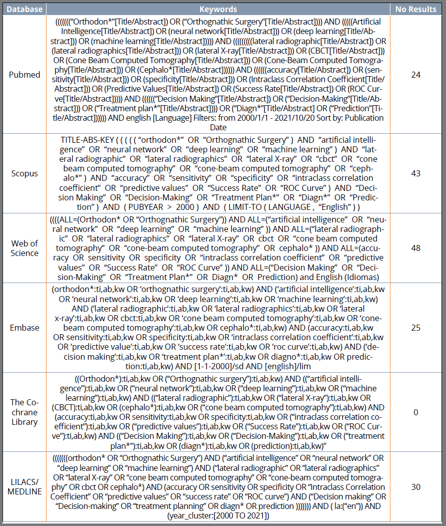
Individual electronic search strategies were developed and conducted on PubMed/Medline, Scopus, Web of Science, Embase, Lilacs, and Cochrane Library published from January 2000 to October 20, 2021, and only English publish articles (Fig. 1).
SELECTION PROCESS
The selection was completed in phases. First, titles, keywords and abstracts were reviewed by two investigators (PHJO and JOLP) separately using the inclusion and exclusion criteria to screen the articles. Duplicated articles were accessed using Mendeley (https://www.mendeley.com)38 and double checked using Microsoft Excel?. Secondly, full texts of the possible eligible studies were assessed and classified as no, maybe, or yes according to the inclusion and exclusion criteria by the two investigators. The ones marked with no for both investigators were excluded, while the ones marked as maybe were resolved through consensus to decide if they would be included or not based on the PICOS and inclusion criteria.
DATA CHARTING PROCESS AND DATA ITEMS
Data charting was conducted by an independent investigator (PHJO) and checked by a second (JOLP). The following data were collected from each article, as follows: author, year of publication, country, study design, sample, mean/range of age, which X-ray based exam was used, aim, algorithm architecture, performance metrics, author suggestion, recommendations and limitation, results, and conclusions.
STUDY RISK OF BIAS ASSESSMENT AND CRITICAL APPRAISAL OF INDIVIDUAL STUDIES
The risk of bias has not assessed, because the aim of the included manuscripts was to assess statistical machine-learning approaches based on pre-selected imaging exams, rather than the clinical aspects of the sample recruitment, or methodologies during the study’s conduction. In AI systems, the BIAS usually is resulted from the algorithm training itself, which there is no specific standard tool to measure this in scoping reviews. Meta-analysis was not conduced since the risk of bias was not assessed. Also, intervention, metrics or outcomes, and study settings were different in the selected studies.39
SYNTHESIS OF RESULTS
For didactic purposes, the included studies were separated in 5 different categories, according to what was evaluated: (1) Orthognathic surgery, (2) TMJ osteoarthritis, (3) Skeletal pattern, (4) Obstructive Sleep Apnea (OSA), and (5) Skeletal maturation/development.
RESULTS
STUDY SELECTION
A total of 170 studies were retrieved in our search following the previously defined strategy. Duplicate removal and screening were performed, and 30 studies were selected for full-text assessment. After eligibility criteria was carefully applied to the full texts, 18 studies were excluded because they were not based only on x-ray images, did not provide quantitative results, could not be applied directly to diagnosis or treatment planning, or aimed to detect cephalometric landmarks. Finally, 12 studies met the eligibility criteria and were included. The PRISMA flowchart showing the process of selection is presented in Figure 2.

Preferred Reporting Items for Systematic Reviews and Meta-Analyses extension for Scoping Reviews (PRISMA-ScR) flow diagram of the study selection process.
STUDY CHARACTERISTICS
The studies were divided into groups according to the AI application and what was evaluated (Fig. 3).

The different X-ray-based images that were used in this scoping review such as lateral radiographs (A), hand-wrist radiographs (B), Posterior radiographs (C), and CBCTs (D). Followed by different machine/deep learning methods that are present in the selected studies and the purposes of the use of AI systems in Orthodontics that are shown in this scoping review.
RESULTS OF INDIVIDUAL STUDIES AND SYNTHESIS OF RESULTS
Orthognathic surgery: Five studies were included regarding the application of AI to predict the need for orthognathic surgery or conventional Orthodontics21-25 (Table 2). Shin et al.21 demonstrated that the DL network proposed by them can standardize the decision process for orthognathic surgery and help orthodontists using posterior and lateral radiographs. Their results for accuracy, sensitivity, and specificity were 0.954, 0.844, and 0.993, respectively. Lin et al.22 was the only study that showed results lower than 90% for accuracy or success rate in predicting the need for orthognathic surgery. However, they evaluated that ANB angle, facial convexity angle, and the maxillary plan to Frankfurt plane angle were the main parameters to define the need for surgery. Lin et al.23 used four different CNN models to predict facial symmetry after orthognathic surgery using CBCT images. Xception model was the most suitable for this purpose with 90% accuracy. Kim et al.24 showed that ResNEt-18 and 34 showed better performance than Resnet-50 and 101 to predict the need for orthognathic surgery using lateral radiographs. The results for the four CNN models test were as follows: Area Under the Curve (AUC): 0.979 (ResNet-18), 0.974 (ResNet-34), 0.945 (ResNet-50), 0.944 (ResNet-101). Accuracy: 0.938 (ResNet-18), 0.936 (ResNet-34), 0.911 (ResNet-50), 0.913 (ResNet-101). Sensitivity: 0.882 (ResNet-18), 0.876 (ResNet-34), 0.806 (ResNet-50), 0.824 (ResNet-101). Specificity: 0.966 (ResNet-18), 0.966 (ResNet-34), 0.964 (ResNet-50), 0.958 (ResNet-101). Lee et al.25 showed that Deep Convolutional Neural Networks (DCNN) can be applied to predict orthognathic surgery and show which structure is the most important for diagnosis. Modified-Alexnet showed higher AUC and accuracy (0.969 and 0.919) than MobileNet (0.908 and 0.838) and ResNet-50 (0.923 and 0.838). The algorithm architecture used for all studies in this section was deep learning based.21-25
Skeletal maturation/development: Four studies regarding the application of AI to assess development or skeletal maturation31,40-42 (Table 2). Kim et al.31 showed that cervical vertebrae (CV) images can be used to predict hand-wrist SMI by presenting a model consisted of eight ML models. Also, age and sex can increase the accuracy. Kök et al.40 concluded that growth development periods and gender can be predicted using ANN. The higher accuracy (0.942) was present by ANN-7 (composed by all 32 linear measurements) followed by ANN-Gender (ANN7+gender). Kök et al.41 results showed that ANN was the most stable algorithm to predict CV stages (CVS1-CVS6) with accuracy results of 93%, 89.7%, 68.8%, 55.6%, 47.4% and 78%, respectively. All studies used lateral radiographs and more than one DL model. However, only Seo et al.42 did not use hand-wrist radiographs as a comparison method. They evaluated and compared six different CNN-based DL models to classify Cervical Vertebral Maturation (CVM). All of them had accuracy higher than 90% and also AUC higher than 90% for all CVM stages. All studies showed that AI can be used to predict skeletal maturation.
Skeletal classification: One study was included regarding the application of AI to classify patients’ skeletal patterns (Table 2). Yu et al.43 evaluated sagittal and vertical discrepancies on lateral radiographs using CNN. It showed results higher than 90% for both skeletal classifications and presented heat maps for a better understanding of the region that influences the most to distinguish skeletal patterns.
TMJ osteoarthritis (TMJOA): One study was included regarding the application of AI to detect TMJ osteoarthritis. Lee et al.44 showed that sagittal CBCT images and deep learning are capable to predict TMJOA. The DL used was a single-shot detector. The authors evaluated accuracy, precision, recall, and F1 score. The results were 0.86, 0.85, 0.84, and 0.84, respectively. The authors suggested that additional imaging modalities should improve the performance of the DL and provide more information on the TMJ inflammation (Table 2).
Obstructive Sleep Apnea (OSA): One study was included regarding the application of AI to detect OSA. Tsuiki et al.45 showed that DCNN model (VGG-19) can accurately identify OSA using lateral radiographs (Table 2). The authors divided the sample images into three groups according to the area that was evaluated as follows: full images (original images), main region (facial profile, the upper airway, and craniofacial soft and hard tissues), and head only (occipital region). The main region had the best results for sensitivity, specificity, and AUC (0.84, 0.81, 0.92) followed by full image (0.9, 0.77, and 0.89), and head only (0.71, 0.63, and 0.7). The authors suggested that demographic characteristics and anthropometric features could improve the diagnostic OSA.
DISCUSSION
SUMMARY OF EVIDENCE
To our knowledge, this is the first scoping review to present X-ray imaging-based AI models applied to diagnosis and treatment planning. As a limitation, we have excluded manuscripts that used panoramic images because the studies were not focused on the Orthodontics’ diagnosis or planning. In most reviewed papers, authors divided AI applications in Orthodontics based on radiographic images into two groups: diagnosis and treatment planning.
Diagnosis. Diagnosis is probably the most important aspect of orthodontic treatment. Assessing development and skeletal maturation is needed for patients still growing and present skeletal alterations. With that is possible to provide the best treatment for each developmental period.46-48 The cervical vertebrae method49 is commonly used to identify maturation stages because it can be assessed in lateral radiographs. However, due to its subjectivity and the chance of distortion depending on the patient positioning, clinicians need to have specific training to use this method. That is one of the reasons why hand-wrist radiographs are still the gold standard for assessing the development stage of a patient.50 However, AI can be used to help orthodontists to identify the CVM stages. All the studies assessed here showed that AI had good results assessing the development stage of patients using lateral radiographs when compared to gold standard or to humans’ evaluation. Furthermore, the different studies showed that ANN model had best results when compared to other AI models.31,40-42
Facial analysis and skeletal classification play an important role in Orthodontics’ diagnosis. All the growth deviations that the patient had will produce a skeletal alteration, and the orthodontist must classify the patient properly in all 3 planes.51-53 However, lateral radiographs can only classify vertical and sagittal relationships. Yu et al.43 aimed to used DL to assess skeletal classification and, unlikely previous studies, they did not use CNN models to perform cephalometric landmarks. The authors justified that eliminating this process would improve the classification performance. For assessing vertical relationships, it was used the Bjork’s Sum and Jarabak’s Ratio and the results showed 96.4% accuracy. For sagittal relationships, the ANB angle and Wits’ appraisal were used and a 95.7% accuracy was obtained.
Patients with OSA usually present a retrognathic mandible and/or a lower hyoid position with causes a more crowded oropharynx. Patients with OSA can be diagnosed in lateral radiographs, mainly when it is a severe case. However, 3D images still provide better and more qualified data. Tsuiki et al.45 evaluated 3 types of images: full lateral radiographs images, the main airway region (including facial profile, teeth, cervical bones, maxilla, and mandible), and the occipital region. Best results were obtained for the main region, and the worst were at the occipital region. Also, their results are comparable with the manual cephalometric analysis. It shows that the use of DL models can be applied as an objective additional diagnosis method to OSA patients.
TMJ osteoarthritis is a multi-system disease. CBCTs allow dentists to diagnose the TMJ bone by finding irregular contour, osseous defects, cortical loss or flattening of the condyles.54 Early diagnosis can be the best chance to minimize the damage caused by osteoarthritis, since there is no treatment available yet. Lee et al.44 evaluated if AI could detect TMJOA in CBCT sagittal images. The results showed that using only the CBCT sagittal images had results higher than 80% like Bianchi et al.,55 that used CBCT, clinical and biological data.
Treatment Planning. Once the diagnosis is placed, treatment planning is the next step in orthodontic treatment. It is important to enhance that treatment planning needs radiographic images, facial evaluation, and models to be made, mainly for borderline patients. The proper decision to treat with orthodontic treatment or orthognathic surgery usually requires more experience from the orthodontist.51,56,57 All studies presented here had results higher than 90% when studied adult patients that need surgical treatment. Lin et al.22 was the only study that used young patients with unilateral cleft lip and palate and had 87.4% accuracy and was the only one to use cephalometric measures. The other studies21,23-25 used DCNN to extract features and identify the structures that play an important role in the decision. The authors justify that by saying that the marking and measurement values can be a bias. This model resembles an orthodontist’s subjective or morphological analysis. Previous studies19,58 compared the orthodontists’ impression and cephalometric values and have concluded that there is the correlation between both analyses.
Limitations. The main limitation of this scoping review was that most studies that apply AI to Orthodontics usually needs multi-source data, rather than x-rays-based image alone. For this reason, we retrieve only 12 manuscripts that meet our PICOs criteria. Also, we have not assessed the BIAS of each AI model due to the lack of standardized tools for this purpose. In addition to that, the lack of general applicability is a major limitation in AI models because most of the data are from the same center. We believe that with a greater number of studies conducted using AI to assist in diagnosis and planning, with more data and preferably multicenter studies, we may observe broader clinical applications and improvements in orthodontic software, thereby enhancing the proposed treatment for our patients.
CONCLUSIONS
In fact, AI has already demonstrated its ability to add valuable information and assist in orthodontic treatment planning, diagnostic, and decision-making processes. While AI models are available to enhance the evaluation of specific issues such as TMJ osteoarthritis, skeletal maturation, skeletal classification, OSA, and the need for orthognathic surgery, it is essential to note that these applications are not universally used in orthodontic treatments. Instead, AI’s role is often context-dependent and can be most helpful in particular situations where detailed image analysis and data-driven decision support can provide significant value. Integrating AI into clinical practice will likely continue to evolve, enhancing treatment planning in selective cases.
REFERENCES
- 1 Raghupathi W, Raghupathi V. Big data analytics in healthcare: promise and potential. Health Inf Sci Syst. 2014 Feb;2:3.
- 2 Park WJ, Park JB. History and application of artificial neural networks in dentistry. Eur J Dent. 2018;12(4):594-601.
- 3 Allareddy V, Rengasamy Venugopalan S, Nalliah RP, Caplin JL, Lee MK, et al Orthodontics in the era of big data analytics. Orthod Craniofac Res. 2019 May;22 Suppl 1:8-13.
- 4 Machoy ME, Szyszka-Sommerfeld L, Vegh A, Gedrange T, Wozniak K. The ways of using machine learning in dentistry. Adv Clin Exp Med. 2020 Mar;29(3):375-84.
- 5 Corbella S, Srinivas S, Cabitza F. Applications of deep learning in dentistry. Oral Surg Oral Med Oral Pathol Oral Radiol. 2021 Aug;132(2):225-38.
- 6 Vinayahalingam S, Xi T, Bergé S, Maal T, Jong G. Automated detection of third molars and mandibular nerve by deep learning. Sci Rep. 2019 Jun;9(1):9007.
- 7 Chen H, Zhang K, Lyu P, Li H, Zhang L, Wu J, et al. A deep learning approach to automatic teeth detection and numbering based on object detection in dental periapical films. Sci Rep. 2019 Mar;9(1):3840.
- 8 Geetha V, Aprameya KS, Hinduja DM. Dental caries diagnosis in digital radiographs using back-propagation neural network. Health Inf Sci Syst. 2020 Jan;8(1):8.
- 9 Valizadeh S, Goodini M, Ehsani S, Mohseni H, Azimi F, Bakhshandeh H. Designing of a computer software for detection of approximal caries in posterior teeth. Iran J Radiol. 2015 Aug;12(4):e16242.
- 10 Orhan K, Bayrakdar IS, Ezhov M, Kravtsov A, Özyürek T. Evaluation of artificial intelligence for detecting periapical pathosis on cone-beam computed tomography scans. Int Endod J. 2020 May;53(5):680-9.
- 11 Okada K, Rysavy S, Flores A, Linguraru MG. Noninvasive differential diagnosis of dental periapical lesions in cone-beam CT scans. Med Phys. 2015 Apr;42(4):1653-65.
- 12 Endres MG, Hillen F, Salloumis M, Sedaghat AR, Niehues SM, Quatela O, et al. Development of a deep learning algorithm for periapical disease detection in dental radiographs. Diagnostics. 2020 Jun;10(6):430.
- 13 Zhao T, Zhou J, Yan J, Cao L, Cao Y, Hua F, et al. Automated adenoid hypertrophy assessment with lateral cephalometry in children based on artificial intelligence. Diagnostics. 2021 Jul;11(8):1386.
- 14 Liu JL, Li SH, Cai YM, Lan DP, Lu YF, Liao W, et al. Automated radiographic evaluation of adenoid hypertrophy based on VGG-Lite. J Dent Res. 2021 Nov;100(12):1337-43.
- 15 Takada K. Artificial intelligence expert systems with neural network machine learning may assist decision-making for extractions in orthodontic treatment planning. J Evid Based Dent Pract. 2016 Sep;16(3):190-2.
- 16 Jung SK, Kim TW. New approach for the diagnosis of extractions with neural network machine learning. Am J Orthod Dentofacial Orthop. 2016 Jan;149(1):127-33.
- 17 Xie X, Wang L, Wang A. Artificial neural network modeling for deciding if extractions are necessary prior to orthodontic treatment. Angle Orthod. 2010 Mar;80(2):262-6.
- 18 Etemad L, Wu TH, Heiner P, Liu J, Lee S, Chao WL, et al. Machine learning from clinical data sets of a contemporary decision for orthodontic tooth extraction. Orthod Craniofac Res. 2021 Dec;24 Suppl 2:193-200.
- 19 Wu BW, Kaban LB, Peacock ZS. Correlation of cephalometric analyses with clinical impression in orthognathic surgery patients: harvold and steiner analyses. J Oral Maxillofac Surg. 2019 Nov;77(11):2308-17.
- 20 Choi HI, Jung SK, Baek SH, Lim WH, Ahn SJ, Yang IH, et al. Artificial intelligent model with neural network machine learning for the diagnosis of orthognathic surgery. J Craniofac Surg. 2019 Oct;30(7):1986-89.
- 21 Shin W, Yeom HG, Lee GH, Yun JP, Jeong SH, Lee JH, et al. Deep learning based prediction of necessity for orthognathic surgery of skeletal malocclusion using cephalogram in Korean individuals. BMC Oral Health. 2021 Mar;21(1):130.
- 22 Lin G, Kim PJ, Baek SH, Kim HG, Kim SW, Chung JH. Early prediction of the need for orthognathic surgery in patients with repaired unilateral cleft lip and palate using machine learning and longitudinal lateral cephalometric analysis data. J Craniofac Surg. 2021;32(2):616-20.
- 23 Lin HH, Chiang WC, Yang CT, Cheng CT, Zhang T, Lo LJ. On construction of transfer learning for facial symmetry assessment before and after orthognathic surgery. Comput Methods Programs Biomed. 2021 Mar;200:105928.
- 24 Kim YH, Park JB, Chang MS, Ryu JJ, Lim WH, Jung SK. Influence of the depth of the convolutional neural networks on an artificial intelligence model for diagnosis of orthognathic surgery. J Pers Med. 2021 Apr;11(5):356.
- 25 Lee KS, Ryu JJ, Jang HS, Lee DY, Jung SK. Deep convolutional neural networks based analysis of cephalometric radiographs for differential diagnosis of orthognathic surgery indications. Appl Sci. 2020;10(6):2124.
- 26 Hwang HW, Park JH, Moon JH, Yu Y, Kim H, Her SB, et al. Automated identification of cephalometric landmarks: part 2-Might it be better than human? Angle Orthod. 2020 Jan;90(1):69-76.
- 27 Park JH, Hwang HW, Moon JH, Yu Y, Kim H, Her SB, et al. Automated identification of cephalometric landmarks: part 1-Comparisons between the latest deep-learning methods YOLOV3 and SSD. Angle Orthod. 2019 Nov;89(6):903-9.
- 28 Ezhov M, Gusarev M, Golitsyna M, Yates JM, Kushnerev E, Tamimi D, et al. Clinically applicable artificial intelligence system for dental diagnosis with CBCT. Sci Rep. 2021 Jul;11(1):15006.
- 29 Takeda S, Mine Y, Yoshimi Y, Ito S, Tanimoto K, Murayama T. Landmark annotation and mandibular lateral deviation analysis of posteroanterior cephalograms using a convolutional neural network. J Dent Sci. 2021 Jul;16(3):957-63.
- 30 Kim H, Shim E, Park J, Kim YJ, Lee U, Kim Y. Web-based fully automated cephalometric analysis by deep learning. Comput Methods Programs Biomed. 2020 Oct;194:105513.
- 31 Kim DW, Kim J, Kim T, Kim T, Kim YJ, Song IS, et al. Prediction of hand-wrist maturation stages based on cervical vertebrae images using artificial intelligence. Orthod Craniofac Res. 2021 Dec;24 Suppl 2:68-75.
- 32 Kim YH, Park JB, Chang MS, Ryu JJ, Lim WH, Jung SK. Influence of the depth of the convolutional neural networks on an artificial intelligence model for diagnosis of orthognathic surgery. J Pers Med. 2021 Apr;11(5):356.
- 33 Lee KS, Ryu JJ, Jang HS, Lee DY, Jung SK. Deep convolutional neural networks based analysis of cephalometric radiographs for differential diagnosis of orthognathic surgery indications. Appl Sci. 2020;10(6):2124.
- 34 Mohammad-Rahimi H, Nadimi M, Rohban MH, Shamsoddin E, Lee VY, Motamedian SR. Machine learning and orthodontics, current trends and the future opportunities: a scoping review. Am J Orthod Dentofacial Orthop. 2021 Aug;160(2):170-92.e4.
- 35 Corbella S, Srinivas S, Cabitza F. Applications of deep learning in dentistry. Oral Surg Oral Med Oral Pathol Oral Radiol. 2021 Aug;132(2):225-38.
- 36 Kudyba SP. Healthcare informatics: improving efficiency and productivity. Boca Raton: CRC Press; 2010.
- 37 Tricco AC, Lillie E, Zarin W, O'Brien KK, Colquhoun H, Levac D, et al. PRISMA Extension for Scoping Reviews (PRISMA-ScR): checklist and explanation. Ann Intern Med. 2018 Oct;169(7):467-73.
- 38 Elston DM. Mendeley. J Am Acad Dermatol. 2019 Nov;81(5):1071.
- 39 Ioannidis JP, Patsopoulos NA, Rothstein HR. Reasons or excuses for avoiding meta-analysis in forest plots. BMJ. 2008 Jun;336(7658):1413-5.
- 40 Kök H, Izgi MS, Acilar AM. Determination of growth and development periods in orthodontics with artificial neural network. Orthod Craniofac Res. 2021 Dec;24 Suppl 2:76-83.
- 41 Kök H, Acilar AM, Izgi MS. Usage and comparison of artificial intelligence algorithms for determination of growth and development by cervical vertebrae stages in orthodontics. Prog Orthod. 2019 Nov;20(1):41.
- 42 Seo H, Hwang J, Jeong T, Shin J. Comparison of deep learning models for cervical vertebral maturation stage classification on lateral cephalometric radiographs. J Clin Med. 2021 Aug;10(16):3591.
- 43 Yu HJ, Cho SR, Kim MJ, Kim WH, Kim JW, Choi J. Automated skeletal classification with lateral cephalometry based on artificial intelligence. J Dent Res. 2020 Mar;99(3):249-56.
- 44 Lee KS, Kwak HJ, Oh JM, Jha N, Kim YJ, Kim W, et al. Automated detection of TMJ osteoarthritis based on artificial intelligence. J Dent Res. 2020 Nov;99(12):1363-7.
- 45 Tsuiki S, Nagaoka T, Fukuda T, Sakamoto Y, Almeida FR, Nakayama H, et al. Machine learning for image-based detection of patients with obstructive sleep apnea: an exploratory study. Sleep Breath. 2021 Dec;25(4):2297-305.
- 46 Hino CT, Cevidanes LH, Nguyen TT, De Clerck HJ, Franchi L, McNamara JA Jr. Three-dimensional analysis of maxillary changes associated with facemask and rapid maxillary expansion compared with bone anchored maxillary protraction. Am J Orthod Dentofacial Orthop. 2013 Nov;144(5):705-14.
- 47 Baccetti T, Franchi L, McNamara JA Jr. Growth in the untreated Class III subject. Semin Orthod. 2007 Sep;13(3):130-42.
- 48 Franchi L, Baccetti T, McNamara JA. Postpubertal assessment of treatment timing for maxillary expansion and protraction therapy followed by fixed appliances. Am J Orthod Dentofacial Orthop. 2004 Nov;126(5):555-68.
- 49 McNamara JA Jr, Franchi L. The cervical vertebral maturation method: a user's guide. Angle Orthod. 2018 Mar;88(2):133-43.
- 50 Cericato GO, Bittencourt MA, Paranhos LR. Validity of the assessment method of skeletal maturation by cervical vertebrae: a systematic review and meta-analysis. Dentomaxillofac Radiol. 2015;44(4):20140270.
- 51 Capelozza Filho L. Diagnóstico em ortodontia. Maringá: Dental Press; 2004.
- 52 Arnett GW, Bergman RT. Facial keys to orthodontic diagnosis and treatment planning. Part I. Am J Orthod Dentofacial Orthop. 1993 Apr;103(4):299-312.
- 53 Arnett GW, Bergman RT. Facial keys to orthodontic diagnosis and treatment planning--Part II. Am J Orthod Dentofacial Orthop. 1993 May;103(5):395-411.
- 54 Wang XD, Zhang JN, Gan YH, Zhou YH. Current understanding of pathogenesis and treatment of TMJ osteoarthritis. J Dent Res. 2015 May;94(5):666-73.
- 55 Bianchi J, Ruellas ACO, Gonçalves JR, Paniagua B, Prieto JC, Styner M, et al. Osteoarthritis of the Temporomandibular Joint can be diagnosed earlier using biomarkers and machine learning. Sci Rep. 2020 May;10(1):8012.
- 56 Proffit WR, Fields HW Jr, Sarver DM, Larson B. Contemporary orthodontics. 6th ed. St Louis: Mosby; 2018.
- 57 Arnett GW, Jelic JS, Kim J, Cummings DR, Beress A, Worley CM Jr, et al. Soft tissue cephalometric analysis: diagnosis and treatment planning of dentofacial deformity. Am J Orthod Dentofacial Orthop. 1999 Sep;116(3):239-53.
- 58 Steiner CC. Cephalometrics in clinical practice. Angle Orthod. 1959 Jan;29(1):8-29.
-
»
Patients displayed in this article previously approved the use of their facial and intraoral photographs.
-
How to cite:
Oliveira PHJ, Gonçalves JR, Gandini Júnior LG, Parizotto JOL, Oliveira MS, Kato RM, Evangelista K, Cevidanes LHS, Bianchi J. Clinical decisions in Orthodontics using x-ray-based images and artificial intelligence approaches: a scoping review. Dental Press J Orthod. 2025;30(4):e2524219.