 Electrolyte-enriched milk is as effective as oral electrolyte solution in correcting imbalances in diarrheal calves
ABSTRACT:
Oral rehydration in calves is traditionally performed by administering an oral electrolyte solution in which the electrolyte concentrate (EC) is diluted in water. Dilution of EC in milk has been used as an alternative method because it encourages voluntary water intake. Although practical, its effectiveness has not been proven consistently. This study compared the effectiveness of these two rehydration methods for correcting imbalances in diarrheal calves. Twenty-four newborn calves with induced osmotic diarrhea were divided into two treatment groups: MG with EC diluted in milk at meals and WG with EC diluted in water (5% body weight at 4 and 12 hours). All calves were fed with milk (4% body weight at 0, 8, and 16 hours) and had free access to water. Venous blood samples were collected at the times: -48 (before induction), -24, 0 (start of treatment), 8, 16, 24, and 48 hours.Packed cell volume (PCV), total plasma protein (TP), pH, pCO2, HCO3 -, BE, Na+, K+, Cl-, L-lactate, creatinine, strong ion difference (SID3), anion gap (AG), total concentration of non-volatile weak acids (Atot), and percentage change in plasma volume (%PV) were measured or calculated. Calves exhibited moderate dehydration, hyponatremia, and mild strong ion acidosis. Both rehydration methods were effective in correcting the imbalances. Plasma volume expansion was faster in the WG and voluntary water intake was higher in the MG. Results based on induced rather than natural diarrhea are the main limitation of the study. Owing to its practicality and effectiveness, dilution of EC in milk can be used to treat non-depressed diarrheal calves with mild to moderate imbalances.
Key words:
neonatal diarrhea; fluid therapy; strong ion acidosis; dehydration
RESUMO:
A reidratação oral em bezerros é tradicionalmente realizada com administração de solução eletrolítica oral na qual o concentrado eletrolítico (CE) é diluído em água. A diluição da CE no leite tem sido utilizada como método alternativo porque incentiva a ingestão voluntária de água. Embora prático, a sua eficácia não foi comprovada de forma consistente. Este estudo comparou a eficácia destes dois métodos de reidratação para corrigir desequilíbrios em bezerros diarreicos. Vinte e quatro bezerros recém-nascidos com diarreia osmótica induzida foram divididos em dois grupos de tratamento: GM com CE diluído em leite nas refeições e GW com CE diluído em água (5% do peso corporal às 4 e 12 horas). Todos os bezerros foram alimentados com leite (4% do peso corporal às 0, 8 e 16 horas) e tiveram livre acesso à água. Amostras de sangue venoso foram coletadas nos horários: -48 (antes da indução), -24, 0 (início do tratamento), 8, 16, 24 e 48 horas. Volume globular (VG), proteína plasmática total (PT), pH, pCO2, HCO3 -, BE, Na+, K+, Cl-, lactato L, creatinina, diferença de íons fortes (SID3), hiato aniônico (AG), concentração total de ácidos fracos não voláties (Atot) e variação do volume plasmático (%PV) foram mensurados ou calculados. Os bezerros exibiram desidratação moderada, hiponatremia e acidose por íons fortes de grau leve. Ambos os métodos de reidratação foram eficazes para a correção dos desequilíbrios. A expansão do volume plasmático foi mais rápida no GW e a ingestão voluntária de água foi maior no GM. A principal limitação do estudo é que os resultados foram baseados em casos de diarreia induzida e não natural. Devido à sua praticidade e eficácia, a diluição da CE no leite pode ser utilizada para tratar bezerros diarreicos não deprimidos e com desequilíbrios leves a moderados.
Palavras-chave:
diarreia neonatal; terapia com fluidos; acidose por íons fortes; desidratação
INTRODUCTION
Neonatal diarrhea is the main cause of death in calves during the first weeks of life and causes considerable economic loss (UMAÑA SEDÓ et al., 2023; USDA, 2014). Calves with diarrhea present varying degrees of imbalances, such as dehydration, hyponatremia, relative hyperchloremia, potassium depletion, hyper-L- and D-lactatemia, hypoglycemia, and metabolic acidosis (CONSTABLE et al., 2005; GOMEZ et al., 2013; 2017; TREFZ et al., 2017).
Regardless of the causative agent, the treatment of calves with diarrhea involves rehydration with electrolyte solutions administered orally or intravenously. Oral electrolyte solutions (OES) are indicated for calves with mild-to-moderate dehydration presenting aboral transit of the ingesta and an active sucking reflex (SMITH, 2009; LORENZ et al., 2011; SMITH & BERCHTOLD, 2014).Treatment with intravenous electrolyte solutions is necessary for calves with severe dehydration or hypovolemic shock, lethargy, or a weak or absent sucking reflex (BERCHTOLD, 2009; CONSTABLE et al., 2021). The OES for neonatal diarrhea treatment must contain electrolytes, such as sodium (90-130 mmol/L), potassium (10-30 mmol/L), and chloride (40-80 mmol/L); alkalizing agents, such as bicarbonate or acetate (60-80 mmol/L); glucose to facilitate intestinal sodium absorption (1-3 glucose-to-sodium ratio); high effective SID3 (60-80 mmol /L); and an osmolarity close to 300 mOsm/L (SMITH & BERCHTOLD, 2014; CONSTABLE et al., 2021).
OES is traditionally prepared by diluting commercial or non-commercial electrolyte concentrate (EC) in water and is administered between meals of milk or milk replacer as one or two extra feedings per day (SMITH & BERCHTOLD, 2014). More recently, an alternative method has been employed in which EC is diluted directly in milk or milk replacer instead of water. This makes the treatment more practical as it facilitates farm management, freeing keepers from providing OES outside of meals, which can contribute to increasing adherence to treatment (GOODELL et al., 2012). Ingestion of milk or milk replacer enriched with electrolytes increases plasma sodium concentration and; consequently, causes thirst. Calves must have free access to water so that their voluntary intake corrects dehydration (BACHMANN et al., 2012; WENGE et al., 2014; WILMS et al., 2020).
Despite the advantages of this alternative method and some manufacturers already recommend diluting commercial EC in milk, few studies have evaluated its effectiveness in correcting water, electrolyte, and acid-base imbalances in calves, comparing it with traditional rehydration with OES. The two rehydration methods were first compared in healthy calves (BACHMANN et al., 2009; 2012). The four studies with diarrheic calves have limitations, such as water deprivation preventing their voluntary intake (KIRCHNER et al., 2014), the lack of measurement of electrolytes and blood gases (WENGE et al., 2014), pretreatment of calves with EC diluted in water or milk, promoting partial correction of imbalances (WENGE-DANGSCHAT et al., 2020), and the use of different ECs diluted in water and milk replacer (MIQUEO et al., 2018). Furthermore, the effects of rehydration methods on blood volume, electrolytes, and blood gases have been monitored for only 4 (BACHMANN et al., 2009; 2012) or 6 hours (KIRCHNER et al., 2014; WENGE-DANGSCHAT et al., 2020) after ingestion.
Therefore, there is no consistent evidence that diluting EC in milk or milk replacer is effective for rehydrating calves. The present study compared the effectiveness of oral administration of a commercial EC diluted in water or milk to reverse water, electrolyte, and acid-base imbalances in neonatal calves with induced osmotic diarrhea and dehydration. We hypothesized that electrolyte-enriched milk is not as efficient as OES for correcting imbalances in diarrheal calves.
MATERIALS AND METHODS
This randomized controlled clinical trial with a repeated measures design was approved by the Ethics Committee on the Use of Animals of the Sociedade Cultural e Educacional de Garça - Faculdade de Ensino Superior e Formação Integral (FAEF), under the process CEUA-FAEF 001/2021, complying with current Brazilian laws. The study was carried out between February 2021 and January 2022, in the experimental shed of the FAEF Veterinary Teaching Hospital in Garça, SP, Brazil.
Animals and experimental procedures
This study included 24 newborn Jersey calves (17 females and 7 males), raised on the same farm. Immediately after birth, they were separated from their dams. All calves received fresh colostrum with Brix degree > 25% through a bottle in a volume corresponding to 15% of body weight (BW), with 10% of the BW ingested within 2 hours of life and 5% of the BW ingested 6 hours later. At 24 hours of age, the concentration of total plasma proteins was 8.30 ± 1.21 g/dL.
The calves were housed in individual pens that were cleaned and covered with sand and wood shavings, where they remained throughout the experimental period. During the first 10 days of adaptation, they received whole milk through a bottle in a volume corresponding to 12% of their BW divided into two feedings per day. Water, Tifton hay (Cynodon dactylon), and commercial pelleted starter feed were provided ad libitum.
When they reached 10 to 15 days of age and had a BW of 28.38 ± 3.09 kg, the calves that remained healthy were subjected to induction of osmotic diarrhea and dehydration for 48 hours, using a previously standardized protocol (LEAL et al., 2008; LEAL et al., 2012; BREGADIOLI et al., 2022): intake of whole milk (16.5 mL/kg) plus sucrose (4 g/kg) diluted at 20% in warm water, every 8 hours, via bottle; and oral administration of spironolactone (Espironolactona 25 mg; Eurofarma Laboratórios S.A., Itapevi, SP, Brazil) and hydrochlorothiazide (Hidroclorotiazida 25 mg; EMS S.A., Hortolândia, SP, Brazil) both at a dose of 2 mg/kg, every 8 hours, immediately after feeding the milk. The calves were deprived of water for 12 hours at night.
The experimental period comprised the induction (-48 to 0 hours), treatment (0 to 24 hours), and follow-up phases (24 to 48 hours). During the treatment and follow-up phases, all calves were fed whole milk through a bottle at a volume corresponding to 12% of their BW, divided into three daily meals (4% of BW every 8 hours). During the treatment phase, water, electrolyte, and acid-base imbalances were corrected over the course of 1 day, using a commercial EC (Glutellac®, Elanco; São Paulo, SP, Brazil). Calves were randomly distributed into two treatment groups (n = 12) according to the dilution of the EC: directly in the milk of meals (milk group; MG; 9 females and 3 males) or in water (water group; WG; 8 females and 4 males).The same EC was used in both groups. In both cases, dilution was performed by adding 25 mL of EC to 1 L of milk or water, following the manufacturer’s recommendations. In MG, the calves received milk enriched with electrolytes (0, 8, and 16 hours). In WG, the prepared OES was administered in bottles in a volume equivalent to 5% of their BW, at two times between meals (4 and 12 hours). From the beginning of the treatment (0 hour), all calves had free access to water available in a bucket. The bucket was replenished as often as necessary to ensure water was always available.
The OES administered in the WG had the following composition: 100.1 mEq/L sodium, 18.5 mEq/L potassium, 60.7 mEq/L chloride, 58 mEq/L acetate, 46 mmol/L glucose, calculated osmolarity of 283 mOsm/L, pH 6.232, and effective SID3 of 58 mEq/L. The electrolyte-enriched milk administered in the MG had this same composition plus the concentrations of sodium, potassium, and chloride originally present in the milk. The final composition was not measured.
Physical examinations
Throughout the experimental period, physical examinations were performed every 8 hours by a single trained bovine practitioner, who was blinded to the group of the calves. The color and tackiness of the mucous membranes, state of hydration, degree of enophthalmos, skin turgor, capillary refill time, appetite, fecal characteristics, attitude, posture, and behavior were evaluated.
Stool consistency, degree of dehydration, and behavior, posture, and sucking reflex were classified using the scoring system previously proposed (SMITH, 2009; WALKER et al., 1998) with some modifications (BREGADIOLI et al., 2023). Adding the defined scores, the overall disease score was determined on a scale of 0-10, where 0 represents healthy and 10 represents the highest disease score.
Sample collection and processing
Calves were weighed and venous blood was collected at -48, -24, 0, 8, 16, 24, and 48 hours, immediately before feeding. The volume of water voluntarily ingested throughout the day was measured at -24, 0, 24, and 48 hours, The total volume offered throughout the day, including the sum of the volumes of replacements needed in the bucket, was subtracted from the volume of water remaining in the bucket at these time points.
Venous blood samples were collected by jugular venipuncture using a disposable needle (30 × 0.8 mm) and vacuum flasks containing EDTA anticoagulant. For blood gas analysis, venous blood samples were collected with heparinized syringes (A-Line Luer Lock, Becton Dickinson Company, BD Brasil, São Paulo, Brazil).
The analyses were performed immediately after collection. Packed cell volume (PCV) was determined using a micro-hematocrit centrifuge, and the concentration of total plasma proteins (TP) was measured by refractometry. Blood gas analyses determined pH, partial pressure of carbon dioxide (pCO2), bicarbonate concentration (HCO3 -), base excess (BE), and concentrations of sodium (Na+), potassium (K+), chloride (Cl-), creatinine, and L-lactate (Epoc; Epocal Inc., Siemens Healthcare Diagnostics, Ottawa, Canada) (RO et al., 2022).
The following variables were calculated using the respective formulas:
a) Anion gap (AG): AG = (Na+ + K+) - (Cl- + HCO3 -)
b) Strong ion difference (SID): SID3 = (Na+ + K+) - (Cl-) (CONSTABLE et al., 2005)
c) Total concentration of non-volatile weak acids (Atot): Atot = TP (g/dL) × 3.43 (CONSTABLE et al., 2005)
d) Percentage change in plasma volume (%PV): %PV = [(TP1/TP2) - 1] × 100, where TP1 is the TP value observed before induction and TP2 is the TP value at subsequent time points (CARLSON & BRUSS, 2008).
Statistical analysis
The Shapiro-Wilk and Brown-Forsythe tests were used to verify the Gaussian distribution and equality of variance, respectively. Two-way repeated measures ANOVA was used to test the effect of time (different time points before and after EC administration), the effect of the EC dilution method (milk × water), and the interaction between these two factors. When the F-statistic was significant, Tukey’s test was used for multiple comparisons. Data are presented as mean ± standard deviations. An error probability of 5% was assumed for all tests. The SigmaPlot for Windows 13.1 (Systat Software Inc., San Jose, California, USA) was used for all analyses.
RESULTS
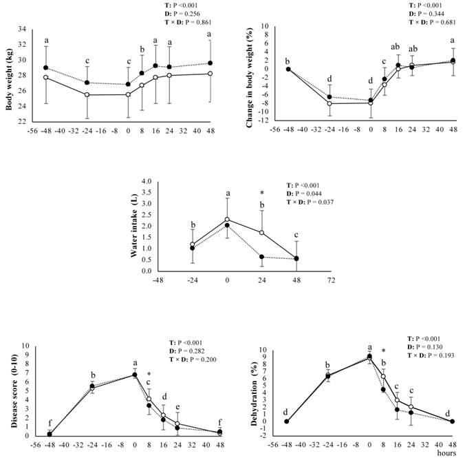
The induction protocol was effective in causing osmotic diarrhea, dehydration, and electrolyte and acid-base imbalances in all calves. The feces were liquid, yellowish in color, and without an unpleasant odor from 8 hours after the start of induction (-40 hours) and maintained these characteristics until the start of treatment (0 hours). During the induction phase, a reduction in BW was observed, and the percentage loss was 7.90 ± 3.45% in MG and 7.27 ± 2.64% in WG (P = 0.547). The disease score gradually increased, reaching maximum values at 0 hour (6.83 ± 0.70 in MG and 6.83 ± 0.38 in WG; P = 0.811), as well as the degree of dehydration, which reached 8.91 ± 0.79% in MG and 9.16 ± 0.71% in WG (P = 0.721). Voluntary water intake was greater on the second day of induction compared to that on the first, reaching values of 2.31 ± 0.95 L in MG and 2.05 ± 0.57 L in WG (P = 0.658) (Figure 1). All calves remained alert in the s standing position, with an active and vigorous sucking reflex throughout the induction phase.
Variations (mean ± standard deviation) in body weight (BW), percentage change in BW, voluntary water intake, disease score, and degree of clinical dehydration observed in neonatal calves with osmotic diarrhea and dehydration induced for 48 hours. The calves were treated with commercial electrolyte concentrate diluted in milk (
 ) and ingested with meals at 0, 8, and 16 hours or diluted in water (
 ) and administered orally, in a volume equivalent to 5% of BW, at 4 and 12 hours. T = time effect; D = effect of the dilution method; T × D = interaction between time and dilution. a,b,c different letters indicate difference between time points (P < 0.05). *indicates difference between dilution methods (P < 0.05).
The changes caused by the induction protocol were characterized by a decrease in venous blood pH, pCO2, HCO3 -, BE, Na+, SID3, and %PV, and an increase in the levels of K+, PCV, TP, Atot, and creatinine (Figure 2 and Figure 3). Cl- levels increased but returned to baseline values at 0 hours, and AG and L-lactate levels remained unchanged. No differences were observed between the groups during the induction phase (P > 0.05).

Variations (mean ± standard deviation) of venous blood pH, pCO2, HCO3 -, BE, Na+, K+, Cl-, and strong ion difference (SID3) in neonatal calves with osmotic diarrhea and dehydration induced for 48 hours. The calves were treated with commercial electrolyte concentrate diluted in milk (
) and ingested with meals at 0, 8, and 16 hours or diluted in water () and administered orally, in a volume equivalent to 5% of BW, at 4 and 12 hours. T = time effect; D = effect of the dilution method; T × D = interaction between time and dilution. a,b,c different letters indicate difference between time points (P < 0.05).

Variations (mean ± standard deviation) of PCV, TP, percentage change in plasma volume (%PV), total concentration of non-volatile weak acids (Atot), creatinine, AG, and L-lactate in neonatal calves with osmotic diarrhea and dehydration induced for 48 hours. The calves were treated with commercial electrolyte concentrate diluted in milk (
) and ingested with meals at 0, 8, and 16 hours or diluted in water () and administered orally, in a volume equivalent to 5% of BW, at 4 and 12 hours. T = time effect; D = effect of the dilution method; T × D = interaction between time and dilution. a,b,c different letters indicate difference between time points (P < 0.05). *indicates difference between dilution methods (P < 0.05).
On the day of treatment, EC was well accepted by all calves, regardless of the dilution method, in milk or water. Sucking was performed actively and vigorously for both enriched milk in the MG and OES in the WG. The two rehydration methods promoted the correction of the induced imbalances with equivalent results and few distinctions between them. Variation over time was observed for all variables studied (P < 0.001), except for AG and L-lactate. In contrast, the effect of the method of EC dilution was only significant for voluntary water intake (P = 0.044). The interaction between the two factors tested was significant for voluntary water intake (P = 0.037), HCO3 - (P = 0.042), BE (P = 0.037), SID3 (P = 0.018), TP (P = 0.018), %PV (P = 0.014), and Atot (P = 0.018).
With the treatments, BW increased, returning to baseline values at 16 hours, disease score decreased, and clinical dehydration was reversed, returning to baseline values at 48 hours (Figure 1). The variation over time analyzed separately for each dilution method showed that the disease score and degree of dehydration returned to baseline values more quickly in the WG (24 hours and 16 hours, respectively) than in the MG (48 hours).
The treatments promoted the correction of pH, pCO2, HCO3 -, and BE from 8 hours onwards. The HCO3 - and BE values exceeded baseline values at 16 hours and remained elevated until 48 hours (Figure 2). When the two dilution methods were analyzed separately, the highest HCO3 - and BE values were observed from 16 hours onwards in WG and from 24 hours onwards in MG. Regarding electrolytes, Na+ concentrations increased with rehydration but returned to baseline values only at 48 hours. K+ concentrations decreased and remained lower than baseline values from 24 hours onwards. The Cl- concentrations remained decreased from 8 hours onwards, and SID3 increased and remained elevated from 16 hours onwards. When the two dilution methods were analyzed separately, the highest SID3 values occurred after 16 hours in WG and 24 hours in MG.
The PCV, TP, %PV, and Atot values returned to baseline at 8 hours, whereas the creatinine values returned to baseline at 16 hours. The PCV and creatinine levels remained unchanged at the following time points. In contrast, from 16 hours onwards, TP and Atot remained low, and %PV remained high (Figure 3). When the two dilution methods were analyzed separately, TP and Atot returned to baseline values more quickly in WG (8 hours) than in MG (16 hours), and %PV assumed higher values earlier in WG (from 16 hours onwards) than in MG (from 24 hours onwards).
No differences were observed between the groups for most of the variables studied, except for the disease score, degree of dehydration, voluntary water intake, and %PV. At 8 hours, the disease score (3.41 ± 0.99) and degree of dehydration (4.50 ± 2.87%) were lower in WG (P = 0.031 and P = 0.009) than in MG (4.16 ± 1.11 and 6.33 ± 2.22%, respectively) (Figure 1). At the same time point, %PV was higher (P = 0.017) in WG (4.44 ± 6.56%) than in MG (-2.28 ± 6.86%) (Figure 3). Water intake did not differ between groups on induction days but was higher in MG (1.72 ± 0.99 L vs. 0.64 ± 0.42 L; P < 0.001) throughout the treatment day (Figure 1).
All calves remained healthy after the experimental period. The fecal consistency returned to normal (scores of 0 or 1) after 24 hours.
DISCUSSION
The protocol used to induce osmotic diarrhea efficiently caused water, electrolyte, and acid-base imbalances that were relatively similar to those found in cases of naturally occurring infectious diarrhea (CONSTABLE et al., 2005; GOMEZ et al., 2017; TREFZ et al., 2015; TREFZ et al., 2017).The imbalances were characterized by moderate dehydration, hyponatremia, relative hyperchloremia, reduction of SID3, and consequently, strong ion metabolic acidosis of mild intensity. The magnitude of the imbalances found in the present study was similar to that observed by other authors who used a similar osmotic diarrhea induction protocol in which dehydration was moderate, with an 8 to 10% reduction in BW, (CONSTABLE et al., 1996; 2001; TAYLOR et al., 2017; BREGADIOLI et al., 2022) and metabolic acidosis was mild (LEAL et al., 2008; 2012; DORÉ et al., 2019).
Unlike cases of naturally occurring infectious diarrhea, there was no increase in AG, and the calves studied remained alert with appetite and vigorous sucking reflex. The absence of behavioral changes is due to the metabolic acidosis caused by this induction method, which is associated with relative hyperchloremia (BREGADIOLI et al., 2022). Hyperchloremia does not cause behavioral changes (GENTILE et al., 2008). Although, D-lactate concentrations were not measured in this study, they were probably not elevated, as hyper-D-lactatemia causes depression, decreased or absent sucking reflex, and postural changes (LORENZ, 2004). The sucrose ingested during the induction phase could have been fermented in the large intestine, together with lactose and other unabsorbed substrates, generating D-lactic acidosis (EWASCHUK et al., 2004). However, as previously demonstrated, the protocol used to induce diarrhea and dehydration does not, in fact, cause an increase in the plasma concentration of D-lacate in calves (BREGADIOLI et al., 2022).
Both types of treatments were able to correct water, electrolyte, and acid-base imbalances, with little distinction between them. This confirmed that the alternative dilution of EC in milk was as effective as the traditional dilution in water. In this study, the effects caused by ingesting OES or milk enriched with electrolytes were similar, thus promoting dehydration correction. This was indicated by the recovery of BW; decreased disease score;reduced PCV, TP, Atot, and creatinine levels; and increased plasma volume. In previous studies with healthy calves (BACHMANN et al., 2009; 2012) or calves with naturally occurring infectious diarrhea, (MIQUEO et al., 2018; WENGE-DANGSCHAT et al., 2020) the results were consistent with those of the present study, as the two dilution methods of EC generated, in general, similar impacts on water balance or imbalance.
Although, the results were equivalent for the two EC dilution methods studied, the disease score, degree of dehydration, and %PV differed between the groups at 8 hours, indicating that rehydration was faster when ingesting OES. This can be explained by the volume of liquid that the WG calves received at 4 hours. After 16hours, these differences were no longer observed, which proved that the dilution of EC in milk caused a greater expansion of plasma volume after the second intake of milk enriched with electrolytes. Similar results have been observed in calves with naturally occurring (WENGE-DANGSCHAT et al., 2020) or induced diarrhea (KIRCHNER et al., 2014). However, the comparison between the results was compromised in the latter case because the calves were deprived of water. Contrary to the differences observed in the present study, hydration methods did not differ in healthy calves when EC was diluted in milk (BACHMANN et al., 2012), or plasma volume expansion was lower with OES intake than when EC was diluted in milk replacer (BACHMANN et al., 2009). This can be explained by the higher final Na+ concentration in the electrolyte-enriched milk replacer, as the original Na+ concentrations low in milk and high in milk replacers (BYERS et al., 2014).
The difference between treatments at 8 hours occurred because WG calves obligatorily ingested a volume corresponding to 5% of BW as OES at 4 hours, which guaranteed faster rehydration. In contrast, the expansion of plasma volume in the MG was determined by the amount of water voluntarily ingested over time. Throughout the treatment day, voluntary water intake was higher in the MG than in the WG, reinforcing previous evidence in diarrheal calves (WENGE et al., 2014; MIQUEO et al., 2018). This is expected when EC is diluted in milk or milk replacer, as the resulting hypertonic solution increases the plasma Na+ concentration, causing thirst. Therefore, for this treatment method to be effective, free access to water is essential because voluntary water intake promotes dehydration correction (BACHMANN et al., 2012; KIRCHNER et al., 2014; WENGE et al., 2014; WILMS et al., 2020). The intake of OES by WG calves should be considered as another influencing factor justifying the lower voluntary water intake throughout the treatment day compared to MG calves.
In contrast, water intake did not differ between diarrheal calves that received OES or EC-enriched milk in a study with a short experimental follow-up period (6 hours) (WENGE-DANGSCHAT et al., 2020). The short experimental period was likely the cause of discrepancy, as this time can be considered insufficient for voluntary water intake to be fully stimulated. Although, not specifically measured, water intake by the MG calves was evidently lower in the morning than in the afternoon or evening. It is possible to assume that a single intake of milk enriched with EC may not adequately stimulate voluntary water intake and that the sum of administrations throughout the day of treatment could probably produce a better effect.
In the present study, milk enriched with EC was administered every 8 hours, totaling three meals on the day of treatment. It is consistent to assume that dividing intake throughout the day may increase voluntary water consumption over 24 hours, thereby guaranteeing the effectiveness of this water replacement method. Adequate voluntary water intake has been observed in calves that ingested EC diluted in milk replacer twice daily (MIQUEO et al., 2018) and in milk three times daily (WENGE et al., 2014). It can be speculated that when milk is used to dilute EC instead of milk replacer, dividing the administration into three doses a day is probably more advantageous for the rehydration of moderately dehydrated calves. This can be confirmed in future studies.
In calves from both groups studied, metabolic acidosis was effectively corrected after 8 hours. This result is due to the composition of the commercial EC used in the present study, which has alkalizing potential as it has a high effective SID3 (58 mmol/L). Its alkalizing capacity has been demonstrated in previous studies. When administered in the form of OES to healthy calves, it did not cause changes in the acid-base balance (BACHMANN et al., 2009) or induced slight alkalinization (BREGADIOLI et al., 2018). In contrast, it was effective in correcting imbalances in dehydrated and acidotic diarrheal calves (BREGADIOLI et al., 2023). When diluted in milk replacer, it did not change the acid-base balance of healthy calves (BACHMANN et al., 2009), but promoted the correction of mild metabolic acidosis in diarrheal calves (MIQUEO et al., 2018).
Regardless of the rehydration method used, electrolyte imbalances were also reversed in the current study. This was characterized by an increase in Na+ concentration with correction of hyponatremia and a decrease in Cl− concentration with reversal of relative hyperchloremia. These changes consequently increased plasma SID3, which promoted alkalinization, according to the strong ion theory (CONSTABLE et al., 2021). Similar effects were observed in previous studies, in which this commercial EC was administered diluted in water in healthy (BREGADIOLI et al., 2018) and diarrheal calves (BREGADIOLI et al., 2023); however, it contradicted the results obtained in healthy calves that received this EC diluted in milk replacer or water (BACHMANN et al., 2009). In the latter case, the calves were monitored for just 4 hours after ingesting the EC, which may explain the differences between the studies. Another commercial EC with an effective SID3 of 49 mmol/L also caused an increase in plasma SID3 and alkalinization when administered in healthy calves diluted in milk or in water (BACHMANN et al., 2012).However, in calves with diarrhea, the same increase was not observed (KIRCHNER et al., 2014; WENGE-DANGSCHAT et al., 2020), probably because these calves did not have reduced plasma SID3 at the time of treatment.
From the results of the previous studies comparing the effects of the two EC dilution methods, it can be inferred that the effects of OES on plasma electrolytes and the variables of acid-base balance do not differ, in general, from those caused by ingesting EC diluted in milk or milk replacer. However, the short monitoring period after ingestion (only 4 or 6 hours) could be considered a limiting factor for the interpretation of observations from most of these previous studies (BACHMANN et al., 2009; 2012; KIRCHNER et al., 2014; WENGE-DANGSCHAT et al., 2020).In the present study, the calves were treated over 24 hours and monitored for 48 hours after the start of treatment and the results confirmed that the effects are, in fact, similar in both rehydration methods.
The results obtained in the present study contradict the hypothesis raised by the authors that diluting EC in milk is not as efficient as diluting it in water for correcting imbalances in calves with diarrhea. It was confirmed that electrolyte-enriched milk was as effective as OES for correcting moderate dehydration and mild strong ion acidosis in calves with osmotically induced diarrhea. Diluting EC in milk is a more practical method than administering OES, as it facilitates the work routine on farms because it does not require the performance of another procedure in addition to offering meals. This may serve as a stimulus for the greater acceptance and adoption of fluid and electrolyte replacement therapy in diarrheal calves (GOODELL et al., 2012). For this alternative method to be effective in reversing dehydration, calves must have free access to water to ensure voluntary water intake. If the calves are deprived of water, therapeutic failure may be accompanied by a risk of hypernatremia, which may lead to signs of neurological dysfunction (BYERS et al., 2014). The risk of this electrolyte imbalance is even greater when EC is diluted in milk replacer instead of milk (KIRCHNER et al., 2014; BYERS et al., 2014; WILMS et al., 2020).
The limitations of the present study were the small number of calves included in each group and the experimentally induced osmotic diarrhea and dehydration, as opposed to cases of naturally occurring infectious diarrhea. The calves studied, despite being moderately dehydrated, presented mild metabolic acidosis and remained alert with a vigorous sucking reflex. As demonstrated, voluntary water intake was sufficient to guarantee the therapeutic success of ingesting milk enriched with electrolytes. Unlike the calves studied, those naturally affected by diarrhea may present varying degrees of depression, which could reduce voluntary water intake and compromise the effectiveness of this alternative rehydration method. The results obtained in the present study are; therefore, most likely applicable to non-depressed diarrheal calves. The effectiveness of ingesting electrolyte-enriched milk to correct dehydration, metabolic acidosis and electrolyte imbalances could be considered uncertain in depressed diarrheal calves and should be further investigated in future studies.
CONCLUSION
Electrolyte-enriched milk is as effective as OES in reversing moderate dehydration in alert responsive suckling calves with induced osmotic diarrhea that have free access to water. Although the ingestion of OES initiated the correction of dehydration faster, the EC diluted in milk increased voluntary water intake, and the results of the two rehydration methods were equivalent in correcting hyponatremia, relative hyperchloremia, and mild strong ion metabolic acidosis. Therefore, due to its practicality and effectiveness, dilution of EC in milk can be used to treat non-depressed diarrheal calves with mild to moderate imbalances.
ACKNOWLEDGEMENTS
This study was funded by the Instituto Nacional de Ciência e Tecnologia para a Cadeia Produtiva do Leite (INCTLeite) and the Conselho Nacional de Desenvolvimento Científico e Tecnológico (CNPq; 465725/2014-7).
REFERENCES
-
BACHMANN, L. et al. Change of plasma volume, osmolality, and acid-base status in healthy calves after feeding of milk and water- and milk-based oral rehydration solutions. Journal of Dairy Science, v.95, n.10, p.6006-6014, 2012. Available from: <Available from: https://pubmed.ncbi.nlm.nih.gov/22863100/ >. Accessed: Mar. 22, 2023. doi: 10.3168/jds.2012-5562.
» https://doi.org/10.3168/jds.2012-5562.» https://pubmed.ncbi.nlm.nih.gov/22863100/ -
BACHMANN, L. et al. Influence of different oral rehydration solutions on abomasal conditions and the acid-base status of suckling calves. Journal of Dairy Science, v.92, n.4, p.1649-1659, 2009. Available from: <Available from: https://pubmed.ncbi.nlm.nih.gov/19307647/ >. Accessed: Mar. 22, 2023. doi: 10.3168/jds.2008-1487.
» https://doi.org/10.3168/jds.2008-1487.» https://pubmed.ncbi.nlm.nih.gov/19307647/ -
BERCHTOLD, J. Treatment of calf diarrhea: intravenous fluid therapy. Veterinary Clinics of North America: Food Animal Practice, v.25, n.1, p.73-99, 2009. Available from: <Available from: https://pubmed.ncbi.nlm.nih.gov/19174284/ >. Accessed: Apr. 07, 2023. doi: 10.1016/j.cvfa.2008.10.001.
» https://doi.org/10.1016/j.cvfa.2008.10.001.» https://pubmed.ncbi.nlm.nih.gov/19174284/ -
BREGADIOLI, G. C. et al. Effectiveness of oral electrolyte solutions with different compositions for the treatment of neonatal calves with induced osmotic diarrhea. Arquivo Brasileiro de Medicina Veterinária e Zootecnia, v.75, n.1, p.1-3, 2023. Available from: <Available from: https://www.scielo.br/j/abmvz/a/fRjBdHv3rYXzXysxQPGMFNF/ >. Accessed: Mar. 22, 2023. doi: 10.1590/1678-4162-12730.
» https://doi.org/10.1590/1678-4162-12730.» https://www.scielo.br/j/abmvz/a/fRjBdHv3rYXzXysxQPGMFNF/ -
BREGADIOLI, G. C. et al. Impacto de diferentes soluções eletrolíticas orais sobre a glicemia e os equilíbrios hídrico, eletrolítico e ácido-base de bezerros neonatos sadios. Arquivo Brasileiro de Medicina Veterinária e Zootecnia, v.70, n.5, p.1433-1442, 2018. Available from: <Available from: https://www.scielo.br/j/abmvz/a/f4Dx6dXtscGsZKzZKrDzjwz/ >. Accessed: Mar. 22, 2023. doi: 10.1590/1678-4162-10211.
» https://doi.org/10.1590/1678-4162-10211.» https://www.scielo.br/j/abmvz/a/f4Dx6dXtscGsZKzZKrDzjwz/ -
BREGADIOLI, G. C. et al. Water intake and feeding with whole milk accentuate metabolic acidosis in calves with induced osmotic diarrhea. Semina: Ciências Agrárias, v.43, n.6, p.2437-2452, 2022. Available from: <Available from: https://pesquisa.bvsalud.org/portal/resource/pt/biblio-1418837 >. Accessed: Mar. 22, 2023. doi: 10.5433/1679-0359.2022v43n6p2437.
» https://doi.org/10.5433/1679-0359.2022v43n6p2437.» https://pesquisa.bvsalud.org/portal/resource/pt/biblio-1418837 -
BYERS, S. R. et al. Sodium balance and the dysnatremias. Veterinary Clinics of North America: Food Animal Practice, v.30, n.2, p.333-350, 2014. Available from: <Available from: https://pubmed.ncbi.nlm.nih.gov/24980725/ >. Accessed: Mar. 22, 2023. doi: 10.1016/j.cvfa.2014.03.003.
» https://doi.org/10.1016/j.cvfa.2014.03.003.» https://pubmed.ncbi.nlm.nih.gov/24980725/ - CARLSON, G. P.; BRUSS, M. L. Fluid, electrolyte, and acid-base balance. In: KANEKO, J. et al. (Eds.). Clinical biochemistry of domestic animals. 6th ed. London: Elsevier, 2008. p.529-559.
-
CONSTABLE, P. D. et al. Comparison of two oral electrolyte solutions for the treatment of dehydrated calves with experimentally-induced diarrhoea. Veterinary Journal, v.162, p.129-140, 2001. Available from: <Available from: https://pubmed.ncbi.nlm.nih.gov/11531397/ >. Accessed: Mar. 22, 2023. doi: 10.1053/tvjl.2000.0553.
» https://doi.org/10.1053/tvjl.2000.0553.» https://pubmed.ncbi.nlm.nih.gov/11531397/ -
CONSTABLE, P. D. et al. Intravenous and oral fluid therapy in neonatal calves with diarrhea or sepsis and in adult cattle. Frontiers in Veterinary Science, n.7, p.1-29, 2021. Available from: <Available from: https://doi.org/10.3389/fvets.2020.603358 >. Accessed: Apr. 07, 2023. doi: 10.3389/fvets.2020.603358.
» https://doi.org/10.3389/fvets.2020.603358.» https://doi.org/10.3389/fvets.2020.603358 -
CONSTABLE, P. D. et al. Use of a quantitative strong ion approach to determine the mechanism for acid-base abnormalities in sick calves with or without diarrhea. Journal of Veterinary Internal Medicine, v.19, n.4, p.581-589, 2005. Available from: <Available from: https://pubmed.ncbi.nlm.nih.gov/16095178/ >. Accessed: May, 10, 2023. doi: 0.1892/0891-6640(2005)19[581:uoaqsi]2.0.co;2.
» https://doi.org/0.1892/0891-6640(2005)19[581:uoaqsi]2.0.co;2.» https://pubmed.ncbi.nlm.nih.gov/16095178/ -
CONSTABLE, P. D. et al. Use of hypertonic saline-dextran solution to resuscitate hypovolemic calves with diarrhea. American Journal of Veterinary Research, v.57, n.1, p.97-104, 1996. Available from: <Available from: https://pubmed.ncbi.nlm.nih.gov/8720246/ >. Accessed: Mar. 22, 2023.
» https://pubmed.ncbi.nlm.nih.gov/8720246/ -
DORÉ, V. et al. Comparison of oral, intravenous, and subcutaneous fluid therapy for resuscitation of calves with diarrhea. Journal of Dairy Science, v.102, n.12, p.11337-11348, 2019. Available from: <Available from: https://www.journalofdairyscience.org/article/S0022-0302(19)30896-3/pdf >. Accessed: Mar. 22, 2023. doi: 10.3168/jds.2019-16970.
» https://doi.org/10.3168/jds.2019-16970.» https://www.journalofdairyscience.org/article/S0022-0302(19)30896-3/pdf -
EWASCHUK, J. B. et al. D-Lactate production and excretion in diarrheic calves. Journal of Veterinary Internal Medicine, v.18, p.744-747, 2004. Available from: <Available from: https://pubmed.ncbi.nlm.nih.gov/15515594/ >. Accessed: Mar. 22, 2023. doi: 10.1892/0891-6640(2004)18<744:dpaeid>2.0.co;2.
» https://doi.org/10.1892/0891-6640(2004)18<744:dpaeid>2.0.co;2.» https://pubmed.ncbi.nlm.nih.gov/15515594/ -
GENTILE, A. et al. Experimentally induced systemic hyperchloremic acidosis in calves. Journal of Veterinary Internal Medicine, v.22, n.1, p.190-195, 2008. Available from: <Available from: https://pubmed.ncbi.nlm.nih.gov/18289309/ >. Accessed: Mar. 22, 2023. doi: 10.1111/j.1939-1676.2008.0028.x.
» https://doi.org/10.1111/j.1939-1676.2008.0028.x.» https://pubmed.ncbi.nlm.nih.gov/18289309/ -
GOMEZ, D. E. et al. Association of unmeasured strong ions with outcome of hospitalized beef and dairy diarrheic calves. Canadian Veterinary Journal, v.58, n.10, p.1086-1092, 2017. Available from: <Available from: https://pubmed.ncbi.nlm.nih.gov/28966359/ >. Accessed: May, 10, 2023. doi: PMC5603936.
» https://doi.org/PMC5603936.» https://pubmed.ncbi.nlm.nih.gov/28966359/ -
GOMEZ, D. E. et al. Contribution of unmeasured anions to acid-base disorders and its association with altered demeanor in 264 calves with neonatal diarrhea. Journal of Veterinary Internal Medicine, v.27, n.6, p.1604-1612, 2013. Available from: <Available from: https://pubmed.ncbi.nlm.nih.gov/24107246/ >. Accessed: May, 10, 2023. doi: 10.1111/jvim.12193.
» https://doi.org/10.1111/jvim.12193.» https://pubmed.ncbi.nlm.nih.gov/24107246/ -
GOODELL, G. M. et al. An alkalinizing oral rehydration solution containing lecithin-coated citrus fiber is superior to a no alkalinizing solution in treating 360 calves with naturally acquired diarrhea. Journal of Dairy Science, v.95, n.11, p.6677-6686, 2012. Available from: <Available from: https://pubmed.ncbi.nlm.nih.gov/22939792/ >. Accessed: Apr. 07, 2023. doi: 10.3168/jds.2012-5605.
» https://doi.org/10.3168/jds.2012-5605.» https://pubmed.ncbi.nlm.nih.gov/22939792/ -
KIRCHNER, D. et al. Dietary influences on the hydration and acid-base status of experimentally dehydrated dairy calves. Veterinary Journal, v.199, n.2, p.251-257, 2014. Available from: <Available from: https://pubmed.ncbi.nlm.nih.gov/24342525/ >. Accessed: Mar. 22, 2023. doi: 10.1016/j.tvjl.2013.11.007.
» https://doi.org/10.1016/j.tvjl.2013.11.007.» https://pubmed.ncbi.nlm.nih.gov/24342525/ -
LEAL, M. L. R. et al. Intravenous hypertonic saline solution (7,5%) and oral electrolytes to treat of calves with noninfectious diarrhea and metabolic acidosis. Journal of Veterinary Internal Medicine, v.26, n.4, p.1042-1050, 2012. Available from: <Available from: https://pubmed.ncbi.nlm.nih.gov/22708669/ >. Accessed: Mar. 22, 2023. doi: 10.1111/j.1939-1676.2012.00960.x.
» https://doi.org/10.1111/j.1939-1676.2012.00960.x.» https://pubmed.ncbi.nlm.nih.gov/22708669/ -
LEAL, M. L. R. et al. Modelo de indução de diarreia osmótica em bezerros holandeses. Ciência Rural, v.38, n.6, p.1650-1657, 2008. Available from: <Available from: https://www.scielo.br/j/cr/a/4tCGKSprrpt3vCW8S5tf6Bm/ >. Accessed: Mar. 22, 2023. doi: 10.1590/S0103-84782008000600024.
» https://doi.org/10.1590/S0103-84782008000600024.» https://www.scielo.br/j/cr/a/4tCGKSprrpt3vCW8S5tf6Bm/ -
LORENZ, I. et al. Calf health from birth to weaning. II Management of diarrhoea in pre-weaned calves. Irish Veterinary Journal, v.64, n.9, p.1, 2011. Available from: <Available from: https://www.ncbi.nlm.nih.gov/pmc/articles/PMC3182126/ >. Accessed: May, 11, 2023. doi: 10.1186/2046-0481-64-9.
» https://doi.org/10.1186/2046-0481-64-9.» https://www.ncbi.nlm.nih.gov/pmc/articles/PMC3182126/ -
LORENZ, I. Investigations on the influence of serum D-lactate levels on clinical signs in calves with metabolic acidosis. Veterinary Journal, v.168, n.3, p.323-327, 2004. Available from: <Available from: https://pubmed.ncbi.nlm.nih.gov/15501151/ >. Accessed: Mar. 22, 2023. doi: 10.1016/j.tvjl.2003.10.021.
» https://doi.org/10.1016/j.tvjl.2003.10.021.» https://pubmed.ncbi.nlm.nih.gov/15501151/ -
MIQUEO, E. et al. Evaluation of different oral rehydration solutions for diarrheic dairy calves. American Journal of Animal and Veterinary Sciences, v.13, n.4, p.143-151, 2018. Available from: <Available from: https://thescipub.com/abstract/ajavsp.2018.143.151 >. Accessed: Mar. 22, 2023. doi: 10.3844/ajavsp.2018.143.151.
» https://doi.org/10.3844/ajavsp.2018.143.151.» https://thescipub.com/abstract/ajavsp.2018.143.151 -
RO, Y. et al. Comparison of the bovine blood gas parameters produced with three types of portable blood gas analyzers. Journal of Veterinary Science, v.23, n.4, p.60, 2022. Available from: <Available from: https://pubmed.ncbi.nlm.nih.gov/35920124/ >Accessed: Dec. 05, 2024. doi: 10.4142/jvs.22050.
» https://doi.org/10.4142/jvs.22050.» https://pubmed.ncbi.nlm.nih.gov/35920124/ -
SMITH, G. W. Treatment of calf diarrhea: oral fluid therapy. Veterinary Clinics of North America: Food Animal Practice, v.25, n.1, p.5-72, 2009. Available from: <Available from: https://pubmed.ncbi.nlm.nih.gov/19174283/ >. Accessed: May, 11, 2023. doi: 10.1016/j.cvfa.2008.10.006.
» https://doi.org/10.1016/j.cvfa.2008.10.006.» https://pubmed.ncbi.nlm.nih.gov/19174283/ -
SMITH, G.; BERCHTOLD, J. Fluid therapy in calves. Veterinary Clinics of North America: Food Animal Practice, v.30, n.2, p.409-427, 2014. Available from: <Available from: https://pubmed.ncbi.nlm.nih.gov/24980729/ >. Accessed: Apr. 07, 2023. doi: 10.1016/j.cvfa.2014.04.002.
» https://doi.org/10.1016/j.cvfa.2014.04.002.» https://pubmed.ncbi.nlm.nih.gov/24980729/ -
TAYLOR, J. D. et al. Comparison of a commercially available oral nutritional supplement and intravenous fluid therapy for dehydration in dairy calves. Journal of Dairy Science, v.100, n.6, p.4839-4846, 2017. Available from: <Available from: https://pubmed.ncbi.nlm.nih.gov/28390725/ >. Accessed: Mar. 22, 2023. doi: 10.3168/jds.2016-12198.
» https://doi.org/10.3168/jds.2016-12198.» https://pubmed.ncbi.nlm.nih.gov/28390725/ -
TREFZ, F. M. et al. Clinical signs, profound acidemia, hypoglycemia, and hypernatremia are predictive of mortality in 1,400 critically ill neonatal calves with diarrhea. PLoS ONE, v.12, n.8, p.1-27, 2017. Available from: <Available from: https://pubmed.ncbi.nlm.nih.gov/28817693/ >. Accessed: May, 11, 2023. doi: 10.1371/journal.pone.0182938.
» https://doi.org/10.1371/journal.pone.0182938.» https://pubmed.ncbi.nlm.nih.gov/28817693/ -
TREFZ, F. M. et al. Quantitative physicochemical analysis of acid-base balance and clinical utility of anion gap and strong ion gap in 806 neonatal calves with diarrhea. Journal of Veterinary Internal Medicine, v.29, p.678-687, 2015. Available from: <Available from: https://pubmed.ncbi.nlm.nih.gov/25818222/ >. Accessed: Mar. 22, 2023. doi: 10.1111/jvim.12556.
» https://doi.org/10.1111/jvim.12556.» https://pubmed.ncbi.nlm.nih.gov/25818222/ -
UMAÑA SEDÓ, S. G. et al. Graduate student literature review: the problem of calf mortality on dairy farms. Journal of Dairy Science, v.106, p.7164-7176, 2023. Available from: <Available from: https://doi.org/10.3168/jds.2022-22795 >. Accessed: Jun. 14, 2024. doi: 0.3168/jds.2022-22795.
» https://doi.org/0.3168/jds.2022-22795.» https://doi.org/10.3168/jds.2022-22795 -
USDA. Dairy 2014: Trends in dairy cattle health and management practices in the United States, 1991-2014. 2014. Available from: <Available from: https://www.aphis.usda.gov/sites/default/files/dairy-trends-hlth-mngmnt-1991-2014.pdf >. Accessed: Jun. 14, 2024.
» https://www.aphis.usda.gov/sites/default/files/dairy-trends-hlth-mngmnt-1991-2014.pdf -
WALKER, P. G. et al. A reliable, practical, and economical protocol for inducing diarrhea and severe dehydration in the neonatal calf. Canadian Journal of Veterinary Research, v.62, n.3, p.205-213, 1998. Available from: <Available from: https://www.ncbi.nlm.nih.gov/pmc/articles/PMC1189477/ >. Accessed: Mar. 22, 2023.
» https://www.ncbi.nlm.nih.gov/pmc/articles/PMC1189477/ -
WENGE-DANGSCHAT, J. et al. Changes in fluid and acid-base status of diarrheic calves on different oral rehydration regimens. Journal of Dairy Science, v.103, n.11, p.10446-10458, 2020. Available from: <Available from: https://pubmed.ncbi.nlm.nih.gov/32981730/ >. Accessed: Mar. 22, 2023. doi: 10.3168/jds.2020-18245.
» https://doi.org/10.3168/jds.2020-18245.» https://pubmed.ncbi.nlm.nih.gov/32981730/ -
WENGE, J. et al. Water and concentrate intake, weight gain and duration of diarrhea in young suckling calves on different diets. Livestock Science, v. 159, p.133-140, 2014. Available from: <Available from: https://pubmed.ncbi.nlm.nih.gov/32362953/ >. Accessed: Mar. 22, 2023. doi: 10.1016/j.livsci.2013.11.004.
» https://doi.org/10.1016/j.livsci.2013.11.004.» https://pubmed.ncbi.nlm.nih.gov/32362953/ -
WILMS, J. N. et al. Hypernatremia in diarrheic calves associated with oral electrolyte administration in water and milk replacer in absence of access to water. Journal of Dairy Science, v.103, n.6, p.5495-5500, 2020. Available from: <Available from: https://pubmed.ncbi.nlm.nih.gov/32307155/ >. Accessed: Mar. 22, 2023. doi: 10.3168/jds.2019-17371.
» https://doi.org/10.3168/jds.2019-17371.» https://pubmed.ncbi.nlm.nih.gov/32307155/
-
CR-2024-0440.R1
-
BIOETHICS AND BIOSECURITY COMMITTEE APPROVAL
This study was previously approved by the Ethics Committee on the Use of Animals of the Sociedade Cultural e Educacional de Garça - Faculdade de Ensino Superior e Formação Integral (FAEF) (process number CEUA-FAEF 001/2021).
-
DATA AVAILABILITY STATEMENT
The data that support the findings of this study are available from the corresponding author upon reasonable request.
-
DECLARATION OF USE OF ARTIFICIAL INTELLIGENCE
No artificial intelligence (AI) tools were used at any stage of this study. The entire research process, including the development of the manuscript, abstract, keywords, hypotheses, analysis, and conclusions, was conducted exclusively by the authors, without the assistance or involvement of AI technologies in writing, revising, or generating content.
Edited by
-
Editors: Rudi Weiblen (0000-0002-1737-9817) Rudiger Daniel Ollhoff (0000-0002-7972-9472)
The data that support the findings of this study are available from the corresponding author upon reasonable request.