 BONE MARROW ASPIRATE IN SPINAL ARTHRODESIS: SYSTEMATIC REVIEW OF EFFICACY AND SAFETY
ABSTRACT
Iliac Crest Bone Graft (ICBG) has historically been recognized as the gold standard in spinal arthrodesis procedures. However, its use is intrinsically associated with donor site complications, including chronic pain, paresthesia, risk of infection, and the limitation of available bone volume for harvesting. These disadvantages have motivated the exploration of biologically compatible and less invasive alternatives, such as Bone Marrow Aspirate (BMA). Although preliminary publications suggest favorable results, uncertainties persist regarding the actual effectiveness and safety of BMA in vertebral arthrodesis. Thus, the present study is a systematic review of the literature, conducted in accordance with the PRISMA Guidelines, whose primary objective was to evaluate the safety and clinical efficacy of BMA in spine arthrodesis procedures. The methodological quality of the identified Randomized Clinical Trials (RCTs) was assessed using the PEDro scale. The bibliographic search was performed by two independent reviewers in the Pubmed, Web Of Science, Scopus, and Embase databases, covering the period from 2005 to 2024. The descriptors “Bone Marrow Aspirate” OR “BMA” AND “Spine Fusion” OR “Spine Arthrodesis” were used, with the application of filters for “Randomized Controlled Trial,” “Meta-Analysis,” and “Systematic Reviews.” After screening and methodological evaluation, the final sample consisted of 14 studies. The consolidated data analysis demonstrated that BMA, notably when used in combination with synthetic grafts, presents satisfactory results concerning fusion rates, postoperative pain levels, and functional outcomes. These findings position BMA as a clinically viable alternative to ICBG. Level of Evidence II; Systematic Review.
Keywords:
Bone Marrow Cells; Spinal Fusion; Bone Graft.
RESUMO
O enxerto ósseo da crista ilíaca (ICBG) é reconhecido historicamente como o padrão-ouro nas artrodeses da coluna vertebral. Contudo, sua utilização está intrinsecamente associada a complicações no sítio doador, incluindo dor crônica, parestesia, risco de infecção e a limitação do volume ósseo disponível para coleta. Tais desvantagens motivaram a exploração de alternativas biologicamente compatíveis e menos invasivas, como o Aspirado de Medula Óssea (BMA). Embora publicações preliminares sugiram resultados favoráveis, persistem incertezas quanto à real efetividade e segurança do BMA nas artrodeses vertebrais. Deste modo, o presente estudo consiste em uma revisão sistemática da literatura, conduzida em conformidade com as Diretrizes PRISMA, cujo objetivo primordial foi avaliar a segurança e a eficácia clínica do BMA em procedimentos de artrodese da coluna. A qualidade metodológica dos Ensaios Clínicos Randomizados (RCTs) identificados foi avaliada utilizando a escala PEDro. A busca bibliográfica foi realizada por dois revisores independentes nas bases de dados Pubmed, Web Of Science, Scopus e Embase, abrangendo o período de 2005 a 2024. Foram empregados os descritores “Bone Marrow Aspirate” OR “BMA” AND “Spine Fusion” OR “Spine Arthrodesis”, com a aplicação de filtros para “Randomized Controlled Trial”, “Meta-Analysis” e “Systematic Reviews”. Após a triagem e avaliação metodológica, a amostra final foi composta por 14 estudos. A análise consolidada dos dados demonstrou que o BMA, notadamente quando utilizado em combinação com enxertos sintéticos, apresenta resultados satisfatórios no que concerne aos índices de artrodese, níveis de dor pós-operatória e desfechos funcionais. Estes achados posicionam o BMA como uma alternativa clinicamente viável ao ICBG. Nível de Evidência II; Revisão Sistemática.
Descritores:
Células de Medula Óssea; Artrodese de coluna; Enxerto Ósseo.
RESUMEN
El injerto óseo de cresta ilíaca (ICBG) ha sido reconocido históricamente como el estándar de oro en las artrodesis de columna vertebral. Sin embargo, su uso está intrínsecamente asociado a complicaciones en el sitio donante, incluyendo dolor crónico, parestesia, riesgo de infección y la limitación del volumen óseo disponible para la recolección. Dichas desventajas motivaron la exploración de alternativas biológicamente compatibles y menos invasivas, como el Aspirado de Médula Ósea (BMA). Aunque las publicaciones preliminares sugieren resultados favorables, persisten incertidumbres en cuanto a la efectividad y seguridad real del BMA en las artrodesis vertebrales. De este modo, el presente estudio consiste en una revisión sistemática de la literatura, llevada a cabo en conformidad con las Directrices PRISMA, cuyo objetivo primordial fue evaluar la seguridad y la eficacia clínica del BMA en procedimientos de artrodesis de columna. La calidad metodológica de los Ensayos Clínicos Aleatorizados (RCTs) identificados fue evaluada utilizando la escala PEDro. La búsqueda bibliográfica fue realizada por dos revisores independientes en las bases de datos Pubmed, Web Of Science, Scopus y Embase, abarcando el período de 2005 a 2024. Se emplearon los descriptores “Bone Marrow Aspirate” OR “BMA” AND “Spine Fusion” OR “Spine Arthrodesis”, con la aplicación de filtros para “Randomized Controlled Trial”, “Meta-Analysis” y “Systematic Reviews”. Tras la selección y evaluación metodológica, la muestra final se compuso de 14 estudios. El análisis consolidado de los datos demostró que el BMA, especialmente cuando se utiliza en combinación con injertos sintéticos, presenta resultados satisfactorios en lo que respecta a los índices de artrodesis, niveles de dolor postoperatorio y resultados funcionales. Estos hallazgos posicionan al BMA como una alternativa clínicamente viable al ICBG. Nivel de Evidencia II; Revisión Sistemática.
Descriptores:
Células de la médula ósea; Fusión espinal; Injerto Óseo.
INTRODUCTION
Spinal arthrodesis is widely used for the management of deformities, instabilities, trauma, and degenerative spinal conditions. Although autologous iliac crest bone graft (ICBG) remains the historical gold standard for achieving fusion, its harvest is associated with significant limitations, including pain, infection, bleeding, neuroma formation, and limited graft availability.1,2
Bone Marrow Aspirate (BMA) has emerged as an alternative. Rich in mesenchymal stem cells (MSCs), cytokines, and growth factors, BMA promotes osteogenesis, angiogenesis, and extracellular matrix deposition.1,2,3,4 Its application, whether alone, in combination with synthetic bone substitutes, or with demineralized bone matrix (DBM), enhances cellular proliferation and biomechanical integration.3 Unlike synthetic grafts, BMA is characterized by biocompatibility and a favorable safety profile,5 even with variations in harvesting, preparation, and application methods.5,6,7,8
This systematic review critically evaluated existing evidence on the clinical efficacy and safety of BMA in spinal arthrodesis procedures.
METHODS
This systematic review followed the PRISMA 2020 Guidelines to synthesize evidence regarding the efficacy and safety of Bone Marrow Aspirate (BMA) in spinal arthrodesis procedures. Randomized clinical trials (RCTs) were assessed using the PEDro scale. The review protocol was prospectively registered in PROSPERO (Registration: CRD420251017360), ensuring methodological transparency and rigor. Only studies with robust designs and clinically relevant outcomes were included.
Search Strategy and Data Extraction
Two independent reviewers conducted a systematic search of the PubMed, Web of Science, Scopus, and Embase databases using the descriptors: “Bone Marrow Aspirate,” “BMA,” “Spine Fusion,” and “Spine Arthrodesis,” combined with filters for “Randomized Controlled Trial,” “Meta-Analysis,” and “Systematic Reviews,” up to December 2024. Search strategies were adapted for each database. Manual searches of reference lists of selected studies were also performed. Grey literature and preprint repositories were not included.
Eligibility Criteria and Study Selection
Inclusion criteria were: (1) studies involving adult humans (≥18 years) undergoing spinal arthrodesis using any type of bone graft; (2) Systematic Reviews, Meta-Analyses, Randomized Controlled Trials, or Prospective Controlled Observational Studies; (3) assessment of outcomes related to spinal fusion, safety profile, and/or clinical efficacy (VAS, ODI, fusion rate); (4) studies published in journals ranked in the first quartile (Q1) of the SCImago Journal Rank (SJR); (5) publication between 2005 and 2024, in any language.
Exclusion criteria were: case reports; case series without a control group; retrospective studies; studies involving pediatric patients, tumors, or infections; inconsistent methodologies; absence of objective data on fusion rates or clinical-functional outcomes; and experimental animal or cadaveric research.
Study selection occurred in two stages: initial screening of titles and abstracts followed by full-text review. Two reviewers (AMMA and DLPLMS) independently applied eligibility criteria, with disagreements resolved by a third reviewer (MACF). Inter-rater reliability was assessed using the kappa coefficient (k). All steps were documented according to the PRISMA flowchart. (Figure 1)

PRISMA 2020 flow diagram for study identification, screening, eligibility, and inclusion.18



The quality of RCTs was evaluated using the PEDro scale (maximum score: 10). Classifications were: high quality (≥7), moderate quality (5-6), or low quality (≤4).
Risk of bias was assessed using RoB2 for randomized trials, examining five domains: randomization bias, deviations from intended interventions, missing outcome data, outcome measurement, and selective reporting. Observational studies were evaluated using ROBINS-I, examining seven domains: confounding, participant selection, intervention classification, deviations, missing data, measurement, and selective reporting.
Due to significant methodological heterogeneity across studies-differences in outcomes, measurement tools, evaluation criteria, and follow-up duration-a qualitative synthesis was performed instead of a meta-analysis. Evidence quality for fusion rate, VAS, ODI, and adverse events was evaluated using the GRADE approach (risk of bias, inconsistency, indirectness, imprecision, publication bias). Funnel plot analysis was not performed due to lack of homogeneous quantitative data.
Data Extraction and Management
Data extraction was performed independently and systematically using a structured spreadsheet (Microsoft Excel®). Extracted variables included: (1) general characteristics (author, year, follow-up period, study design); (2) patient profile (sample size, age, sex, diagnosis, attrition); (3) intervention details (graft type, fusion technique, spinal region, number of levels); (4) BMA specifics (harvesting site, volume, concentration, application, combination with biomaterials); (5) outcomes (fusion rate via X-ray or CT, complications, and validated clinical scales such as VAS, ODI, LBOS, SF-36, Prolo).
RESULTS
Methodological Quality Analysis and Extracted Data
Study Identification
The systematic search identified 2,631 records, which were screened for duplication, study design, and relevance based on predefined eligibility criteria. After screening, 60 studies were selected for full-text reading, of which 42 were excluded for not meeting inclusion criteria. Inter rater agreement between reviewers was high (k = 0.891). A total of 14 studies were included in the qualitative synthesis, as represented in the PRISMA flowchart. (Figures 1-6)

General Characteristics
The included studies comprised 318 patients undergoing spinal arthrodesis, predominantly in the lumbar spine. Only one study assessed the use of BMA in cervical fusion. The mean patient age was 61 years (range 46-74). Follow up durations ranged from 12 to 36 months.
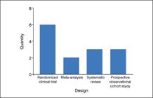
Study designs included 6 RCTs, 3 prospective controlled cohort studies, 3 systematic reviews, and 2 meta analyses. Table 1 summarizes the PEDro scores of the included clinical trials.
Methodological Quality
Of the included RCTs, 33% (n = 2) were classified as high quality (PEDro = 7), while 67% (n = 4) were classified as moderate quality (5-6 points). Risk of bias analysis using RoB2 (for RCTs) and ROBINS I (for observational studies) found RoB2: 3 studies with low risk of bias, 2 with moderate risk, and 1 with high risk; ROBINS I: 1 study with low risk of bias, 2 with moderate risk.
GRADE evidence quality was categorized as high for 2 meta analyses and 1 systematic review, moderate for 6 studies (2 reviews, 3 RCTs, 1 cohort study), and low for 5 remaining studies.
Radiographic Outcomes
Fusion rates varied from 45.5% to 100%, influenced by BMA concentration, type of scaffold, and length of follow up.5,6,9,12
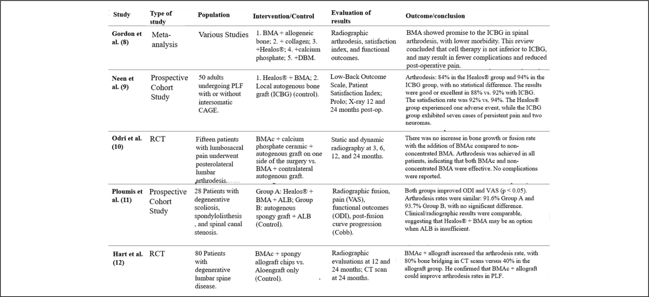
García et al.5 reported superior fusion rates with concentrated BMA (BMAc) + allograft compared with ICBG on radiography and CT at 6 and 12 months. Yamada et al.3 achieved a 93.5% 24 month fusion rate using porous β TCP + BMA. Hart et al.12 observed unilateral fusion in 80% of cases with BMAc vs. 40% with allograft. Já Johnson et al.6 e Neen et al.9 reported fusion rates between 84% and 94%, with no significant differences between BMA and ICBG groups.
Radiographic assessment varied considerably: 11 studies used serial radiographs, while eight incorporated CT with objective fusion criteria. Imaging timing and fusion definitions differed significantly among studies.
Clinical and Functional Outcomes
Validated assessment tools included VAS, ODI, LBOS, SF 36, and JOA. Both BMA and ICBG groups demonstrated significant postoperative improvements in pain and function, with no statistically significant differences in most trials.
García de Frutos et al.5 Johnson et al.6 Neen et al.9 and Odri et al.10 found no significant group differences in VAS, ODI, LBOS, or SF 36 outcomes, although all studies showed clinical improvement. Only the BMA group showed significant improvement in the SF 36. Ploumis et al.11 reported a mean JOA recovery of 76.1% using MSCs derived from BMA.11
Adverse Events and Safety Profile
Across studies, BMA presented lower morbidity. Reported adverse events included: BMA: 1 radiculopathy, 1 superficial infection, 2 hematomas; ICBG: 7 cases of donor site chronic pain, 2 neuromas, 1 wound infection. Among included RCTs, low back pain, radiculopathy, and surgical site infection accounted for 49% of all adverse events, regardless of treatment.3,4,5,6,7,8,9,10,11,12,13,14,15,16, 17
García de Frutos et al.5 observed adverse events in 9% of BMA patients vs. 15% in ICBG patients. Neen et al.9 found higher donor site pain in ICBG patients (14%), whereas only one BMA patient exhibited a complication. Hart et al.12 reported a favorable safety profile for BMAC. Johnson et al.6 Niu et al.13 Yamada et al.3 and Odri et al.10 reported no BMA related complications. Overall, BMA demonstrated lower morbidity, clinical viability, and a superior safety profile compared with ICBG, with no severe adverse events reported.3,4,5,9,10,11,12,13,16
DISCUSSION
This systematic review compiled highand moderate-quality evidence regarding the use of Bone Marrow Aspirate (BMA) as an alternative to Iliac Crest Bone Graft (ICBG) in spinal arthrodesis. Analysis of 14 studies - including randomized controlled trials, prospective cohort studies, systematic reviews, and meta-analyses - demonstrates that BMA, particularly when combined with osteoconductive scaffolds, yields satisfactory clinical and radiographic outcomes, with a superior safety profile compared to ICBG.
Bone Fusion
Fusion rates varied according to cellular concentration, graft type, and scaffold. Garcia et al.5 reported superior fusion with BMA + allograft (77%; p < 0.05), and Hart et al.12 observed 80% fusion at 24 months. Gonzalez-Tartiere et al.14 demonstrated significantly higher fusion in the BMA + allograft group based on CT at 6 months (96% vs. 55%) and radiographs at 12 months (96% vs. 52%). Imam et al.17 reported 95% fusion at 3 years using concentrated BMA + β TCP.
Conversely, Neen et al.9 and Niu13 found lower fusion rates when BMA was combined with Healos® or calcium sulfate, highlighting the importance of scaffold characteristics and preparation methods.
Hsieh et al.15 identified fusion rates ranging from 76.2%-100% in cervical procedures, increasing to 84%-100% when BMA was added. Khashan et al.7 highlight that the combination of BMA with osteoconductive scaffolds may outperform ICBG, although methodological heterogeneity limits the strength of these conclusions.
BMA Combined with Different Scaffolds
BMA efficacy varied with scaffold osteoconductive properties. Higher quality trials such as Johnson et al. and Hart et al.12 reported similar fusion rates between BMA assisted grafts (86% and 80%) and ICBG (91% and 84%) (p > 0.05). In contrast, Niu observed lower fusion with calcium sulfate (45.5%).13 García de Frutos et al.5 demonstrated superior fusion in 360° lumbar arthrodesis using BMA + allograft (71% vs. 51%), with significant differences at 6 months (83% vs. 46%) and a trend at 12 months.
Meta analyses support these findings. Gordon et al.8 observed BMA + allograft superiority in two RCTs. Feng et al.16 reported enhanced fusion with rhBMP 2 + BMA (p < 0.001), and gains with allograft (p = 0.010) and hydroxyapatite + local bone when combined with BMA.16
Methodological variability limits definitive conclusions. Nevertheless, the available data indicate that BMA, particularly when combined with appropriate scaffolds, may match or even surpass ICBG.
Radiographic Analysis and Follow up
Radiographic evaluation lacked standardization across studies. CT demonstrated higher sensitivity than radiography, as noted by Gordon et al.8 and Feng et al.16 complicating comparisons. Fusion criteria also varied: Neen et al.9 emphasized trabecular continuity and segmental stability; Hart et al.12 accepted unilateral bridging bone. Hsieh et al.15 required intervertebral and peri implant trabeculae.
Follow up duration influenced fusion detection. Hsieh et al.15 compared groups with different follow up times (13 vs. 32 months), favoring longer follow-up. Garcia et al.5 and Hart et al.12 used 12 and 24 month follow-up periods.
Another limitation involves imaging rater blinding: while Neen et al.9 and Johnson et al.6 used independent blinded evaluators, Odri et al.10 used the Arthrodese™ software. Feng et al.16 estimated predictive value below 70% for some radiographic methods, cautioning against overreliance on imaging alone, especially given asymptomatic pseudarthroses.
BMA Cell Concentration and Efficacy
Cell concentration appears to influence fusion outcomes. Johnson et al.6 reported a positive correlation between CD34+ cell count and lumbar fusion. García de Frutos et al.5 documented MSC expansion to 8.4 × 10⁵ MSCs/cm3 with superior fusion (82% vs. 46%; p = 0.0001). Hart et al.12 obtained 80% fusion using concentrated BMA (1.74 × 10⁴ MSCs/L). Imam et al.17 confirmed the relationship between callus volume and BMA cell concentration (p = 0.01).
However, Odri et al.10 found no significant difference between concentrated and non-concentrated BMA regarding fusion, although concentrated BMA produced slightly larger bone volume (117 mm3 vs. 99 mm3, p = 0.009. Ploumis et al.11 . reported 91.6% fusion with non-concentrated BMA but with longer consolidation time (11 vs. 8 months; p < 0.05. Gordon et al.8 found non concentrated BMA comparable to ICBG.8
The heterogeneity in BMA concentration protocols limited the scope of this review, as well as that of Hsieh et al.15 Moreover, BMA cellular concentration may vary among study participants according to age, harvesting site, volume, BMI, and smoking status, which further complicates standardization and analysis.1,2,3
Clinical and Functional Outcomes
The clinical and functional outcomes show a certain consistency, with improvements in pain and function after the use of BMA in spinal arthrodesis. Hsieh et al.15 reported significant improvements in pain (VAS) and functional recovery in 96% of patients, as well as relevant gains in the Nurick, JOA, and NDI scores. Hart et al.12 observed an 88% reduction in ODI. Ploumis et al.11 reported significant improvements in ODI and VAS in both the Healos® + BMA and the allograft + LBG groups (p < 0.05).
Neen et al.9 found higher LBOS, PSI, and Prolo scores for the ICBG group, although without statistical significance. Johnson et al.6 also reported no differences between groups, likely due to the small sample size (24 patients). This hypothesis is supported by larger studies, which report favorable outcomes with BMA. Yamada et al.3 demonstrated a 76.1% JOA recovery rate, while García de Frutos et al.5 observed significant improvements in VAS and ODI. Gonzalez-Tar et al.14 reported early functional improvement and higher fusion rates in the BMA group (70.6% at 3 months and 67.7% at 6 months).
The anti-inflammatory effect of BMA1,2,3 may have contributed to reduced pain and improved function in the cited studies; however, methodological heterogeneity and inconsistent follow-up compromise the strength of these findings.
BMA Application, Harvesting, and Processing Methods
The heterogeneity in BMA harvesting, preparation, and application techniques hindered consistent analysis and comparison across studies. Niu et al.13 applied 10 mL of non-concentrated BMA to autologous bone chips or CaSO4 granules, reporting fusion rates lower than those of ICBG (86% vs. 91%), although no cellularity analysis was performed.
Johnson et al.6 aspirated 100 mL of BMA and increased cellularity fourfold through centrifugation. Neen et al.9 harvested 36 mL of BMA using a Jamshidi needle, maximizing osteoprogenitor yield while minimizing dilution. García de Frutos et al.5 collected 150 mL of BMA and expanded MSCs ex vivo, achieving a concentration of 1×10⁶ MSCs/cm3 and superior fusion rates compared with (82% vs. 46%; p = 0.0001). Hart et al.12 concentrated BMA, obtaining 1.74×10⁴ MSCs/L and an 80% fusion rate (p = 0.011).
The posterior iliac crest was the most common harvest site and was considered by Imam et al.17 to be more efficient than the anterior crest in terms of cell concentration. Data on vertebral BMA harvest are insufficient for valid comparisons.
Overall, techniques involving greater processing and higher cellular concentration tend to improve fusion outcomes in spinal arthrodesis; however, the lack of methodological uniformity and the limited number of robust studies restrict the strength of these conclusions. 5,6,9,10,11,12,13
Safety and Complications
Most studies reported lower morbidity with BMA. Johnson et al.6 and Niu et al.13 did not report any relevant adverse events associated with BMA, whereas ICBG resulted in persistent pain (28%), hematoma (9.5%), seroma (14.3%), infection (9.5%), and paresthesia (24%), according to the meta-analysis by Gordon et al.8
Conversely, the meta-analysis by Feng et al.16 reported severe complications with rhBMP-2 + BMA, including heterotopic calcification, osteolysis, and malignancy. Even so, ICBG exhibited the second-highest risk of adverse events (p = 0.030).
Neen et al.9 reported persistent donor-site pain in 14% of patients treated with ICBG, whereas only one patient in the BMAc group experienced mild, self-limited pain. Feng et al.16 Hart et al.12 and García de Frutos et al.5 indicated that BMA demonstrates comparable or superior efficacy to ICBG, with a more favorable safety profile. However, the lack of methodological standardization underscores the need for more rigorous studies.
The limitations of this review include substantial methodological heterogeneity among the studies, which precluded performing a meta-analysis and creating funnel-plot assessments of publication bias. Additionally, the limited number of high-quality randomized clinical trials-and of studies specifically addressing cervical arthrodesis-restricted the overall strength of the evidence. Finally, by restricting inclusion to Q1 journals in the SJR ranking, the review increased the robustness of selected studies but potentially introduced selection bias.
CONCLUSION
This systematic review demonstrates that BMA, particularly when combined with osteoconductive scaffolds, represents an effective and safe alternative to ICBG in spinal arthrodesis. The findings indicate bone fusion rates that are comparable to or greater than those achieved with ICBG, significant clinical improvement in pain and functional scores, and a lower incidence of donor-site complications. However, methodological heterogeneity across available studies-including variations in BMA harvesting, preparation, and application protocols, as well as the lack of standardization in radiographic fusion criteria-limits the strength of these recommendations. Although promising, the current body of evidence still requires multicenter randomized clinical trials with larger sample sizes, standardized techniques, and longer follow-up.
-
Study conducted by the Hospital Getúlio Vargas, Recife, PE, Brazil.
DATA AVAILABILITY DECLARATION
The data supporting the conclusions of this Systematic Review are fully contained within the manuscript. As this study is a synthesis of evidence, it did not generate primary data. The data sources used are publicly available and can be accessed through the original databases (PubMed, Web of Science, Scopus, and Embase), referenced in the “References” section.
REFERENCES
-
1 Alsaleh KA, Tougas CA, Roffey DM, Wai EK. Osteoconductive bone graft extenders in posterolateral thoracolumbar spinal fusion: a systematic review. Spine (Phila Pa 1976). 2012;37(16):E993-1000. doi: 10.1097/BRS.0b013e3182518859.
» https://doi.org/10.1097/BRS.0b013e3182518859. -
2 Ajiboye RM, Hamamoto JT, Eckardt MA, Wang JC. Clinical and radiographic outcomes of concentrated bone marrow aspirate with allograft and demineralized bone matrix for posterolateral and interbody lumbar fusion in elderly patients. Eur Spine J. 2015;24(11):2567-72. doi: 10.1007/s00586-015-4117-5.
» https://doi.org/10.1007/s00586-015-4117-5. -
3 Yamada T, Yoshii T, Sotome S, Yuasa M, Kato T, Arai Y, et al. Hybrid grafting using bone marrow aspirate combined with porous β-tricalcium phosphate and trephine bone for lumbar posterolateral spinal fusion: a prospective, comparative study versus local bone grafting. Spine (Phila Pa 1976). 2012;37(3):E174-9. doi: 10.1097/BRS.0b013e3182269d64.
» https://doi.org/10.1097/BRS.0b013e3182269d64. -
4 Buser Z, Brodke DS, Youssef JA, Meisel HJ, Myhre SL, Hashimoto R, et al. Synthetic bone graft versus autograft or allograft for spinal fusion: a systematic review. J Neurosurg Spine. 2016;25(4):509-516. doi: 10.3171/2016.1.SPINE151005.
» https://doi.org/10.3171/2016.1.SPINE151005. -
5 García de Frutos A, González-Tartière P, Coll Bonet R, Ubierna Garcés MT, Del Arco Churruca A, Rivas García A, et al. Randomized clinical trial: expanded autologous bone marrow mesenchymal cells combined with allogeneic bone tissue, compared with autologous iliac crest graft in lumbar fusion surgery. Spine J. 2020;20(12):1899-1910. doi: 10.1016/j.spinee.2020.07.014.
» https://doi.org/10.1016/j.spinee.2020.07.014. -
6 Johnson RG. Bone marrow concentrate with allograft equivalent to autograft in lumbar fusions. Spine (Phila Pa 1976). 2014;39(9):695-700. doi: 10.1097/BRS.0000000000000254.
» https://doi.org/10.1097/BRS.0000000000000254. -
7 Khashan M, Inoue S, Berven SH. Cell based therapies as compared to autologous bone grafts for spinal arthrodesis. Spine (Phila Pa 1976). 2013;38(21):1885-91. doi: 10.1097/BRS.0b013e3182a3d7dc.
» https://doi.org/10.1097/BRS.0b013e3182a3d7dc. -
8 Gordon A, Newsome F, Ahern DP, McDonnell JM, Cunniffe G, Butler JS. Iliac crest bone graft versus cell-based grafts to augment spinal fusion: a systematic review and meta-analysis. Eur Spine J. 2024;33(1):253-263. doi: 10.1007/s00586-023-07941-9.
» https://doi.org/10.1007/s00586-023-07941-9. -
9 Neen D, Noyes D, Shaw M, Gwilym S, Fairlie N, Birch N. Healos and bone marrow aspirate used for lumbar spine fusion: a case controlled study comparing healos with autograft. Spine (Phila Pa 1976). 2006;31(18):E636-40. doi: 10.1097/01.brs.0000232028.97590.12.
» https://doi.org/10.1097/01.brs.0000232028.97590.12. -
10 Odri GA, Hami A, Pomero V, Seite M, Heymann D, Bertrand-Vasseur A, et al. Development of a per-operative procedure for concentrated bone marrow adjunction in postero-lateral lumbar fusion: radiological, biological and clinical assessment. Eur Spine J. 2012;21(12):2665-72. doi: 10.1007/s00586-012-2375-z.
» https://doi.org/10.1007/s00586-012-2375-z. -
11 Ploumis A, Albert TJ, Brown Z, Mehbod AA, Transfeldt EE. Healos graft carrier with bone marrow aspirate instead of allograft as adjunct to local autograft for posterolateral fusion in degenerative lumbar scoliosis: a minimum 2-year follow-up study. J Neurosurg Spine. 2010;13(2):211-5. doi: 10.3171/2010.3.SPINE09603.
» https://doi.org/10.3171/2010.3.SPINE09603. -
12 Ploumis A, Albert TJ, Brown Z, Mehbod AA, Transfeldt EE. Healos graft carrier with bone marrow aspirate instead of allograft as adjunct to local autograft for posterolateral fusion in degenerative lumbar scoliosis: a minimum 2-year follow-up study. J Neurosurg Spine. 2010;13(2):211-5. doi: 10.3171/2010.3.SPINE09603.
» https://doi.org/10.3171/2010.3.SPINE09603. -
13 Ploumis A, Albert TJ, Brown Z, Mehbod AA, Transfeldt EE. Healos graft carrier with bone marrow aspirate instead of allograft as adjunct to local autograft for posterolateral fusion in degenerative lumbar scoliosis: a minimum 2-year follow-up study. J Neurosurg Spine. 2010;13(2):211-5. doi: 10.3171/2010.3.SPINE09603.
» https://doi.org/10.3171/2010.3.SPINE09603. -
14 Gonzalez-Tartiere P, García de Frutos A, Ubierna Garcés MT, Rivas García A, Bagó Granell J, Càceres Palou E. et al. Randomized, prospective clinical trial to evaluate efficacy and safety in lumbar fusion surgery of implantation of autologous bone marrow mesenchymal cells expanded ex vivo and combined with allogeneic bone tissue, compared with autologous iliac crest graft; part II: clinical findings. The Spine Journal. 2019;19(9):S220-1. doi: 10.1016/j.spinee.2019.05.561.
» https://doi.org/10.1016/j.spinee.2019.05.561. -
15 Hsieh PC, Chung AS, Brodke D, Park JB, Skelly AC, Brodt ED, et al. Autologous Stem Cells in Cervical Spine Fusion. Global Spine J. 2021;11(6):950-965. doi: 10.1177/2192568220948479.
» https://doi.org/10.1177/2192568220948479. -
16 Feng JT, Yang XG, Wang F, He X, Hu YC. Efficacy and safety of bone substitutes in lumbar spinal fusion: a systematic review and network meta-analysis of randomized controlled trials. Eur Spine J. 2020;29(6):1261-1276. doi: 10.1007/s00586-019-06257-x.
» https://doi.org/10.1007/s00586-019-06257-x. -
17 Imam MA, Holton J, Ernstbrunner L, Pepke W, Grubhofer F, Narvani A, et al. A systematic review of the clinical applications and complications of bone marrow aspirate concentrate in management of bone defects and nonunions. Int Orthop. 2017;41(11):2213-2220. doi: 10.1007/s00264-017-3597-9.
» https://doi.org/10.1007/s00264-017-3597-9. -
18 Moher D, et al. The PRISMA 2020 statement: an updated guideline for reporting systematic reviews. BMJ. 2021;372:n71. doi: 10.1136/bmj. n71.
» https://doi.org/10.1136/bmj.n71
Edited by
-
Reviewed by:
Alexandre Fogaça