
 ELIGIBILITY CRITERIA FOR OBLIQUE LUMBAR INTERBODY FUSION ACCORDING TO MORPHOLOGICAL ASSESSMENT OF THE SAFETY CORRIDOR BY IMAGING EXAMS: SYSTEMATIC REVIEW
ABSTRACT
Objective: To evaluate the safety corridors for performing oblique lumbar interbody fusion (OLIF), identifying anatomical parameters that influence its feasibility and establishing criteria for indication and contraindication based on imaging tests.
Methods: A systematic review was carried out according to the PRISMA guidelines, searching databases such as PubMed, Scopus, Web of Science, and Embase. After applying the exclusion criteria, 11 articles were selected. Statistical analysis included descriptive and inferential measures, taking into account the morphology of the oblique corridor, the position of the Psoas muscle and great vessels, and the viability of muscle retraction.
Results: The data indicated that the oblique corridor narrows in a caudal direction, especially at L4-L5, where ineligibility for OLIF was highest (19%). The combined operative window (bare window + Psoas window) was a determining factor for the viability of the technique. Anatomical differences between the right and left sides were also observed, influencing surgical accessibility.
Conclusion: The OLIF technique is a promising alternative, but its applicability depends on a detailed preoperative assessment of the anatomical corridors. Standardizing indication criteria and unifying nomenclature could optimize patient selection and reduce complications. Further studies are needed to consolidate guidelines on the minimum safety parameters for performing the procedure. Level of Evidence I; Systematic Review.
Keywords:
Orthopedics; Surgery; Spine; Magnetic Resonance Imaging.
RESUMO
Objetivo: Avaliar os corredores de segurança para a realização da fusão intersomática lombar oblíqua (OLIF), identificando parâmetros anatômicos que influenciem sua viabilidade e estabelecendo critérios de indicação e contraindicação com base em exames de imagem.
Métodos: Foi realizada uma revisão sistemática conforme as diretrizes PRISMA, com busca em bases de dados como PubMed, Scopus, Web of Science e Embase. Após aplicação dos critérios de exclusão, 11 artigos foram selecionados. A análise estatística incluiu medidas descritivas e inferenciais, considerando a morfologia do corredor oblíquo, a posição do músculo Psoas e dos grandes vasos, e a viabilidade da retração muscular do referido músculo.
Resultados: Os dados indicaram que o corredor oblíquo se estreita em direção caudal, especialmente em L4-L5, onde a inelegibilidade para OLIF foi maior (19%). A janela operatória combinada (bare window + Psoas window) foi um fator determinante para a viabilidade da técnica. Diferenças anatômicas entre os lados direito e esquerdo também foram observadas, influenciando a acessibilidade cirúrgica.
Conclusão: A técnica OLIF é uma alternativa promissora, mas sua aplicabilidade depende de uma avaliação pré-operatória detalhada dos corredores anatômicos. A padronização dos critérios de indicação e a unificação da nomenclatura podem otimizar a seleção dos pacientes e reduzir complicações. São necessários estudos adicionais para consolidar diretrizes sobre os parâmetros mínimos de segurança para a realização do procedimento. Nível de Evidência I; Revisão Sistemática.
Descritores:
Ortopedia; Cirurgia; Coluna Vertebral; Ressonância Magnética.
RESUMEN
Objetivo: Evaluar los corredores de seguridad para la realización de la fusión lumbar intercorporal oblicua (OLIF), identificando los parámetros anatómicos que influyen en su viabilidad y estableciendo criterios de indicación y contraindicación basados en pruebas de imagen.
Métodos: Se realizó una revisión sistemática siguiendo las directrices PRISMA, buscando en bases de datos como PubMed, Scopus, Web of Science y Embase. Tras aplicar los criterios de exclusión, se seleccionaron 11 artículos. El análisis estadístico incluyó medidas descriptivas e inferenciales, teniendo en cuenta la morfología del corredor oblicuo, la posición del músculo Psoas y los grandes vasos, y la viabilidad de la retracción muscular de este músculo.
Resultados: Los datos indicaron que el corredor oblicuo se estrecha en dirección caudal, especialmente en L4-L5, donde la inelegibilidad para OLIF fue mayor (19%). La ventana operatoria combinada (ventana desnuda + ventana del Psoas) fue un factor determinante para la viabilidad de la técnica. También se observaron diferencias anatómicas entre los lados derecho e izquierdo, que influyeron en la accesibilidad quirúrgica.
Conclusión: La técnica OLIF es una alternativa prometedora, pero su aplicabilidad depende de una evaluación preoperatoria detallada de los corredores anatómicos. La estandarización de los criterios de indicación y la unificación de la nomenclatura podrían optimizar la selección de pacientes y reducir las complicaciones. Se necesitan más estudios para consolidar las directrices sobre los parámetros mínimos de seguridad para realizar el procedimiento. Nivel de Evidencia I: Revisión sistemática.
Descriptores:
Ortopedia; Cirugía; Columna Vertebral; Resonancia Magnética.
INTRODUCTION
Lumbar interbody fusion has been performed as a surgical alternative for the treatment of degenerative disc disease and other related pathologies, aiming to stabilize the painful segment during movement, provide direct and indirect decompression of neural elements, restore the angle of the segment closer to physiological, and correct deformities.1-3 In an attempt to preserve the integrity of the posterior elements of the spine, which are fundamental for spinal stability, anterolateral interbody fusion techniques have been developed.
Although they spare the posterior elements, resulting in decreased postoperative pain and reduced likelihood of developing adjacent segment disease,3 anterolateral approach techniques carry potential complications, such as the risk of injury to major vessels and important nerve structures that may cause sensory, motor deficits, and pain in the thigh region.4
Lateral oblique interbody fusion (Oblique lumbar interbody fusion - OLIF), as an oblique anterior access route to the Psoas, was initially described by Mayer.5 This technique would theoretically reduce the occurrence of neurological injuries compared to transpsoas techniques, as well as the occurrence of injuries resulting from manipulation of the abdominal wall, in addition to allowing access at all lumbar levels.2
Potential complications of the technique include peritoneal violation, permanent motor neurological deficit, transient motor weakness of the Psoas muscle, sympathetic plexus injury, urological injuries, and vascular injuries.2 Major vessel injuries have a low incidence of approximately 0.3 - 2.4%. However, they must be considered because they invariably present potentially catastrophic outcomes, such as the occurrence of deaths.2,4,6
The anatomical knowledge of the region to be addressed is essential to minimize adverse events during and after the procedure. In this context, the Oblique Corridor was described for the performance of OLIF. This corridor was documented in a study that used magnetic resonance imaging, showing a safety region for the anterior approach to the Psoas, and validating this finding with the aim of enabling the preoperative evaluation of patients candidates for the OLIF7 procedure.7 Such evaluation is fundamental, as the inadequate selection of patients candidates for this treatment is directly related to an increase in the number of complications.
In light of the above, it is understood that the anatomical knowledge of the Oblique Corridor is essential for the spine surgeon to develop an appropriate surgical plan based on the preoperative evaluation, so that risks and complications are reduced. Thus, this Systematic Review aims to identify imaging parameters that guide the preoperative evaluation for the most appropriate selection of patients candidates for treatment with OLIF. It seeks to identify, in particular, patients who are ideal candidates for the procedure because they have a favorable Oblique Corridor.
METHOD
A systematic review study was conducted with the aim of evaluating the dimensions of safety corridors for performing oblique interbody lumbar fusion, considering the anatomical evaluation of the lumbar region based on magnetic resonance imaging regarding parameters mentioned in Ng et al.8 In the analysis of this work, the fixed model of Mantel-Haenszel was chosen,9 which analyzes multiple strata and compares them statistically with the analytical outcomes.
The systematic review was conducted in accordance with PRISMA guidelines Protocol (Preferred Reporting Items for Systematic Reviews and Meta-Analyses) and aimed to answer the following research question: which patients are suitable for the safe performance of OLIF, based on the preoperative imaging evaluation?
The search strategy was developed in a structured manner to ensure the retrieval of studies relevant to the guiding question of the review. This stage was supported by a librarian specialized in the development and refinement of search terms. The descriptors and their synonyms were identified in the following controlled terminologies: Health Sciences Descriptors (DeCS), Medical Subject Headings (MeSH) and Emtree. In addition, keywords and terminological variations were included to broaden the scope of the search.
Initially, an exploratory search was conducted in PubMed. As a final strategy, the following descriptors and their respective synonyms and free terms were used: (“transforaminal lumbar interbody fusion” OR “posterior lumbar interbody fusion” OR “oblique lumbar interbody fusion” OR “anterior lumbar interbody fusion” OR “extreme lateral interbody fusion” OR “XLIF”) AND (“Magnetic Resonance Imaging” OR “Magnetic Resonance” OR “Radiological Evaluation” OR “Preoperative Evaluation” OR “Oblique Corridor” OR “Operative Window”). Any article that responded to the mentioned search strategy was initially included in the sample. Exclusion criteria were defined as articles that evaluated patients with malformations or vascular pathologies, spinal deformities, or a history of abdominal, lumbar, or retroperitoneal surgery.
The systematic search was conducted in the following databases and portals: PubMed, Scopus, Web of Science, and Embase. The 414 articles retrieved were imported into the Rayyan QCRI tool (Qatar Computing Research Institute, Doha, Qatar), where 196 duplicate articles were identified and removed before the initial screening. In the first phase of screening, two independent reviewers analyzed the titles and abstracts of the 218 articles, applying the previously established inclusion and exclusion criteria. The 17 articles that met the eligibility criteria had their full texts retrieved for detailed evaluation, with six articles excluded in this phase of the assessment.
During the reading of the full text, the evaluation remained independent and blinded. Any disagreements between the reviewers were resolved through discussion and, when necessary, with the mediation of a third reviewer. The entire process of searching, screening, and selecting studies was documented in detail and presented in the PRISMA flowchart. (Figure 1)
To assess heterogeneity and improve the analytical consistency of the articles, Cochran’s Q test was used,10 whose null hypothesis posits that the included systematic review studies do not exhibit heterogeneity regarding randomized analyses and proposed therapeutic interventions (safety corridor assessments for performing OLIF), in addition to the clinical and methodological heterogeneity observed in the selected articles. For this, the I2 index was used, according to the methodology described by Thompson and Higgins.11
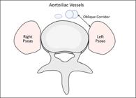
Descriptive statistical analyses of the selected articles were performed, which evaluated anatomy with the advent of imaging tests, considering hypothesis tests with a significance level of 0.05%. The following anatomical parameters were evaluated: The Oblique corridor is defined as the region between the anteromedial edge of the Psoas muscle and the lateral edge of the major vessels. A schematic figure of the oblique corridor is presented in Figure 1.
Additionally, some important anatomical regions must be considered. The following parameters were evaluated: the Vascular Window, which is determined by the distance between the left edge of the abdominal aorta or the left iliac vessels and the median sagittal plane. The window of the Psoas Major muscle is determined by the distance between the anterior edge of the Psoas Major muscle and a point located in the mid-transverse plane of the intervertebral disc. Finally, the surgical window consists of the sum of the anatomical regions of the Naked Window and the Psoas Major Window.12 This is the area anatomically used for performing OLIF.
Furthermore, the realization of OLIF requires adequate knowledge of Moro’s classification. Knowledge of Moro’s regions applies to the anatomical evaluation of the vasculature. The positioning of the large vessels in the regions referred to as AII, AIII, and AIV could be considered a contraindication for the realization of OLIF.
All statistical analyses were conducted using the software JASP, version 0.19.2 (2024). These analyses were assessed alongside the variables linked to the positioning of the vertebrae, in addition to mean comparisons and confidence intervals, which were analyzed by hypothesis tests based on the parametricity of the data (Student’s t test), considering an alpha of 0.05%.
RESULTS
After applying the exclusion criteria, 11 articles were included in the statistical analyses, considering the description of the evaluation methodologies for performing lumbar intersomatic fusion procedures. Based on the analysis of a total sample of 2795 patients included in these studies, it was found that the age of the analyzed individuals ranged from 18 to 80 years (mean age of 49 years), with a slight predominance of the male population (52%). (Figure 2) demonstrates the selection process of the studies that constituted the final sample of this review.

The selected studies showed good heterogeneity when grouped (Table 1). The descriptive analysis and sensitivity assessment of imaging methods in the anatomical corridors are in Table 2.
Q Test values and I2 test for analysis of articles published between 2016 and 2023, which evaluated safety parameters of the anatomical corridors.
Temporal analysis and diagnostic sensitivity of imaging exams along the different anatomical corridors.
The characteristics involving data collection for each evaluated study, its sampling design, and the statistical comparison between surgical viabilities across vertebral levels are described in Table 3.
Data collection and hypothesis testing for the difference in surgical viability associated with the L2 and L5 vertebrae.
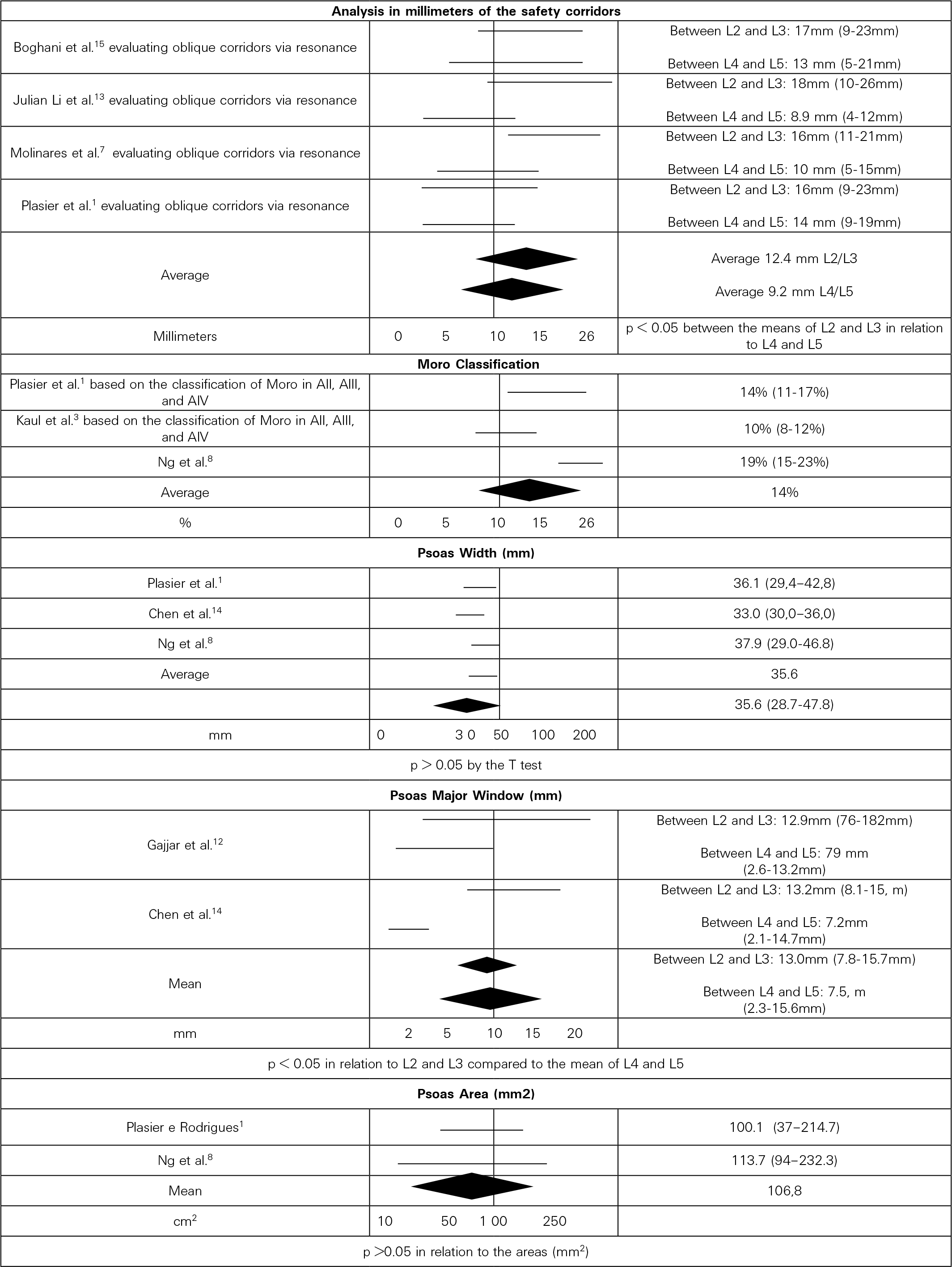
Figure 3 presents the Forest Plot that gathers the articles that performed the direct comparison between the safety corridors, measured in millimeters, the different vertebral levels, and the comparison by Moro’s classification and parameters related to the Psoas muscle.
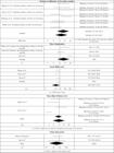
Forest plot related to comparative analysis between anatomical parameters of the Psoas muscle and vertebral levels.
Table 4 shows data on non-eligibility for the OLIF technique according to Ng et al.8 where it is seen that intersomatic fusion is not possible for oblique corridor values less than 10 millimeters.
DISCUSSION
The results of the statistical analyses revealed that the size of the corridors decreases in the caudal direction (p < 0.05 regarding L2 and L3), regardless of age groups and sex (p > 0.05). No statistical differences were observed between the variation in millimeters of the corridors, although a difference of more than 34% of their size was identified when comparing the more cranial levels (L2 and L3) to the more caudal ones (L4 and L5). Regarding the anatomical variations related to the width and area of the Psoas, no differences were observed in the quantitative assessments among the evaluated studies. The same observation cannot be made when comparing the Psoas window and the vertebral levels, where significance is seen between L2 and L3, compared to L4 and L5 (p < 0.05).
There was no significant difference between ineligibility and sex, but rather concerning age group (over 65 years). In relation to the vertebral levels, differences were observed between levels L2 and L3, which are smaller compared to L4 and L5, where greater ineligibility is seen, with an average of 19% (16.5 - 25.0%).
OLIF has been established as a viable alternative to traditional lumbar fusion techniques, offering benefits such as less compromise of the posterior elements, reduced postoperative pain, and lower risk of lumbar plexus injury when compared to the transpsoas approach (LLIF).14 However, its applicability depends on specific anatomical factors, making detailed evaluation through imaging essential in surgical planning.
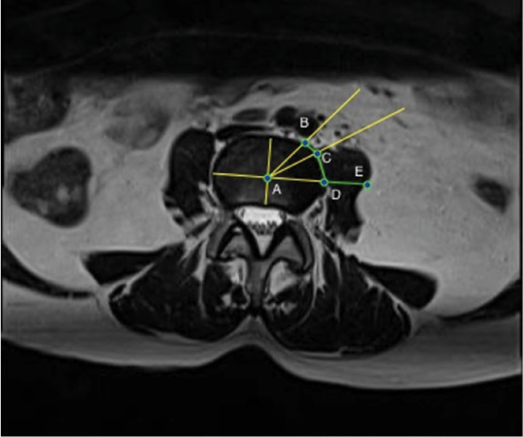
The literature suggests that an oblique corridor with at least 5 mm may be suitable for OLIF, provided that the Psoas muscle can be retracted to expand the total operative window to 10 mm.12 This view is supported by Chen et al,14 who propose that the actual operative window be defined as the sum of the bare window with the Psoas window. A representation of the actual operative window can be seen in Figure 4.

Graphical representation. Oblique Corridor BC. Psoas Window CD. Actual Operative Window BD. Width of the Psoas DE.
Gajjar et al.12 classify the oblique corridor into three distinct stages: stage 0 (non-existent oblique corridor, making OLIF extremely risky), stage 1 (corridor between 0-5 mm, allowing OLIF with retraction of the Psoas) and stage 2 (corridor greater than 5 mm, facilitating the execution of the technique).12 Additionally, Boghani et al.15 documented that the maximum dilation of the corridor can reach 26 mm, reinforcing the importance of detailed preoperative evaluation to determine the feasibility of the procedure.12
Even if the Oblique Corridor is not detected, OLIF can still be performed in some cases, as the presence of a Psoas window may allow for muscle retraction and enable access to the intervertebral disc.17 However, the width of the Psoas must be documented, as when excessive, it hinders retraction and may compromise the safety of the procedure. Furthermore, there are significant anatomical variations in other anatomical parameters of the oblique corridor across different lumbar levels, including Vascular Window, Width and Thickness of the Psoas, and the total operative window. These factors should be taken into account in surgical planning.12
The anatomy of the Psoas and the positioning of the major vessels vary as the vertebral levels become more caudal. The Psoas progressively becomes more anterior, thick, and wide, while the oblique corridor narrows, reducing the available space for the OLIF approach.14 It is also observed that the pre-vertebral vessels between L4 and S1 tend to be located more dorsally, close to the ventral surface of the Psoas muscle, increasing the need for vascular retraction at these levels.14 Additionally, there are differences between access sides, with the Oblique Corridor tending to be smaller on the right side compared to the left.14
Terminological standardization is also a relevant point, as the literature presents a great variation in the nomenclature attributed to the Oblique Corridor, being referred to in at least seven distinct ways in previous works. Unifying these terms would contribute to a better understanding and clinical applicability of the anatomical findings.12
The analysis of complications reinforces that OLIF has a lower incidence of adverse events compared to other intersomatic approaches. It is known that the technique is associated with a lower risk of vascular injury compared to ALIF and reduces the likelihood of lumbar plexus injury when compared to LLIF.13 However, the anatomy of the Psoas can be a limiting factor, especially in cases of elevated Psoas (Rising Psoas), classified as AII, AIII, and AIV in the Moro classification.8
OLIF has shown high rates of clinical success, with bone consolidation ranging from 84-100%, surpassing the average of other lumbar intersomatic fusion techniques.1 However, potential intra and postoperative complications should still be considered. Vascular injury, although rare, is the most serious complication. Other complications include injury to the vertebral endplate, fracture, ureteral injury, and subsidence of the intersomatic cage.1 In the postoperative period, the most frequent complication is transient weakness of the Psoas muscle or the Quadriceps Muscle, with an incidence of 6-22%.1 In a sample of 137 patients undergoing OLIF, Woods et al.18 reported an overall complication rate of 11.7%, with the most frequent being subsidence (4.4%), adynamic ileus (2.9%), and vascular injury (2.9%), especially at L5-S1.1
The anatomy of the oblique corridor also presents specific challenges depending on the vertebral level to be addressed. The L4-L5 level is often the most challenging, as in addition to the progressive narrowing of the oblique corridor, the presence of the High Psoas can further hinder surgical access.3,8 It is known that the absence of a measurable Oblique Corridor occurs in approximately 10.5% of cases, while the presence of elevated Psoas is observed in 19.4% of patients. About 4.7% present both conditions, which makes the procedure unfeasible.8
In light of the above, OLIF appears to be a promising surgical technique, associated with less tissue injury, less blood loss, faster recovery, and high rates of bone fusion.14 However, its applicability must be carefully planned based on a detailed preoperative assessment, considering the anatomical parameters of the Oblique Corridor, the positioning of the Psoas and major vessels, and the feasibility of muscle retraction of the Psoas to enlarge the corridor. Future studies should aim to standardize the terminologies used, improve patient selection guidelines, and develop strategies to minimize complications associated with individual anatomical variations.
CONCLUSION
As discussed earlier, factors such as a non-existent or very reduced Oblique Corridor (≤ 10 mm) and the presence of an elevated Psoas represent potential contraindications for the procedure. Through the analysis of a total sample of 2795 patients, an average of 15.43% inaptitude was found for the L2-L3 level. Additionally, the anatomical complexity of the L4-L5 level, where the Oblique Corridor tends to be narrower and the Psoas bulkier, can increase surgical risks and make the approach unfeasible with a total of 27.1% inaptitude. One must also consider the average of 14% of vessels located in the AII-AIV positions of Moro as a potential additional contraindication.
These parameters should be considered to ensure the most accurate selection of patients who have adequate anatomical parameters for the safe performance of OLIF.
-
Study conducted by the State University of Campinas (UNICAMP), Zeferino Vaz University City - Barão Geraldo, Campinas, SP, Brazil, 13083-970.
DATA AVAILABILITY DECLARATION
The contents underlying the research are available in the manuscript.
REFERENCES
-
1 Plasier EJL, Rodríguez JLU. Left oblique corridor for pre-psoas approach: image study. Coluna/Columna. 2023;22(1):e269183. doi: 10.1590/S1808-185120222201269183.
» https://doi.org/10.1590/S1808-185120222201269183. -
2 Batista MC, Amaral RAD, Melo Filho FAD, Pokorny G, Daher M, Pratali R, et al. Study of the diameter of the oblique corridor in lateral decubitus and dorsal decubitus: radiological study. Coluna/Columna. 2022;21(3):e249402. doi: 10.1590/S1808-185120222103249402.
» https://doi.org/10.1590/S1808-185120222103249402. -
3 Kaul R, Kumar K, Jeyaraman M, Goswami B, Choudhary MC, Chhabra HS, et al. MRI-Based Morphometric Study Regarding Operative Windows of Oblique Lumbar Interbody Fusion in Indian Population. Indian J Orthop. 2021;55(Suppl 2):366-373. doi: 10.1007/s43465-021-00393-7.
» https://doi.org/10.1007/s43465-021-00393-7. -
4 Liu L, Liang Y, Zhang H, Wang H, Guo C, Pu X, et al. Imaging Anatomical Research on the Operative Windows of Oblique Lumbar Interbody Fusion. PLoS One. 2016;11(9):e0163452. doi: 10.1371/journal.pone.0163452.
» https://doi.org/10.1371/journal.pone.0163452. -
5 Mayer HM. A new microsurgical technique for minimally invasive anterior lumbar interbody fusion. Spine (Phila Pa 1976). 1997;22(6):691-9; discussion 700. doi: 10.1097/00007632-199703150-00023.
» https://doi.org/10.1097/00007632-199703150-00023. -
6 Wang Z, Liu L, Xu XH, Cao MD, Lu H, Zhang KB. The OLIF working corridor based on magnetic resonance imaging: a retrospective research. J Orthop Surg Res. 2020;15(1):141. doi: 10.1186/s13018-020-01654-1.
» https://doi.org/10.1186/s13018-020-01654-1. -
7 Molinares DM, Davis TT, Fung DA. Retroperitoneal oblique corridor to the L2-S1 intervertebral discs: an MRI study. J Neurosurg Spine. 2016;24(2):248-255. doi: 10.3171/2015.3.SPINE13976.
» https://doi.org/10.3171/2015.3.SPINE13976. -
8 Ng JP, Kaliya-Perumal AK, Tandon AA, Oh JY. The Oblique Corridor at L4-L5: A Radiographic-Anatomical Study Into the Feasibility for Lateral Interbody Fusion. Spine (Phila Pa 1976). 2020;45(10):E552-E559. doi: 10.1097/BRS.0000000000003346.
» https://doi.org/10.1097/BRS.0000000000003346. -
9 Rayner JCW. Cochran-Mantel-Haenszel tests for the completely randomised design. J Korean Stat Soc. 2021;50(1):185-97. doi: 10.1007/s42952-020-00068-3.
» https://doi.org/10.1007/s42952-020-00068-3. - 10 Aslam M. Cochran’s Q test for analyzing categorical data under uncertainty. J Big Data. 2023;10(1):147.
-
11 Thompson SG, Higgins JP. How should meta-regression analyses be undertaken and interpreted? Stat Med. 2002;21(11):1559-73. doi: 10.1002/sim.1187.
» https://doi.org/10.1002/sim.1187. -
12 Gajjar S, Jhala A, Kiri S, Mistry M. MRI Study to Evaluate Anatomic Oblique Lumbar Interbody Fusion Corridor in Indian Population. J Minim Invasive Spine Surg Tech. 2021:6(2):90-97. doi: 10.21182/jmisst.2021.00157.
» https://doi.org/10.21182/jmisst.2021.00157. -
13 Julian Li JX, Mobbs RJ, Phan K. Morphometric MRI Imaging Study of the Corridor for the Oblique Lumbar Interbody Fusion Technique at L1-L5. World Neurosurg. 2018;111:e678-e685. doi: 10.1016/j.wneu.2017.12.136.
» https://doi.org/10.1016/j.wneu.2017.12.136. -
14 Chen X, Chen J, Zhang F. Imaging Anatomic Research of Oblique Lumbar Interbody Fusion in a Chinese Population Based on Magnetic Resonance. World Neurosurg. 2019;128:e51-e58. doi: 10.1016/j.wneu.2019.03.244.
» https://doi.org/10.1016/j.wneu.2019.03.244. -
15 Boghani Z, Steele WI, Barber SM, Lee JJ, Sokunbi O, Blacklock JB, et al. Variability in the size of the retroperitoneal oblique corridor: A magnetic resonance imaging-based analysis. Surg Neurol Int. 2020;11:54. doi: 10.25259/SNI_438_2019.
» https://doi.org/10.25259/SNI_438_2019. -
16 Tannoury C, Das A, Saade A, Bhale R, Chen K, Tannoury T. The Antepsoas (ATP) Surgical Corridor for Lumbar and Lumbosacral Arthrodesis: A Radiographic, Anatomic, and Surgical Investigation. Spine (Phila Pa 1976).;47(15):1084-1092. doi: 10.1097/BRS.0000000000004360.
» https://doi.org/10.1097/BRS.0000000000004360. -
17 Chen KT, Wei ST, Tseng C, Ou SW, Sun LW, Chen CM. Transforaminal Endoscopic Lumbar Discectomy for L5-S1 Disc Herniation With High Iliac Crest: Technical Note and Preliminary Series. Neurospine. 2020;17(Suppl 1):S81-S87. doi: 10.14245/ns.2040166.060.
» https://doi.org/10.14245/ns.2040166.060. -
18 Woods KR, Billys JB, Hynes RA. Technical description of oblique lateral interbody fusion at L1-L5 (OLIF25) and at L5-S1 (OLIF51) and evaluation of complication and fusion rates. Spine J. 2017;17(4):545-553. doi: 10.1016/j.spinee.2016.10.026.
» https://doi.org/10.1016/j.spinee.2016.10.026.
Edited by
-
Reviewed by:
Alexandre Fogaça

 Source: Service Collection.

 Source: Service Collection.