 CONTRALATERAL PAIN IN RECURRENT LUMBAR DISC HERNIATION: CASE REPORT
ABSTRACT
Contralateral radiculopathy due to disc herniation (DH) is a rare and challenging condition. Contralateral symptoms may result from nerve root traction or local inflammation, complicating surgical decision-making. This case report describes the endoscopic treatment of a recurrent lumbar DH with contralateral symptoms. A 48-year-old woman with low back pain radiating to the left lower limb (LLL) was diagnosed with L4-L5 DH. After conservative treatment failure, endoscopic discectomy was performed with significant improvement. Five months later, the patient presented with recurrent DH and intense pain in the right lower limb (RLL). A new left extra-foraminal endoscopic procedure was performed, resulting in symptom resolution and functional recovery. Contralateral DH may result from preexisting stenosis or inflammation. The endoscopic approach offers a less invasive and effective treatment option with rapid recovery. Endoscopic surgery was effective in treating recurrent DH with contralateral symptoms, but further studies are needed to determine the best approach. Level of Evidence IV; Case Report.
Keywords:
Intervertebral Disc Displacement; Endoscopic Surgical Procedures; Radiculopathy; Recurrence.
RESUMO
A radiculopatia contralateral à hérnia de disco (HD) é uma condição rara e desafiadora. Os sintomas contralaterais podem ser causados por tração da raiz contralateral ou por processo inflamatório, complicando a escolha do tratamento cirúrgico. Este relato descreve um caso de HD lombar recidivada com sintomas contralaterais tratados por técnica endoscópica. Uma mulher de 48 anos com dor lombar e irradiação para o membro inferior esquerdo (MIE), foi diagnosticada com HD L4-L5. Após falha no tratamento conservador, foi realizada discectomia endoscópica com melhora significativa. Após 5 meses, a paciente apresentou recidiva com dor intensa no membro inferior direito (MID). Foi realizado novo procedimento endoscópico extra-foraminal esquerdo, com resolução dos sintomas e recuperação funcional. A HD contralateral pode ser resultado de fatores como estenose preexistente ou inflamação. A técnica endoscópica oferece uma abordagem menos invasiva e eficaz, com recuperação rápida. A técnica endoscópica foi eficaz no tratamento da recidiva de HD com sintomas contralaterais, porém, mais estudos são necessários para definir a melhor abordagem. Nível de Evidência IV; Relato de Caso.
Descritores:
Deslocamento do Disco Intervertebral; Procedimentos Cirúrgicos Endoscópicos; Radiculopatia; Recidiva.
RESUMEN
La radiculopatía contralateral debido a hernia de disco (HD) es una condición rara y desafiante. Los síntomas contralaterales pueden ser causados por tracción de la raíz nerviosa o inflamación local, lo que complica la toma de decisiones quirúrgicas. Este informe describe el tratamiento endoscópico de una HD lumbar recurrente con síntomas contralaterales. Mujer de 48 años con dolor lumbar irradiado al miembro inferior izquierdo (MII), diagnosticada con HD L4-L5. Tras el fracaso del tratamiento conservador, se realizó una discectomía endoscópica con mejora significativa. Cinco meses después, la paciente presentó recidiva con dolor intenso en el miembro inferior derecho (MID). Se realizó un nuevo procedimiento endoscópico extra-foraminal izquierdo, con resolución de los síntomas y recuperación funcional. La HD contralateral puede deberse a estenosis preexistente o inflamación. La cirugía endoscópica ofrece una opción de tratamiento menos invasiva y eficaz con recuperación rápida. La cirugía endoscópica fue eficaz en el tratamiento de HD recurrente con síntomas contralaterales, pero se necesitan más estudios para determinar el mejor enfoque. Nivel de Evidencia IV; Reporte de Caso.
Descriptores:
Desplazamiento del Disco Intervertebral; Procedimientos Quirúrgicos Endoscópicos; Radiculopatía; Recurrencia.
INTRODUCTION
Patients with disc herniation (DH) may present contralateral symptoms that sometimes confuse surgeons during surgical planning1,2. In this situation, the pathophysiology of radiculopathy may not be associated with direct compression of the root but rather with traction of the contralateral root or by the local inflammatory process3-5.
The lack of clinical-radiological correlation between DH and its symptoms can raise doubts about the real origin of the radiculopathy or the side to be addressed in cases where surgical treatment is needed1,2,6. In these cases, a bilateral decompression or a unilateral decompression on the side of the DH or the symptom6,7 can be performed. In cases of DH recurrence, contralateral symptoms are even rarer8.
In this context, we will report a case of lumbar DH recurrence with contralateral symptoms treated using an endoscopic technique.
CASE REPORT
The patient signed the informed consent form (ICF) and the Research Ethics Committee approved the report (CAAE 70416223.8.0000.5487, opinion number 6.339.106).
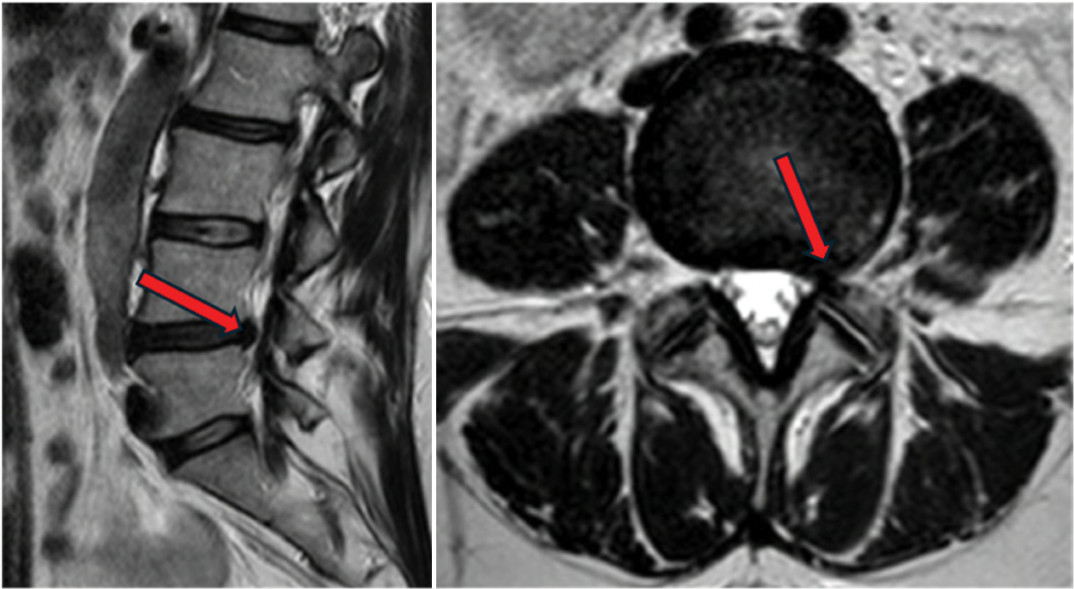
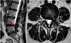
48-year-old female patient, complaining of low back pain for four months, with radiation to the left lower limb (LLL) and worsening with physical exertion. On physical examination, there was a sensory, neurological alteration in the L4 dermatome territory on the left, muscle strength grade V in the lower limbs, and positive Lasegue on the left. The pain intensity was assessed using the Visual Analog Scale for pain (VAS), with 6/10 in the lumbar region and 8/10 in the LLL. Functional disability was assessed by the Oswestry Disability Index (ODI), indicating a severe disability (60%). Simple X-rays without significant changes; dynamic X-rays in flexion and extension without signs of instability; and computed tomography (CT) without evidence of disc or yellow ligament calcifications. The lumbar spine’s magnetic resonance imaging (MRI) revealed the presence of DH L4-L5 disc herniation with compression of the L4 root (Figure 1).
With the failure of clinical treatment after six weeks and the persistence of the painful condition and sensory neurological deficit, surgical treatment was chosen. A left L4-L5 posterolateral endoscopic discectomy with foraminoplasty was performed, with the removal of the herniated disc fragment. The procedure was performed without complications under general anesthesia and intraoperative neurophysiological monitoring (IONM). Discharge from the hospital occurred 10 hours after the procedure. In the immediate postoperative period, the patient reported a significant improvement in pain, with a lumbar and LLL VAS of 1/10. There was an improvement in functional capacity, with an ODI of 10% (minimal disability).
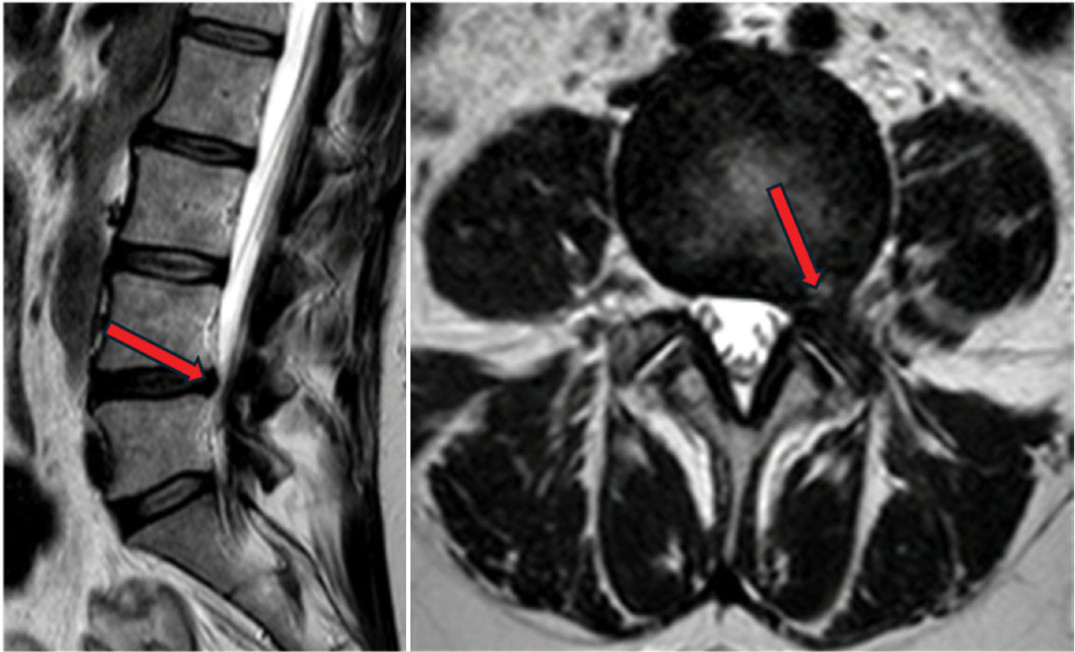
After five months of follow-up, the patient returns with a complaint of intense pain (VAS 8/10) in the lower back and right lower limb (RLL) of sudden onset, related to minor exertion, associated with progressive sensory neurological deficit in the right L5 dermatome. ODI at the time was 80%. MRI showed new DH herniation on the left at L4-L5, larger in volume than the previous DH (Figure 2).
A new surgical procedure was performed, with a posterolateral extraforaminal endoscopic approach on the left, contralateral to the symptomatic side, and removal of a large herniated disc fragment. The option was made for extra-foraminal access at the base of the L5 pedicle to avoid injury to the descending root due to previous foraminoplasty and direct access to foraminal fibrosis (Figure 3).9 The procedure was performed again under general anesthesia and IONM without complications. Eight hours after the procedure, the patient was discharged from the hospital.
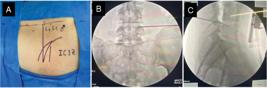
Marking of the entry point of the left extra-foraminal postero-lateral access L4-L5 (A) and puncture at the base of the L5 pedicle guided by radioscopy (B, C).
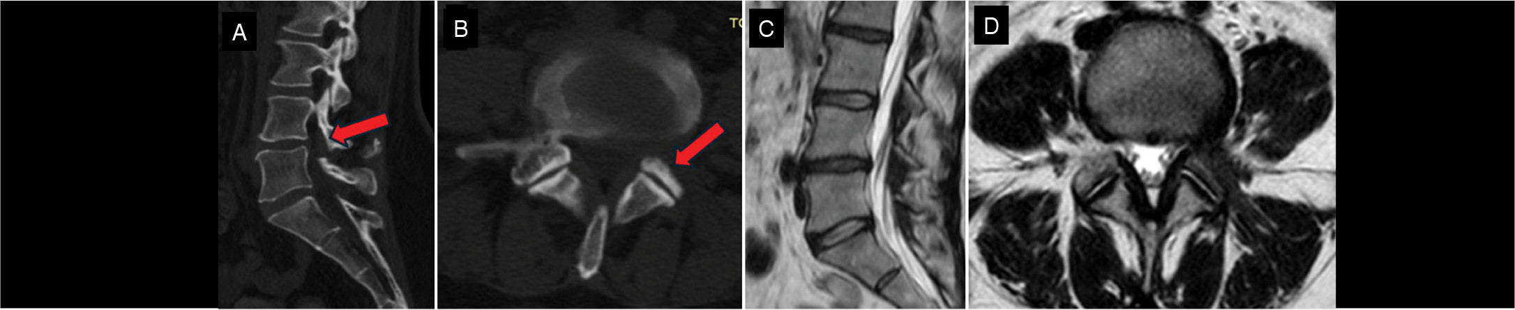
The VAS in the immediate postoperative period in the lumbar region went to 2/10 and in the RLL to 1/10. After six months of the procedure, the ODI was 10%, which remained until 12 months of follow-up. Postoperative CT shows adequate foraminotomy (Figures 4A/4B). MRI with four months of follow-up shows residual disc protrusion at L4-L5, but the patient is asymptomatic (Figures 4C/4D).

Sagittal (A) and axial (B) CT scans show adequate foraminotomy (arrow); MRI shows residual L4-L5 protrusion (C, D).
DISCUSSION
MRI is an exam with high sensitivity and specificity in identifying disc diseases of the spine, but it may not demonstrate the exact cause of symptoms of radiculopathy or neurogenic claudication10. Most lumbar radiculopathies are caused by direct nerve root compression, usually caused by HD or stenosis10-12. However, other causes, such as traction of the contralateral root, pre-existing stenoses, epidural adhesions, or the local inflammatory process itself, can generate contralateral symptoms8,13. Choudhury et al. reported three cases of contralateral radiculopathy to DH and concluded that the syndrome may be caused by spondylotic or stenotic changes14.
Several theories have already been proposed to explain the reasons for contralateral radiculopathy in patients with DH5. One of them is that the symptomatology is attributed to a pre-existing bone stenosis in the context of spondylosis. The authors of this theory suggested that DH can cause displacement and impaction of the dural sac with the emerging nerve roots against a stenosed contralateral lateral recess5,14.
Another theory is that at some levels, without dural fixation to the posterior longitudinal ligament, the ipsilateral nerve root can be easily displaced posteriorly without being significantly compressed in the case of a DH. However, the contralateral descending nerve root can be displaced laterally against the edge of the lateral recess and compressed with contact force. The pressure applied as a result of the contact force can lead to sciatic pain syndrome5,7,15.
Similar to the theory of pre-existing bone stenosis, Karabekir et al. considered the possibility that contralateral pain is caused by thickening or hypertrophy of the yellow ligament5,16. This narrowing allows for less tolerance of contralateral root displacement, which can cause symptoms of radiculopathy, especially in cases of large DH16.
It is still possible that contralateral symptomatology may occur due to DH obstruction of venous circulation. This obstruction causes dysfunction of the contralateral root even in the absence of disc material in contact with the root5,17.
Yang et al. proposed, furthermore, that the increase in epidural fat could impose a greater compression pressure on the contralateral root in the case of DH5,18.
Sucu and Gelal reported a series of cases similar to our study. They claimed that the contralateral symptoms may be related to the traction of the roots on the opposite side. Your descriptive study suggested that surgical intervention on the disc side alone was sufficient1. The mechanism of contralateral side symptomatology seems to be similar to Kernohan’s notch syndrome, in which an uncal herniation causes displacement of the cerebral peduncle against the edge of the tentorial notch, resulting in a false localizing sign19.
Hayashi et al. observed in previous studies that mechanical stress of the nerve root occurs in the anatomical structure of the ipsilateral and contralateral lateral recess, which may thus generate contralateral sciatic pain. The hernia can displace the dural sac to the opposite side, and the friction of the contralateral descending nerve root against the lateral recess can cause radiculitis and pain in the contralateral lower limb2.
In cases of recurrent DH, another cause to be considered is the presence of fibrosis or adhesions, even after minimally invasive procedures such as spinal endoscopy6,20.
Finally, contralateral pain can be generated only by the local inflammatory process8. The injury of the annulus fibrosus can release inflammatory substances, such as prostaglandins, and irritate the ipsilateral or contralateral root21.
In the reported case, we suggest that the contralateral pain is more associated with a local inflammatory process due to the release of new disc fragments, formation of fibrosis, or adhesion due to more medial manipulation of the posterior edge of the disc in the previous surgery. There is also a possible dynamic stenosis with increased diffuse disc bulging under axial compression. The previous foraminal opening may have contributed to the absence of ipsilateral pain since the nerve root had more room to move without being compressed.
CONCLUSION
Contralateral radiculopathy to lumbar DH is an uncommon clinical condition rarely found in the literature. In our case, the endoscopic technique was effective in treating recurrent DH with contralateral symptoms, but further studies should be encouraged to evaluate the best approach in these cases.
-
Study conducted by the Clínica Atualli Spine Care, São Paulo, SP, Brazil.
REFERENCES
-
1 Sucu HK, Gelal F. Lumbar disk herniation with contralateral symptoms. Eur Spine J. 2006;15(5):570-4. doi: 10.1007/s00586-005-0971-x.
» https://doi.org/10.1007/s00586-005-0971-x. -
2 Hayashi N, Iba H, Ohnaru K, Nakanishi K, Hasegawa T. Radiculopathy Contralateral to the Side of Disc Herniation -Microendoscopic Observation. Spine Surg Relat Res. 2018;2(4):304-8. doi: 10.22603/ssrr.2017-0062.
» https://doi.org/10.22603/ssrr.2017-0062. - 3 Kawakami M, Tamaki T, Hayashi N, Hashizume H, Nishi H. Possible mechanism of painful radiculopathy in lumbar disc herniation. Clin Orthop Relat Res. 1998;(351):241-51.
-
4 Modic MT, Ross JS, Obuchowski NA, Browning KH, Cianflocco AJ, Mazanec DJ. Contrast-enhanced MR imaging in acute lumbar radiculopathy: a pilot study of the natural history. Radiology. 1995;195(2):429-35. doi: 10.1148/radiology.195.2.7724762.
» https://doi.org/10.1148/radiology.195.2.7724762. -
5 Ruschel LG, Agnoletto GJ, Aragão A, Duarte JS, de Oliveira MF, Teles AR. Lumbar disc herniation with contralateral radiculopathy: a systematic review on pathophysiology and surgical strategies. Neurosurg Rev. 2021;44(2):1071-81. doi: 10.1007/s10143-020-01294-3.
» https://doi.org/10.1007/s10143-020-01294-3. -
6 Park JH, Jang JW, Park WM, Park CW. Contralateral Keyhole Biportal Endoscopic Surgery for Ruptured Lumbar Herniated Disc: A Technical Feasibility and Early Clinical Outcomes. Neurospine. 2020;17(Suppl 1):S110-9. doi: 10.14245/ns.2040224.112.
» https://doi.org/10.14245/ns.2040224.112. -
7 Akdeniz T, Kaner T, Tutkan I, Ozer AF. Unilateral surgical approach for lumbar disc herniation with contralateral symptoms. J Neurosurg Spine. 2012;17(2):124-7. doi: 10.3171/2012.4.SPINE11365.
» https://doi.org/10.3171/2012.4.SPINE11365. -
8 Gao Q, Yang H, Masood U, Zhou C, Cen Y, Song Y. Lumbar Disc Herniation with Contralateral Symptoms: A Case-Series of 11 Patients and Literature Review. Orthop Surg. 2023;15(11):2839-47. doi: 10.1111/os.13849.
» https://doi.org/10.1111/os.13849. -
9 Bergamaschi JP, de Oliveira Teixeira K, Soares TQ, de Araújo FF, Depieri GV, Lugão AF, et al. Extraforaminal Full-Endoscopic Approach for the Treatment of Lateral Compressive Diseases of the Lumbar Spine. J Pers Med. 2023;13(3):453. doi: 10.3390/jpm13030453.
» https://doi.org/10.3390/jpm13030453. -
10 Hancock MJ, Koes B, Ostelo R, Peul W. Diagnostic accuracy of the clinical examination in identifying the level of herniation in patients with sciatica. Spine (Phila Pa 1976). 2011;36(11):E712-9. doi: 10.1097/BRS.0b013e3181ee7f78.
» https://doi.org/10.1097/BRS.0b013e3181ee7f78. -
11 Kim HS, Patel R, Paudel B, Jang JS, Jang IT, Oh SH, et al. Early Outcomes of Endoscopic Contralateral Foraminal and Lateral Recess Decompression via an Interlaminar Approach in Patients with Unilateral Radiculopathy from Unilateral Foraminal Stenosis. World Neurosurg. 2017;108:763-73. doi: 10.1016/j.wneu.2017.09.018.
» https://doi.org/10.1016/j.wneu.2017.09.018. -
12 Abdul Jalil MF, Lam MF, Wang YY. Is that lumbar disc symptomatic? Herniated lumbar disc associated with contralateral radiculopathy. BMJ Case Rep. 2014;2014:bcr2013202726. doi: 10.1136/bcr-2013-202726.
» https://doi.org/10.1136/bcr-2013-202726. -
13 Van Boxem K, Rijsdijk M, Hans G, de Jong J, Kallewaard JW, Vissers K, et al. Safe Use of Epidural Corticosteroid Injections: Recommendations of the WIP Benelux Work Group. Pain Pract. 2019;19(1):61-92. doi: 10.1111/papr.12709.
» https://doi.org/10.1111/papr.12709. -
14 Choudhury AR, Taylor JC, Worthington BS, Whitaker R. Lumbar radiculopathy contralateral to upper lumbar disc herniation: report of 3 cases. Br J Surg. 1978;65(12):842-4. doi: 10.1002/bjs.1800651205.
» https://doi.org/10.1002/bjs.1800651205. -
15 Kornberg M. Sciatica contralateral to lumbar disk herniation. Orthopedics. 1994;17(4):362-4. doi: 10.3928/0147-7447-19940401-12.
» https://doi.org/10.3928/0147-7447-19940401-12. -
16 Karabekir HS, Yildizhan A, Atar EK, Yaycioglu S, Gocmen-Mas N, Yazici C. Effect of ligamenta flava hypertrophy on lumbar disc herniation with contralateral symptoms and signs: a clinical and morphometric study. Arch Med Sci. 2010;6(4):617-22. doi: 10.5114/aoms.2010.14477.
» https://doi.org/10.5114/aoms.2010.14477. -
17 Kalemci O, Kizmazoglu C, Ozer E, Arda MN. Lumbar disc herniation associated with contralateral neurological deficit: can venous congestion be the cause?. Asian Spine J. 2013;7:60-2. doi: 10.4184/asj.2013.7.1.60.
» https://doi.org/10.4184/asj.2013.7.1.60. - 18 Yang JS, Zhang DJ, Hao DJ. Lumbar disc herniation with contralateral radiculopathy: do we neglect the epidural fat? Pain Physician. 2015;18(2):E253-6.
- 19 Pearce JM. Kernohan’s notch. Eur Neurol. 2006;55:230-2.
-
20 O’Sullivan MG, Connolly AE, Buckley TF. Recurrent lumbar disc protrusion. Br J Neurosurg. 1990;4(4):319-25. doi: 10.3109/02688699008992741.
» https://doi.org/10.3109/02688699008992741. -
21 Richardson SM, Doyle P, Minogue BM, Gnanalingham K, Hoyland JA. Increased expression of matrix metalloproteinase-10, nerve growth factor and substance P in the painful degenerate intervertebral disc. Arthritis Res Ther. 2009;11(4):R126. doi: 10.1186/ar2793.
» https://doi.org/10.1186/ar2793.
Edited by
-
Reviewed by:
Aluizio Arantes