
 COMPLEX REGIONAL PAIN SYNDROME AFTER LUMBAR ENDOSCOPIC DISCECTOMY: CASE REPORT
ABSTRACT
Endoscopic spine surgery has been widely employed in the treatment of compressive conditions such as disc herniations, facet cysts, and stenoses. Despite being a minimally invasive technique, the increased number of procedures is accompanied by a rise in complications, especially during the learning curve. We report the case of a 48-year-old female patient who underwent lumbar endoscopic discectomy and developed Complex Regional Pain Syndrome (CRPS) postoperatively, a rare complication not previously described in the literature for this type of surgery. The patient had chronic lumbar radiculopathy, which had worsened over the last six months, and after failing conservative treatment, surgery was chosen. Postoperatively, the patient developed intense pain, edema, allodynia, and vasomotor signs, leading to the diagnosis of CRPS. Treatment included physiotherapy and sympathetic block, resulting in significant improvement. The development of CRPS may be associated with factors such as manipulation of the dorsal root ganglion and nociceptive sensitization. This case highlights the importance of recognizing CRPS as a possible complication in endoscopic spine surgery and the need for a multidisciplinary approach for proper management. Level of Evidence IV; Case Report.
Keywords:
Endoscopic Surgical Procedure; Complex Regional Pain Syndromes; Intraoperative Complications; Spine; Sympathetic Nerve Block.
RESUMO
A cirurgia endoscópica da coluna tem sido amplamente empregada no tratamento de condições compressivas, como hérnias de disco, cistos facetários e estenoses. Apesar de ser uma técnica minimamente invasiva, o aumento do número de procedimentos vem acompanhado de um aumento nas complicações, especialmente durante a curva de aprendizagem. Relatamos o caso de uma paciente de 48 anos submetida à discectomia lombar endoscópica (DLE) que desenvolveu Síndrome da Dor Complexa Regional (SDCR) no pós-operatório, uma complicação rara não descrita anteriormente na literatura para esse tipo de cirurgia. A paciente apresentava lombociatalgia crônica, com piora nos últimos seis meses, e após falha no tratamento conservador, optou-se pelo tratamento cirúrgico. No pós-operatório, a paciente desenvolveu dor intensa, edema, alodínia, e sinais vasomotores, sendo diagnosticada com SDCR. O tratamento incluiu fisioterapia e bloqueio simpático, resultando em melhora significativa. O desenvolvimento da SDCR pode estar associado a fatores como manipulação do gânglio da raiz dorsal e sensibilização nociceptiva. Este caso destaca a importância de reconhecer a SDCR como uma possível complicação na cirurgia endoscópica de coluna, e a necessidade de abordagem multidisciplinar para o manejo adequado. Nível de Evidência IV; Relato de Caso.
Descritores:
Procedimentos Cirúrgicos Endoscópicos; Síndrome da Dor Complexa Regional; Complicações Intraoperatórias; Coluna Vertebral; Bloqueio Nervoso Simpático.
RESUMEN
La cirugía endoscópica de columna se ha empleado ampliamente en el tratamiento de condiciones compresivas, como hernias de disco, quistes facetarios y estenosis. A pesar de ser una técnica mínimamente invasiva, el aumento del número de procedimientos va acompañado de un aumento en las complicaciones, especialmente durante la curva de aprendizaje. Reportamos el caso de una paciente de 48 años sometida a discectomía lumbar endoscópica que desarrolló el Síndrome de Dolor Regional Complejo (SDRC) en el postoperatorio, una complicación rara no descrita previamente en la literatura para este tipo de cirugía. La paciente presentaba radiculopatía lumbar crónica, que había empeorado en los últimos seis meses, y después de fallar el tratamiento conservador, se optó por el tratamiento quirúrgico. En el postoperatorio, la paciente desarrolló dolor intenso, edema, alodinia y signos vasomotores, diagnosticándose SDRC. El tratamiento incluyó fisioterapia y bloqueo simpático, con una mejora significativa. El desarrollo del SDRC puede estar asociado a factores como la manipulación del ganglio de la raíz dorsal y la sensibilización nociceptiva. Este caso destaca la importancia de reconocer el SDRC como una posible complicación en la cirugía endoscópica de columna y la necesidad de un enfoque multidisciplinario para un manejo adecuado. Nivel de Evidencia IV; Reporte de Caso.
Descriptores:
Procedimientos Quirúrgicos Endoscópicos; Síndromes de Dolor Regional Complejo; Complicaciones Intraoperatorias; Columna Vertebral; Bloqueo Nervioso Simpático.
INTRODUCTION
Endoscopic spinal surgery has been widely used for compressive spinal syndromes, especially in cases of disc herniations, facet cysts, and stenosis. An increase in the number of complications accompanies the exponential increase in the number of endoscopic lumbar spine surgeries1. Many of these complications occur during the learning curve of the technique, such as incomplete discectomy, insufficient decompression, dural injury, and dysesthesias. Other neurological complications may occur due to increased epidural pressure caused by saline solution and excessive manipulation of the nerve root or dorsal root ganglion. The increase in epidural and intracranial pressure caused by the continuous irrigation of saline solution can cause headache, neck pain, dizziness, tinnitus, transient amnesia, and, in more severe cases, seizure and death1. The literature shows that inadvertent manipulation of the root can cause root injury, neuropraxia or transient paralysis, and dysesthesias. However, the literature still does not report cases of complex regional pain syndrome (CRPS) as a complication of endoscopic lumbar discectomy (ELD). In this context, this study aims to report a case of a patient undergoing ELD evolving with CRPS in the postoperative period2,3.
CASE REPORT
The patient signed the informed consent form (ICF), and the report was approved by the Ethics Committee (CAAE 70416223.8.0000.5487, opinion number 6.339.106).
48-year-old female patient with chronic low back pain, experiencing progressive worsening in the last six months. Burning pain in the lumbar region is associated with paresthesia radiating to the anterior aspect of the left thigh, corresponding to the L3 dermatome. The pain intensity was assessed using the visual analog scale (VAS), showing 8 in the lumbar region and in the left thigh, with hypoesthesia in the left L3 territory besides a preserved muscle strength (grade V). Clinical drug treatment and rehabilitation for more than three months without improvement. The patient had a history of posterior arthrodesis from L4 to S1 15 years ago. Radiological study of computed tomography (CT) of the lumbar spine and magnetic resonance imaging (MRI) showed disc herniation and left L3-L4 foraminal stenosis (Figure 1).
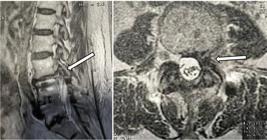
Sagittal and axial cut of the preoperative MRI, showing extruded disc herniation and left L3-L4 foraminal stenosis.
Surgical treatment was chosen, and the technique selected was lumbar discectomy via endoscopic approach with foraminoplasty through the left L3-L4 transforaminal access.
The procedure was performed under general anesthesia. The puncture was performed 8 centimeters from the midline. The outside-in technique used an endoscope with a 4.3mm working channel and 30 degrees of angulation. After access, the lateral portion of the superior articular process of L4 was identified, and foraminoplasty was performed with a cutting bone burr. After bone opening and partial removal of the yellow ligament from the lateral recess, a discectomy was performed, removing the extruded fragment.
On the first day post-operative, the patient developed intense and intermittent pain, shock-like, throughout the entire left lower limb without defining a dermatome associated with allodynia throughout the entire left lower limb.
On the second day postoperative, the patient developed significant swelling in the left lower limb, mainly in the left knee, associated with warmth and redness of the entire limb. A radiological study of lumbar CT and MRI was performed, with adequate foraminoplasty and no evidence of hernia recurrence (Figure 2). Two electromyographies of the lower limbs were performed in different centers, both being normal. Vascular examination ruled out deep vein thrombosis in the lower limbs.

Clinical aspect of the lower limbs, with increased volume and redness of the left lower limb (A); sagittal MRI section (B) and axial CT section (C) post-operative, showing improvement of the left L3-L4 foraminal space (red circle and asterisk).
Conservative physical rehabilitation measures were adopted with physiotherapy, in addition to drug treatment with analgesic (Dipyrone), opioid (Tramadol), anti-inflammatory (NSAID) Ketoprofen, anticonvulsant (Pregabalin 450mg/day), and dual antidepressant (Duloxetine 120mg/day), without improvement.
Due to the persistence of symptoms, a left L3-L4 foraminal block with dexamethasone and ropivacaine was chosen without improvement. The evolution of the condition led to the diagnostic hypothesis of complex regional pain syndrome affecting the left lower limb, with Budapest criteria present. A lumbar sympathetic block was then performed at the level of L2, guided by radioscopy, with 10ml of Ropivacaine 0.2mg/ml and 10mg of dexamethasone, with significant improvement in symptoms. Adjuvant medications and physical rehabilitation were maintained for another six weeks, with complete improvement of symptoms.
DISCUSSION
Manipulation of the emerging root or dorsal root ganglion can cause neurological complications in spinal endoscopy, especially during the learning curve. The main complications during this period are dysesthesia, root injury, and dural injury. Due to the increase in the number of surgeries performed, new complications will be reported. There is still no description in the literature of CRPS as a complication of lumbar endoscopic discectomy4.
CRPS was initially reported in war veterans in the United States during the American Civil War, with traumatic injuries to peripheral nerves being diagnosed as Causalgia4. The American Civil War doctor Weir Mitchell observed that about 10% of patients with partial traumatic peripheral injury in the distal extremities had a dramatic impact whose clinical syndrome consisted of prominent spontaneous distal burning pain, exceeding in intensity and clinical evolution of the causal event. In addition, patients reported skin hypersensitivity to light and mechanical stimulation5.
The diagnosis of CCRPS is clinical, based on history and physical examination. Its incidence ranges from 5 to 26 per 100,000 individuals/year6-9. It seems more common in the upper extremities, more frequent in females, and in the age group of 50 to 70 years3.
It can still be divided into two subtypes, based on the absence (CRPS type I, formerly known as reflex sympathetic dystrophy) or presence (CRPS type II, formerly known as causalgia) of a nerve injury10.
Currently, no single pathophysiological mechanism has been identified as responsible for CRPS. It is understood to be an elaborate combination of different factors that begin to occur at the time of the initial injury, including sensitization of the central nervous system (CNS), autonomic dysfunction, and inflammatory changes10.
The development of CRPS can occur through direct traumatic injury or insidious events, leading to changes in the peripheral nervous system (PNS). Nociceptive sensitization occurs through the release of inflammatory mediators, such as Tumor Necrosis Factor (TNF-alpha) and Prostaglandins E2, leading to a decrease in the depolarization threshold locally, probably contributing to the hyperalgesia of these patients. It is also believed that during traumatic injury, there is degeneration of alpha-type fibers and maintenance of gamma-type fibers, resulting in an imbalance in nerve conduction and increased nociceptive afference of gamma-type fibers11.
The sensitization of the CNS is fundamental for the emergence of CRPS. Continuous activation of the peripheral nerve after injury has been shown to increase the efficacy of synaptic nociceptors in the dorsal horn mediated by the neuropeptides Glutamate and substance P, decreasing the response threshold to mechanical and thermal stimuli, which can lead to hyperpathy and allodynia11.
It is believed that adrenergic and nociceptive neurons are mutually involved, leading to increased pain after sympathetic stimulation. For example, there is an increase in alpha-1 adrenergic receptors in limbs with CRPS and increased pain after intradermal injection of phenylephrine. Other aspects may be associated with the autonomic response, such as a change in the temperature of the involved limbs10.
Substance P and calcitonin gene-related peptides are related to increased inflammatory mediators such as TNF alpha, Interleukin 1, 6, and Nerve Growth Factor (NGF) potentiating nociceptive stimuli11.
There is evidence that patients with compromised emotional states are more likely to develop CRPS. Patients with post-traumatic stress have higher incidences of CRPS when compared to the control group12. There is also a great influence of stress or anxiety on the progression of the Syndrome12.
Our report considers a multifactorial cause of CRPS: mechanical manipulation of the L3 dorsal root ganglion by the working sheath, transient thermal injury to the emerging root by the bipolar, previous chronic pain, and psychological factors.
Diagnostic Criteria for CRPS
There is no specific diagnostic test for CRPS. However, diagnostic criteria were developed in 1994 by the International Association for the Study of Pain (IASP)13. Due to their low specificity and diagnostic errors, the 1994 criteria were used less frequently13. In 2003, new diagnostic criteria were developed, the Budapest Criteria, which have greater sensitivity and specificity and are more widely used today14.
Imaging tests can be used to rule out complementary diagnoses. More recently, thermography has emerged as an auxiliary method in diagnosis, where showing a temperature difference between the limbs greater than 1ºC is observed in positive cases of CRPS15.
In the reported case, we observed the presence of all the Budapest criteria, which aided in the diagnosis. Postoperative imaging exams ruled out new disc hernias and showed adequate foraminal decompression. Dysesthesia represents a differential diagnosis of CRPS, but it was ruled out in our case due to vasomotor, motor, and sudomotor signs such as edema, warmth in the leg, and redness15.
Treatment
Currently, the treatment of CRPS is divided into five pillars: pain medications (analgesics, NSAIDs, opioids), physical therapy (occupational therapy and physiotherapy), psychological support, neuro-modulating medications, and interventional procedures10,13,16.
The use of analgesic medications and NSAIDs has not proven effective in neuropathic diseases17; however, they have a considerable role in CRPS, especially in the acute phase of the disease. Steroid anti-inflammatories can also be employed, especially in the acute phase of CRPS, with good results17,18.
Physical therapy plays a fundamental role in the treatment of CRPS16,19. In addition to conventional physiotherapy, graded motor imagery (GMI) and mirror therapy are important in improving these patients21. Smart et al.20 showed the benefits of GMI, with functional improvement and pain reduction at six months in patients with CRPS type I20.
Many patients with CRPS have chronic pain and exhibit functional disability of the limb. In these cases, it is common to identify some level of stress, anxiety, or even depression. Psychotherapy plays a very important role in these cases and should always be adopted as the first line of treatment16.
The use of neuromodulatory medications in patients with CRPS has the same basis as for other types of neuropathic pain21. Few randomized clinical studies compare the efficacy of these medications in CRPS. The most commonly used medications in our environment are gabapentinoids (Gabapentin and Pregabalin) and antidepressants (tricyclic-amitriptyline; or dual-duloxetine)22,23.
Interventional procedures to treat CRPS may include sympathetic block, spinal stimulation, and dorsal ganglion stimulation23. Part of the pathophysiology of this syndrome is explained by autonomic dysfunction, with an exacerbated response of the adrenergic system, with sympathetic blockade being used for this purpose. Patients with affected upper limbs undergo stellate ganglion block, and in cases of lower limb pain, lumbar sympathetic block22.
Visnjevac et al.24, in a systematic study from 2017, showed the effectiveness of spinal stimulation in patients with CRPS, with evidence of pain improvement and patient satisfaction24. In the same study, symptoms in CRPS were improved with dorsal ganglion stimulation24.
In our case, multidisciplinary treatment was performed, with satisfactory evolution mainly after the lumbar sympathetic block. Physical rehabilitation therapy with physiotherapy was the measure adopted since the onset of symptoms, with progressive improvement after the third week of treatment.
CONCLUSION
Endoscopic spine surgery has been widely used and is not without complications. As the number of cases performed increases, new complications arise. CRPS is rarely reported in endoscopic spinal procedures, but it should be remembered as a possible complication. Your understanding, identification, pharmacological, and interventional handling are important tools in the patient’s clinical improvement.
-
Study conducted by the Atualli Spine Care Clinic, São Paulo, SP, Brazil.
REFERENCES
- 1 Pan M, Li Q, Li S, Mao H, Meng B, Zhou F, et al. Percutaneous Endoscopic Lumbar Discectomy: Indications and Complications. Pain Physician. 2020;23(1):49-56.
- 2 Hernández-Porras BC, Plancarte-Sánchez R, Alarcón-Barrios S, Sámano-García M. Síndrome doloroso regional complejo: revisión. Cir Cirujanos. 2017;85(4):366-74.
- 3 Bonica J, Ballantyne JC. Bonica`s management of pain. In: Massey MT, Harden RN, ed. Complex regional pain syndrome. 5nd ed. Philadelphia: Wolters Kluwer; 2019. p. 1177-1245.
- 4 Kessler A, Yoo M, Calisoff R. Complex regional pain syndrome: An updated comprehensive review. NeuroRehabilitation. 2020;47(3):253-64.
- 5 McMahon S, Koltzenburg M, Tracey I, Turk D. Wall and Melzack´s text book of pain. In: Binder A, Baron R. Complex regional pain syndromes. 6nd ed. Philadelphia: Elsevier Saunders; 2013. p. 1276-1295.
- 6 Sandroni P, Benrud-Larson LM, McClelland RL, Low PA. Complex regional pain syndrome type I: incidence and prevalence in Olmsted county, a population-based study. Pain. 2003;103(1-2):199-207.
- 7 Mos M, Bruijn AG, Huygen FJ, Dieleman JP, Stricker BH, Sturkenboom MC. The incidence of complex regional pain syndrome: A population-based study. Pain. 2007;129(1-2):12-20.
- 8 Elsharydah A, Loo NH, Minhajuddin A, Kandil ES. Complex regional pain syndrome type 1 predictors epidemiological perspective from a national database analysis. J Clin Anesth. 2017;39:34-7.
- 9 Ott S, Maihofner C. Signs and symptoms patients with complex regional pain syndrome. J Pain. 2018;19(6):599-611.
- 10 Shim H, Rose J, Halle S, Shekane P. Complex regional pain syndrome: a narrative review for the practising clinician. Br J Anaesth. 2019;123(2):e424-33.
- 11 Woolf CJ. Central sensitization: implications for the diagnosis and treatment of pain. Pain. 2011;152(3 Suppl):S2-S15.
- 12 Speck V, Schlereth T, Birklein F, Maihofner C. Increased prevalence of posttraumatic stress disorder in CRPS. Eur J Pain. 2017;21(3):466-73.
- 13 Urits I, Shen AH, Jones MR, Viswanath O, Kaye AD. Complex regional pain syndrome, current concepts and treatment options. Curr Pain Headache Rep. 2018;22(2):10.
- 14 Harden RN, Bruehl S, Perez RS, Birklein F, Marinus J, Maihofner C, et al. Validation of proposed diagnostic criteria (the “Budapest Criteria”) for complex regional pain syndrome. Pain. 2010;150(2):268-74.
- 15 Harden RN, Bruehl S, Perez RS, Birklein F, Marinus J, Maihofner C, et al. Development of a severity score for CRPS. Pain. 2010;151(3):870-6.
- 16 Harden RN, Oaklander ALBurton AW, Perez RSGM, Richardson K, Swan M, et al. Complex regional pain syndrome: practical diagnostic and treatment guidelines. 4th edition. Pain Med. 2013;14(2):180-229.
- 17 Rasmussen-Barr E, Held U, Grooten WJ, Roelofs PDDM, Koes BW, van Tulder MW, et al. Nonsteroidal anti-inflammatory drugs for sciatica: an updated cochrane review. Spine (Phila Pa 1976). 2017;42(8):586-94.
- 18 Kalita J, Misra U, Kumar A, Bhoi SK. Long-term prednisolone in post-stroke complex regional pain syndrome. Pain Physician. 2016;19(8):565-74.
- 19 Perez RS, Zollinger PE, Dijkstra PU, Thomassen-Hilgersom IL, Zuurmond WW, Rosenbrand KC, et al. Evidence based guidelines for complex regional pain syndrome type 1. BMC Neurol. 2010;10:20.
- 20 Smart KM, Wand BM, O’Connell NE. Physiotherapy for pain and disability in adults with complex regional pain syndrome (CRPS) types I and II. Cochrane Database Syst Rev. 2016;2(2):CD010853.
- 21 Dworkin RH, O’Connor AB, Backonja M, Farrar JT, Finnerup NB, Jensen TS, et al. Pharmacologic management of neuropathic pain: evidence-based recommendations. Pain. 2007;132(3):237-51.
- 22 Serpell MG. Neuropathic pain study group. Gabapentin in neuropathic pain syndromes: a randomised, double-blind, placebo-controlled trial. Pain. 2002;99(3):557-66.
- 23 Van de Vusse AC, Stomp-van den Berg SG, Kessels AH, Weber WE. Randomised controlled rial of gabapentin in complex regional pain syndrome type 1 [ISRCTN84121379]. BMC Neurol. 2004;4:13.
- 24 Visnjevac O, Costandi S, Patel BA, Azer G, Agarwal P, Bolash R, et al. A comprehensive outcome-specific review of the use of spinal cord stimulation for complex regional pain syndrome. Pain Pract. 2017;17(4):533-45.
Edited by
-
Reviewed by:
Aluizio Arantes


