
 Aggressive hemangiomas of the spine: differential diagnosis and treatment strategies
ABSTRACT
Objective: To describe a case series of aggressive vertebral hemangiomas and hemangioendotheliomas treated at a referral center, highlighting differential diagnosis and therapeutic strategies.
Methods: A retrospective case series and literature review including symptomatic patients with imaging and/or histopathological confirmation, treated over the past 25 years at a Latin American spine surgery center.
Results: Six patients (five females, one male; mean age 46.3 years) were treated with vertebroplasty, intralesional resection, or en bloc vertebrectomy. Most presented with severe pain, and half had neurological deficits. Preoperative embolization was performed in three cases and radiotherapy in two. All patients showed satisfactory clinical improvement.
Conclusions: Aggressive vertebral hemangiomas and hemangioendotheliomas require precise diagnosis and individualized management. A multimodal approach combining embolization, surgery, and radiotherapy proved effective in symptom control and recurrence prevention. Level of Evidence IV; Retrospective Case Series.
Keywords:
Hemangioma; Spinal Neoplasms; Hemangioendothelioma; Bone Neoplasms.
RESUMO
Objetivo: Descrever uma série de casos de hemangiomas vertebrais agressivos e hemangioendoteliomas tratados em um centro de referência, com ênfase no diagnóstico diferencial e nas estratégias terapêuticas empregadas.
Métodos: Estudo retrospectivo de casos e revisão da literatura, com inclusão de pacientes sintomáticos e confirmação diagnóstica por imagem e/ou histopatologia, tratados nos últimos 25 anos em um centro latino-americano especializado em cirurgia da coluna.
Resultados: Seis pacientes (cinco mulheres, um homem; média de 46,3 anos) foram tratados com técnicas como vertebroplastia, ressecção intralesional ou vertebrectomia em bloco. A maioria apresentou dor intensa e metade tinha déficit neurológico. Foram realizados embolização pré-operatória em três casos e radioterapia em dois. Todos apresentaram evolução clínica satisfatória.
Conclusões: Hemangiomas vertebrais agressivos e hemangioendoteliomas exigem diagnóstico cuidadoso e abordagem individualizada. A estratégia multimodal, combinando embolização, cirurgia e radioterapia, mostrou-se eficaz no controle dos sintomas e na prevenção de recidiva. Nível de Evidência IV; Estudo Retrospectivo de Série De Casos.
Descritores:
Hemangioma; Neoplasias da Coluna Vertebral; Hemangioendotelioma; Neoplasias Ósseas.
RESUMEN
Objetivo: Describir una serie de casos de hemangiomas vertebrales agresivos y hemangioendoteliomas tratados en un centro de referencia, destacando el diagnóstico diferencial y las estrategias terapéuticas empleadas.
Métodos: Serie de casos retrospectiva y revisión de la literatura que incluyó pacientes sintomáticos con diagnóstico confirmado por imágenes y/o histopatología, tratados durante los últimos 25 años en un centro latinoamericano especializado en cirugía de columna.
Resultados: Seis pacientes (cinco mujeres, un hombre; edad media de 46,3 años) fueron tratados con vertebroplastia, resección intralesional o vertebrectomía en bloque. La mayoría presentó dolor intenso y la mitad tenía déficit neurológico. Se realizó embolización preoperatoria en tres casos y radioterapia en dos. Todos evolucionaron con mejoría clínica satisfactoria.
Conclusiones: Los hemangiomas vertebrales agresivos y los hemangioendoteliomas requieren diagnóstico preciso y manejo individualizado. El enfoque multimodal, que combina embolización, cirugía y radioterapia, fue eficaz en el control de los síntomas y la prevención de recurrencias. Nivel de Evidencia IV; Estudio Retrospectivo de Series de Casos.
Descriptores:
Hemangioma; Neoplasias de la Columna Vertebral; Hemangioendotelioma; Neoplasias Óseas.
INTRODUCTION
Vascular tumors are usually divided into three groups: hemangiomas, hemangioendotheliomas, and angiosarcomas. Hemangiomas and their subtypes are considered benign tumors, hemangioendotheliomas are intermediate tumors, and angiosarcomas are classified as malignant tumors. Despite this, considerable divergence remains in their classification due to overlapping histological features.1
Vertebral hemangiomas (VH) are the most common benign tumors of the spinal column. These tumors are frequently discovered incidentally, with a reported prevalence of 10-12%, although some studies report a higher prevalence (26-41%).2-5 Most VHs are benign, but a small subset exhibit aggressive behavior and are classified as aggressive hemangiomas, accounting for approximately 1% of cases.6 The main clinical presentations include pain, spinal instability, and neurological deficits. There is currently no standardized treatment protocol for non-malignant vascular tumors. Treatment options may include radiotherapy, embolization, vertebroplasty, and surgical resection.7
The present study describes a series of cases of aggressive vertebral hemangiomas, presenting other differential diagnoses and discussing the main therapeutic strategies based on the literature and our 25 years of institutional experience.
METHODS
A retrospective case series and literature review were conducted using a database of patients diagnosed with aggressive vertebral hemangiomas and hemangioendotheliomas, treated at a Latin American Spine Surgery referral center over the past 25 years.
Patients were considered eligible for inclusion if they had a confirmed diagnosis based on imaging and/or histopathology, the presence of significant symptoms, and had undergone some form of therapeutic intervention. Exclusion criteria were: incomplete clinical or imaging data, as well as loss to follow-up.
Collected data included demographic information, clinical presentation, imaging findings, type of treatment, and post-treatment outcomes. All procedures were conducted according to ethical guidelines for human research. This study was approved by the Ethics and Research Committee of the institution (registration number - CAAE: 56429322.7.0000.5273).
RESULTS
Six patients underwent surgical treatment (five females, one male), with a mean age of 46.3 years (range: 38-64). Tumor locations included cervical spine (one case), thoracic spine (two cases), lumbar spine (two cases), and sacrum (one case).
Regarding clinical presentation, 83% of patients presented with severe pain, and 50% had neurological deficits. The most prevalent histopathological diagnosis was hemangioma (n=4), followed by hemangioendothelioma (n=1) and epithelioid hemangioendothelioma (EHE) (n=1).
Preoperative embolization was performed as an adjuvant treatment to reduce intraoperative bleeding in 3 cases. The surgical techniques used were: vertebroplasty (n=1), intralesional resection (n=1), and en bloc vertebrectomy (n=4). Adjuvant radiotherapy was used in two cases (one due to positive margins, another due to local recurrence).
Case presentation
Case 1
A 46-year-old female with severe low back pain (no neurological deficit) and diagnosed with aggressive L3 hemangioma. The patient was treated with preoperative embolization and sequential percutaneous vertebroplasty, presenting 40% of pain relief. (Figure 1)
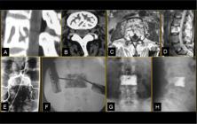
A, B - Sagittal and axial CT scan showing lytic stretch mark lesion at L3 vertebra. C, D - Axial and sagittal MRI demonstrating high signal in T2 sequence and aggressive behavior with epidural extension. E - Angiography and embolization of feeding arteries. F - Percutaneous vertebroplasty of L3. G, H - Postoperative radiographs of lumbar spine showing bone cement without leakage.
Case 2
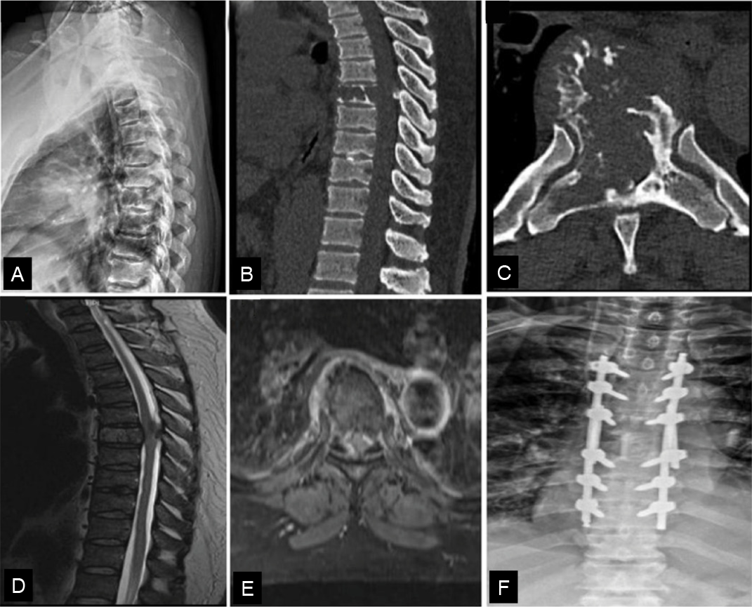
A 64-year-old male with 3-month paraparesis was diagnosed with hemangioendothelioma at T7. The patient underwent posterior en bloc vertebrectomy with compromised margins and posterior spinal reconstruction using four rods for increased stability. An allograft was used to reduce radiation artifacts and to improve recurrence assessment. (Figure 2)
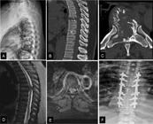
A- Lateral radiograph of the thoracic spine showing a lytic lesion in T7. B, C - Sagittal and axial CT scan showing lytic lesion with cortical erosion. D, E - Sagittal and axial MRI demonstrating spinal cord compression and soft tissue involvement. F - Postoperative radiograph in anteroposterior showing spine reconstruction of T7 with strut allograft and posterior instrumentation with multiple rods.
Case 3
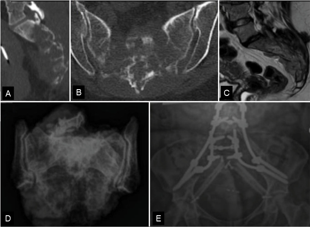
A 38-year-old female with 5-year sacral pain and sphincter dysfunction. The patient was diagnosed with epithelioid hemangioendothelioma and underwent total sacrectomy with clear margins. Reconstruction was performed using pedicle and iliac screws, allograft, and vascularized fibula graft. The patient presented substantial pain relief. (Figure 3)

A, B - CT scan (sagittal, coronal) revealing multiple expansile lytic lesions with few polka-dot signs. C - Sacral MRI showing tumor expansion into the epidural space and retrorectal fascia (Waldeyer fascia). D - Intraoperative digital radiograph showing specimens with free margins. E - Radiograph demonstrating spinopelvic reconstruction with multiple rods and Cathedral technique with vascularized fibular graft.
Case 4
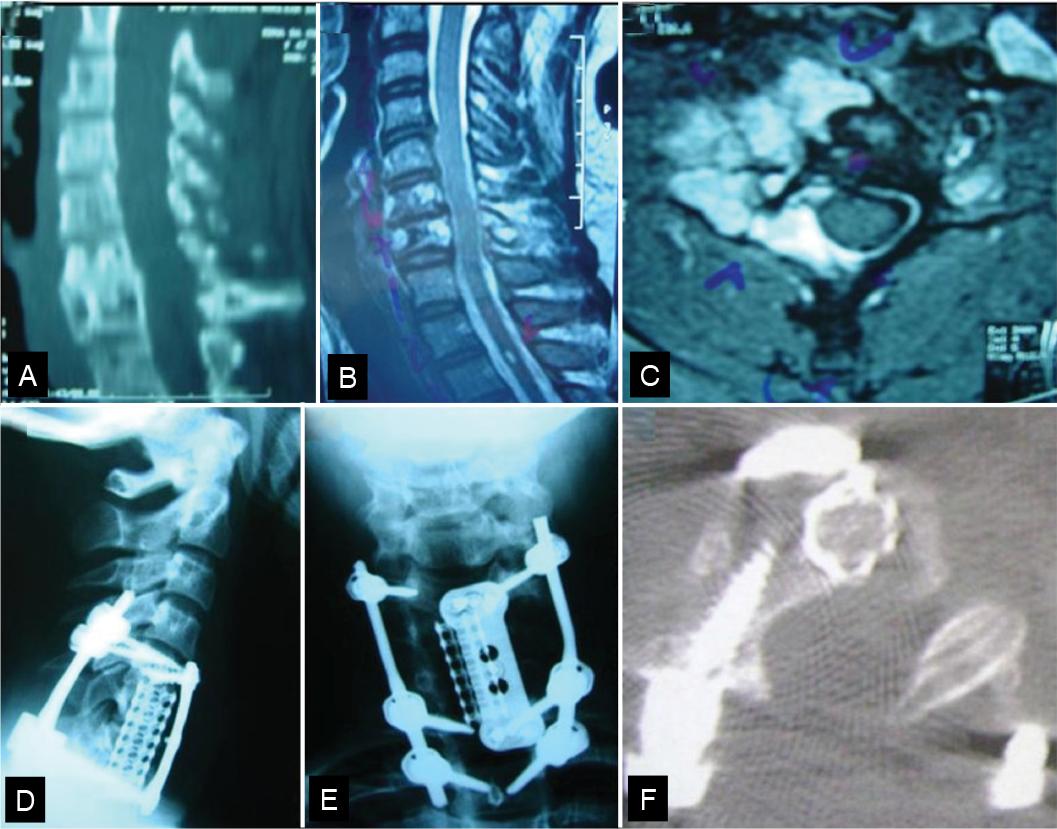
A 48-year-old female with atypical hemangioma of C6-C7 underwent corpectomy, decompression, intralesional resection, anterior and posterior cervical fusion with pedicle screws. Local recurrence at 5-year follow-up was managed with radiotherapy. (Figure 4)

A - CT scan in sagittal view with lytic cortical bulging in C6 and C7. B and C - MRI showing high signal invading the epidural space and surrounding the right vertebral artery. D and F - Lateral and anteroposterior cervical radiographs showing C6 and C7 corpectomy and reconstruction with Harms cage and posterior instrumentation with pedicle screws and rods. F - Postoperative CT axial view.
Case 5
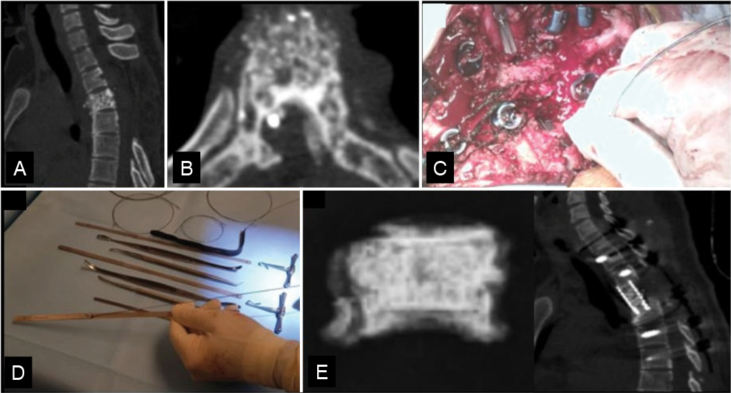
A 48-year-old female diagnosed with an aggressive hemangioma of T3 and previously submitted to a laminectomy. The patient was classified as Frankel C, presenting with tumor progression and spinal instability. The patient underwent revision en bloc spondylectomy and reconstruction using an anterior cage and posterior pedicle screw instrumentation. The patient presented partial neurological recovery. (Figure 5)

A, B - CT scan revealing T3 hemangioma with polka-dolt sign progression after previous laminectomy in another institution. C - Intraoperative image of posterior vertebral column resection with T-saw. D - Malleable retractor with T-saw attachment. E - Digital intraoperative radiograph demonstrating T3 vertebral body with adjacent endplates of superior and inferior vertebrae.
Case 6
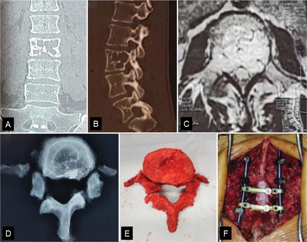
A 34-year-old female with radicular pain from aggressive L3 hemangioma underwent en bloc vertebrectomy via a combined approach. The patient experienced complete pain relief and is currently asymptomatic. (Figure 6)

A, B - CT views showing lytic lesion in L3 with cortical expansion. C - Axial MRI demonstrating aggressive tumor mass and nerve root compression. D, E - Digital radiograph and intraoperative specimen. F - Intraoperative image with spinal reconstruction.
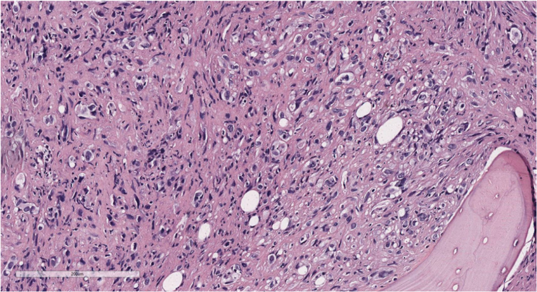
Similarly, histological findings are important for the classification of vertebral hemangiomas. (Figures 7 and 8)

Epithelioid hemangioma. Benign neoplasm composed of well-formed blood vessels, with endothelium showing abundant eosinophilic cytoplasm, vesicular and rounded nuclei, and infiltration of eosinophils between the vessels. H&E staining, 200x magnification.

Epithelioid hemangioendothelioma. Malignant neoplasm composed of nests and cords of atypical epithelioid cells (abundant eosinophilic cytoplasm, atypical vesicular or hyperchromatic nuclei). Some epithelioid cells exhibit intracytoplasmic vacuoles. H&E staining, 200x magnification.
DISCUSSION
Vertebral hemangiomas are the most common tumors of the spine. The literature estimates their prevalence between 10-12%, but more recent studies report a higher prevalence, ranging from 26-41%.2-5 This apparent increase is likely attributable to the superior sensitivity of computed tomography (CT) and magnetic resonance imaging (MRI). However, symptomatic vertebral hemangiomas have a low prevalence, accounting for 0.9% to 1.2% of all cases.8,9
Prevalence tends to increase with age, regardless of sex or spinal segment involved.4 Prevalence tends to increase with age, regardless of sex or spinal segment involved. These tumors are commonly observed in patients over 58 years old. In the present study, we observed symptomatic patients with a mean age of 46 years and a female predominance. However, conflicting studies are reported in the literature regarding sex predominance 4,7,10,11
Hemangiomas are commonly found in the thoracolumbar spine, most often in the vertebral body. However, they may also present involvement of the lamina and pedicle. Less frequently, these tumors can affect the cervical spine.8 In our series, we found vascular vertebral tumors in all spinal regions from the cervical spine to the sacrum, with the vast majority involving both anterior and posterior vertebral elements.
Although benign, a small percentage of vertebral hemangiomas can be symptomatic (0.9%-1.2%) and potentially exhibit axial pain, radicular pain, or myelopathy.2,3,8 Some studies report that less than 5% of hemangiomas are symptomatic.12,13 Aggressive vertebral hemangiomas (AVHs) account for approximately 1% of hemangiomas and are characterized by bone destruction, pathological fracture, invasion of adjacent tissues, spinal deformity, and severe neurological deficits such as paraplegia.5,11,13,14 Multilevel vertebral involvement has been reported in up to 30% of cases.9,15 In our series of cases, all patients who underwent surgical treatment presented at least pain and/or neurological deficits, associated with bone destruction, neural structure compression, or biomechanical instability.
Differential diagnosis of vertebral lesions is challenging, as they may mimic primary malignant tumors or metastases.16 Therefore, differential diagnoses include multiple myeloma, lymphoma, hematologic malignancies, and Paget’s disease. Additionally, clinical findings such as spastic gait and imaging characteristics such as hypointense lesions on MRI should be carefully evaluated.5,17
Data on benign vascular tumors are limited, with most available literature consisting of case series or case reports, and therefore, no consensus on treatment strategies has been established. Treatment options available include radiotherapy, embolization, vertebroplasty, and en bloc resection.7 Treatment of AVHs is particularly challenging due to their potential for uncontrollable bleeding. There is no universal consensus on the management of these tumors, but a wide range of therapeutic approaches is reported.11 In asymptomatic or mildly symptomatic cases, observation may be sufficient, or minimally invasive treatments can be used for pain relief. Decompressive surgery may be needed for more aggressive cases. For patients with neurological deficits, open surgical treatment is generally indicated.18,19 Electrochemotherapy has been a treatment option used in cases of therapeutic failure.20
Intralesional injection of ethanol or monoethanolamine causes thrombosis and destruction of the vascular endothelium, resulting in devascularization, lesion reduction, and subsequent decompression of neural elements.21,22 Sclerotherapy is not used as a definitive therapy and can serve as a neoadjuvant treatment to control bleeding.10,18 Nevertheless, complications are reported and include vertebral collapse, osteonecrosis, asystole, and spinal cord infarction.11
Arterial embolization is another strategy that can be used as an isolated treatment in patients with pain attributed to the hemangioma. However, it is not recommended for patients with acute neurological deficits.8 Moreover, long-term efficacy is not yet well established, albeit delayed symptomatic relief has been observed.23 Embolization is contraindicated when angiography reveals a connection with the artery of Adamkiewicz (arteria radicularis magna).2,5,18,24 Preoperative embolization is highly recommended to reduce intraoperative bleeding.7,8,10-12,15,18,24,25 In the present study, preoperative embolization was performed in 50% of the cases.
Percutaneous vertebroplasty using bone cement is described as an effective treatment for AVH, offering spinal stabilization, pain relief, and allowing rapid recovery.14,26 In lytic lesions of the C1 lateral mass, a transoral approach can also be performed.27 The local heat generated by the cement reaction contributes to local tumor control.14 This technique is effective in the treatment of asymptomatic AVHs, being also used combined with preoperative embolization and surgical decompression.8,28 We employed this combined approach in one of our cases in this study. Vertebroplasty can be feasible to reduce intraoperative bleeding (by up to 57%) combined with decompression compared to decompression alone.7 Cement leakage occurs in 20-35% of cases, but it is rarely symptomatic.29,30 Kyphoplasty presents a lower risk of cement leakage.14 In one of our cases, we performed a combined strategy involving embolization followed by vertebroplasty, achieving favorable outcomes with no complications.
Radiofrequency ablation has been described as an effective method for disease and symptom control, although it is rarely used for the treatment of AVHs.31,32 This technique destroys tissue using thermal energy (60°C-100°C) to induce protein denaturation and coagulative tumor necrosis.31,33 It has been reported that the use of robot-assisted radiofrequency is effective in tumors located in anatomically complex areas. Radiofrequency has been used for hemorrhage control combined with other methods, such as hemostatic agents, decompression, and pedicle screw fixation.34,35
Open surgical treatment is indicated in cases involving spinal instability or severe/ progressive neurological deficits.2,6,11,13,18,19,28,36 Surgical approaches can range from isolated decompression to en bloc vertebral resection.25,37 Despite their benign nature, recurrence rates of AVHs range from 3% to 30% following minimally invasive treatment, whereas en bloc resection offers lower recurrence rates and may eliminate the need for adjuvant radiotherapy.18,38
Decompression may be performed with or without instrumentation for cervical stabilization. In cases involving lesions confined to the posterior elements of the spine, decompression without instrumentation may be sufficient. However, this is typically reserved for tumors affecting only the lamina and spinous process, without facet joint involvement, or even for palliative patients with high risk.18 Lesions affecting the anterior and middle spinal columns are better addressed with corpectomy. For extensive lesions involving all three columns, en bloc resection is preferred.18 Complete en bloc spondylectomy offers high local disease control and improves long-term survival. However, it is a technically complex procedure, associated with significant morbidity and intraoperative blood loss. Therefore, subtotal resection is commonly employed, despite its higher local recurrence rate.7 In the present study, we also prioritized en bloc resection whenever feasible, despite the technical challenges, especially for aggressive hemangiomas, hemangioendotheliomas, and the epithelioid subtype, aiming to reduce bleeding, prevent local recurrence, and avoid adjuvant radiotherapy.
Radiotherapy plays an important role as an adjuvant treatment in the postoperative setting, particularly in cases of incomplete resection or more aggressive tumors, minimizing the risk of recurrence.2,6 Although standard radiation protocols are effective, higher radiation doses can be administered using modern planning and delivery techniques.39 The incidence of radiation-induced sarcoma is low (<1%).40,41 Radiotherapy requires caution due to the risks of necrosis and myelitis.10
Emerging technologies such as carbon fiber implants have the advantage of reduced radiographic artifacts on CT and MRI, allowing more precise and safer radiotherapy.42-44 Structural bone grafts are also used to reduce metal-induced imaging artifacts. Although high-level evidence remains limited, radiotherapy is widely recognized for its potential deleterious effects on tissues and may impair bone integration. Recent studies suggest that radiotherapy may be associated with delays in fusion time up to two months.45 In our center, carbon fiber vertebral body cages are not yet available, and we use titanium and allografts. Some studies suggest that the use of autologous grafts is a significant predictor of successful bone fusion (75% vs. 41%). Among the differential diagnoses for rare vascular tumors are hemangioendotheliomas and EHE, which exhibit varying degrees of malignancy.46,47
EHE is a very rare neoplasm considered an intermediate-grade malignancy with histological and clinical characteristics between aggressive hemangioma and high-grade angiosarcoma. It can mimic an epithelial tumor.40 Similar to other vascular tumors, it often presents with multiple noncontiguous lesions in up to 50% of cases. It remains unclear whether these represent multicentric lesions or metastases. High intraoperative blood loss has been reported.45,48 Radiographically, EHE appears as an expansive lytic lesion without sclerotic margins and may be associated with cortical disruption, occasionally demonstrating a “polka-dot” or “soap bubble” matrix pattern, and sometimes has sclerotic margins.49 Due to its rarity and unpredictable clinical behavior, no standardized treatment exists. However, preoperative embolization followed by wide-margin surgical resection yields better outcomes and often avoids the need for adjuvant therapy.25,40,46,50-52 In our serie cases, a patient with sacral EHE underwent total en bloc sacrectomy, achieving negative margins and satisfactory functional outcome, despite short-term follow-up.
CONCLUSION
Aggressive vertebral hemangiomas require special attention due to their potential for neurological compromise, spinal instability, and recurrence. Treatment decisions should be individualized and must consider differential diagnoses, as well as the clinical presentation, radiological characteristics, and histopathological findings.
-
Study conducted by the Instituto Nacional de Traumatologia e Ortopedia Jamil Haddad, located at Av. Brasil, 500, Caju, Rio de Janeiro, RJ, Brazil. 20940-070.
-
Reviewed by:
Marcelo Risso
ACKNOWLEDGMENTS
We gratefully acknowledge Dr. Anabela Caruso for generously providing the histopathological images for this work.
REFERENCES
-
1 Hart JL, Edgar MA, Gardner JM. Vascular tumors of bone. Semin Diagn Pathol. 2014;31(1):30-8. doi: 10.1053/j.semdp.2014.01.003.
» https://doi.org/10.1053/j.semdp.2014.01.003. -
2 Luz LXD, Simões MS, Oliveira BDA, Miotto GJ, Abreu EVD. AGGRESSIVE VERTEBRAL HEMANGIOMAS - CASE SERIES AND LITERATURE REVIEW. Coluna/Columna. 2020;19(4):293-6. doi: 10.1590/S1808-185120201904223670.
» https://doi.org/10.1590/S1808-185120201904223670. -
3 Fox MW, Onofrio BM. The natural history and management of symptomatic and asymptomatic vertebral hemangiomas. J Neurosurg. 1993;78(1):36-45. doi: 10.3171/jns.1993.78.1.0036. PMID: 8416240.
» https://doi.org/10.3171/jns.1993.78.1.0036. -
4 Abul-Kasim K, Persson E, Levinsson A, Strömbeck A, Selariu E, Ohlin A. Vertebral Hemangiomas: Prevalence, new classification and natural history. magnetic resonance imaging-based retrospective longitudinal study. Neuroradiol J. 2023;36(1):23-30. doi: 10.1177/19714009221098115.
» https://doi.org/10.1177/19714009221098115. -
5 Serdio A, Barral I, Esnal IG, Beristain M, Goienetxea A, Badiola J. Comprehensive Management Review of Aggressive Vertebral Hemangiomas. [Internet]. 2023. [accessed 2025 May 1]. Available at: https://www.essr2023.org/formulario-comunicaciones/posters/14d72faf42b82eee4dae07172c244398.pdf
» https://www.essr2023.org/formulario-comunicaciones/posters/14d72faf42b82eee4dae07172c244398.pdf -
6 Pereira Duarte M, Camino Willhuber G, Kido G, Bassani J, Petracchi M, Solá C, et al. Acute Paraparesis due to Aggressive Vertebral Hemangioma. Report of Two Cases and Literature Review. Rev Asoc Argent Ortop Traumatol. 2021;86(3):398-406. doi: 10.15417/issn.1852-7434.2021.86.3.1161.
» https://doi.org/10.15417/issn.1852-7434.2021.86.3.1161. -
7 Diarra MD, Zhang Z, Wang Z, Yinwang E, Li H, Wang S, et al. Surgical Options for Aggressive Vertebral Hemangiomas A case series, literature review and treatment recommendations. J Bone Oncol. 2023;43:100515. doi: 10.1016/j.jbo.2023.100515.
» https://doi.org/10.1016/j.jbo.2023.100515. -
8 Mousavi SR, Akbari S, Rasekhi A, Kazeminezhad A, Motlagh MAS, Taherpour S. A less aggressive approach to the treatment of aggressive vertebral hemangioma of the thoracic spine: A case report and literature review. Int J Surg Case Rep. 2023;105:108027. doi: 10.1016/j.ijscr.2023.108027.
» https://doi.org/10.1016/j.ijscr.2023.108027. -
9 Santos RHP, França CNL, Guimarães VD, Prigenzi CKC, Fontoura EAF, Botelho RV. Aggressive vertebral hemangiomas in asymptomatic patients: A review. Surg Neurol Int. 2020;11:453. doi: 10.25259/SNI_683_2020.
» https://doi.org/10.25259/SNI_683_2020. -
10 Kato K, Teferi N, Challa M, Eschbacher K, Yamaguchi S. Vertebral hemangiomas: a review on diagnosis and management. J Orthop Surg Res. 2024;19(1):310. doi: 10.1186/s13018-024-04799-5.
» https://doi.org/10.1186/s13018-024-04799-5. -
11 Qiu B, Joo P, Ajabnoor R, Boyce B, Mesfin A. Surgical management of aggressive hemangiomas of the spine. J Clin Neurosci. 2020;78:67-72. doi: 10.1016/j.jocn.2020.06.012.
» https://doi.org/10.1016/j.jocn.2020.06.012. -
12 Teferi N, Abukhiran I, Noeller J, Helland LC, Bathla G, Ryan EC, Nourski KV, Hitchon PW. Vertebral hemangiomas: Diagnosis and management. A single center experience. Clin Neurol Neurosurg. 2020;190:105745. doi: 10.1016/j.clineuro.2020.105745.
» https://doi.org/10.1016/j.clineuro.2020.105745. -
13 Kuo A, Ahorukomeye P, Gordon ZL. Aggressive thoracic vertebral hemangioma: case report and literature review. Spinal Cord Ser Cases. 2023;9(1):20. doi: 10.1038/s41394-023-00577-3.
» https://doi.org/10.1038/s41394-023-00577-3. -
14 Giorgi P, Compagnone D, Gallazzi E, Schirò GR. Early percutaneous treatment of an aggressive vertebral hemangioma: A case report with a 5-year follow-up. J Craniovertebr Junction Spine. 2020;11(2):139-142. doi: 10.4103/jcvjs.JCVJS_31_20.
» https://doi.org/10.4103/jcvjs.JCVJS_31_20. -
15 Girardo M, Zenga F, Bruno LL, Rava A, Massè A, Maule M, et al. Treatment of Aggressive Vertebral Hemangiomas with Poly Vinyl Alcohol (PVA) Microparticles Embolization, PMMA, and Short Segment Stabilization: Preliminary Results with at Least 5 Years of Follow-up. World Neurosurg. 2019;128:e283-e288. doi: 10.1016/j.wneu.2019.04.138.
» https://doi.org/10.1016/j.wneu.2019.04.138. -
16 Winfield JM, Poillucci G, Blackledge MD, Collins DJ, Shah V, Tunariu N, et al. Apparent diffusion coefficient of vertebral haemangiomas allows differentiation from malignant focal deposits in whole-body diffusion-weighted MRI. Eur Radiol. 2018;28(4):1687-1691. doi: 10.1007/s00330-017-5079-2.
» https://doi.org/10.1007/s00330-017-5079-2. - 17 Zafeiris CP, Lewkonia P, Jacobs WB. Atypical vertebral hemangioma: an aggressive form of a benign disease. Case Report and Literature Review. J Musculoskelet Neuronal Interact. 2021;21(2):317-321.
-
18 Swaminathan G, Jonathan GE, Mani SA, Keshava SN, Moses V, Prabhu K. Surgical strategies in the management of aggressive spinal haemangiomas: Retrospective case series with literature review and a practical treatment algorithm. Brain Spine. 2023;4:102736. doi: 10.1016/j.bas.2023.102736.
» https://doi.org/10.1016/j.bas.2023.102736. -
19 Patel S, Ansari D, Patil SN, Burch TG, Chaker AN, Rosinski CL, et al. High-Grade Spinal Hemangioma: A National Cancer Database Analysis. World Neurosurg. 2021;148:e527-e535. doi: 10.1016/j.wneu.2021.01.020.
» https://doi.org/10.1016/j.wneu.2021.01.020. -
20 Tedesco G, Noli LE, Griffoni C, Ghermandi R, Facchini G, Peta G, et al. Electrochemotherapy in Aggressive Hemangioma of the Spine: A Case Series and Narrative Literature Review. J Clin Med. 2024;13(5):1239. doi: 10.3390/jcm13051239.
» https://doi.org/10.3390/jcm13051239. -
21 Doppman JL, Oldfield EH, Heiss JD. Symptomatic vertebral hemangiomas: treatment by means of direct intralesional injection of ethanol. Radiology. 2000;214(2):341-8. doi: 10.1148/radiology.214.2.r00fe46341.
» https://doi.org/10.1148/radiology.214.2.r00fe46341. -
22 Takeda C, Tanishima S, Ohuchi Y, Mihara T, Yamaga K, Yoshida M, et al. Sclerotherapy for Aggressive Vertebral Hemangioma with Severe Bone Destruction: A 5-Year Analysis. Spine Surg Relat Res. 2024;8(5):548-551. doi: 10.22603/ssrr.2024-0023.
» https://doi.org/10.22603/ssrr.2024-0023. -
23 Eichberg DG, Starke RM, Levi AD. Combined surgical and endovascular approach for treatment of aggressive vertebral haemangiomas. Br J Neurosurg. 2018;32(4):381-388. doi: 10.1080/02688697.2017.1344619.
» https://doi.org/10.1080/02688697.2017.1344619. -
24 Chalamgari A, Valle D, Palau Villarreal X, Foreman M, Liu A, Patel A, et al. Vertebral Primary Bone Lesions: Review of Management Options. Curr Oncol. 2023;30(3):3064-3078. doi: 10.3390/curroncol30030232.
» https://doi.org/10.3390/curroncol30030232. -
25 Brunette-Clément T, Weil AG, Shedid D. Total en bloc spondylectomy of locally aggressive vertebral hemangioma in a pediatric patient. Childs Nerv Syst. 2021;37(6):2115-2120. doi: 10.1007/s00381-020-04954-3.
» https://doi.org/10.1007/s00381-020-04954-3. -
26 Gupta AK, Phukan P, Bodhey N. Percutaneous vertebroplasty for the treatment of symptomatic vertebral hemangioma with long-term follow-up. Interdiscip Neurosurg. 2021;23:100968. doi: 10.1016/j.inat.2020.100968.
» https://doi.org/10.1016/j.inat.2020.100968. -
27 Clarençon F, Shotar E, Cormier E, Premat K, Drir M, Lahlou G, et al. Transoral vertebroplasty for the C1 lateral mass. J Neurointerv Surg. 2020;12(9):879-885. doi: 10.1136/neurintsurg-2020-015853.
» https://doi.org/10.1136/neurintsurg-2020-015853. -
28 Papadakos D, Boulieris S, Theofanopoulos A, Fermeli D, Constantoyannis C. Tubular laminectomy and percutaneous vertebroplasty for aggressive vertebral hemangioma. Surg Neurol Int. 2021;12:27. doi: 10.25259/SNI_888_2020.
» https://doi.org/10.25259/SNI_888_2020. -
29 Kim BS, Hum B, Park JC, Choi IS. Retrospective review of procedural parameters and outcomes of percutaneous vertebroplasty in 673 patients. Interv Neuroradiol. 2014;20(5):564-75. doi: 10.15274/INR-2014-10080.
» https://doi.org/10.15274/INR-2014-10080. - 30 Layton KF, Thielen KR, Koch CA, Luetmer PH, Lane JI, Wald JT, Kallmes DF. Vertebroplasty, first 1000 levels of a single center: evaluation of the outcomes and complications. AJNR Am J Neuroradiol. 2007;28(4):683-9.
-
31 Erşen Ö, Tüzün HY, Kilinç NS, BiLekli AB, Ege T. RADIOFREQUENCY ABLATION PLUS VERTEBROPLASTY IN THE TREATMENT OF SYMPTOMATIC VERTEBRAL HEMANGIOMAS WITHOUT NEUROLOGICAL DEFICIT. J Turk Spinal Surg. 2020;31(2):85-7. doi: 10.4274/jtss.galenos.2020.120.
» https://doi.org/10.4274/jtss.galenos.2020.120. -
32 Tomasian A, Jennings JW. Vertebral Hemangioma: Percutaneous Minimally Invasive Image-Guided Radiofrequency Ablation. J Vasc Interv Radiol. 2020;31(11):1949-1952.e1. doi: 10.1016/j.jvir.2020.06.015.
» https://doi.org/10.1016/j.jvir.2020.06.015. -
33 Sharma S, Kamal R, Rathi AK. Vertebral hemangioma - the current radiation therapy perspective. Rep Pract Oncol Radiother. 2023;28(1):93-101. doi: 10.5603/RPOR.a2023.0009.
» https://doi.org/10.5603/RPOR.a2023.0009. -
34 Kaoudi A, Capel C, Chenin L, Peltier J, Lefranc M. Robot-Assisted Radiofrequency Ablation of a Sacral S1-S2 Aggressive Hemangioma. World Neurosurg. 2018;116:226-229. doi: 10.1016/j.wneu.2018.05.060.
» https://doi.org/10.1016/j.wneu.2018.05.060. -
35 Canbay S, Kayalar AE, Gel G, Sabuncuoğlu H. A novel surgical technique for aggressive vertebral hemangiomas. Neurocirugia (Engl Ed). 2019;30(5):233-237. English, Spanish. doi: 10.1016/j.neucir.2018.08.003.
» https://doi.org/10.1016/j.neucir.2018.08.003. -
36 Goraya GS, Singhal S, Paul BS, Paul G. Aggressive Vertebral Hemangioma: The Mystery of Spastic Legs Unveiled by a Purple Shoulder. Cureus. 2022;14(1):e21568. doi: 10.7759/cureus.21568.
» https://doi.org/10.7759/cureus.21568. -
37 Ge W, Qu Y, Hou T, Zhang J, Li Q, Yang L, et al. Case report: Surgical treatment of a primary giant epithelioid hemangioendothelioma of the spine with total en-bloc spondylectomy. Front Oncol. 2023;13:1109643. doi: 10.3389/fonc.2023.1109643.
» https://doi.org/10.3389/fonc.2023.1109643. -
38 Vasudeva VS, Chi JH, Groff MW. Surgical treatment of aggressive vertebral hemangiomas. Neurosurg Focus. 2016;41(2):E7. doi: 10.3171/2016.5.FOCUS16169.
» https://doi.org/10.3171/2016.5.FOCUS16169. -
39 Aich RK, Deb AR, Banerjee A, Karim R, Gupta P. Symptomatic vertebral hemangioma: treatment with radiotherapy. J Cancer Res Ther. 2010;6(2):199-203. doi: 10.4103/0973-1482.65248.
» https://doi.org/10.4103/0973-1482.65248. -
40 Albakr A, Schell M, Drew B, Cenic A. Epithelioid hemangioendothelioma of the spine: case report and review of the literature. J Spine Surg. 2017;3(2):250-259. doi: 10.21037/jss.2017.05.05.
» https://doi.org/10.21037/jss.2017.05.05. -
41 Ma J, Wang L, Mo W, Yang X, Xiao J. Epithelioid hemangioendotheliomas of the spine: clinical characters with middle and long-term follow-up under surgical treatments. Eur Spine J. 2011;20(8):1371-6. doi: 10.1007/s00586-011-1798-2.
» https://doi.org/10.1007/s00586-011-1798-2. -
42 Ho D, Corde S, Chen C, Saade G, Betteridge C, Mobbs R. The use of carbon fiber/polyetheretherketone (CF/PEEK) in pedicle screw fixation for spinal neoplasms-potential advantages in postoperative imaging and radiotherapy planning. J Spine Surg. 2024;10(1):8-21. doi: 10.21037/jss-23-93.
» https://doi.org/10.21037/jss-23-93. -
43 Cofano F, Di Perna G, Monticelli M, Marengo N, Ajello M, Mammi M, et al. Carbon fiber reinforced vs titanium implants for fixation in spinal metastases: A comparative clinical study about safety and effectiveness of the new “carbon-strategy”. J Clin Neurosci. 2020;75:106-111. doi: 10.1016/j.jocn.2020.03.013.
» https://doi.org/10.1016/j.jocn.2020.03.013. -
44 Lam FC, Guru S, AbuReesh D, Hori YS, Chuang C, Liu L, et al. Use of Carbon Fiber Implants to Improve the Safety and Efficacy of Radiation Therapy for Spine Tumor Patients. Brain Sci. 2025;15(2):199. doi: 10.3390/brainsci15020199.
» https://doi.org/10.3390/brainsci15020199. -
45 Elder BD, Ishida W, Goodwin CR, Bydon A, Gokaslan ZL, Sciubba DM, et al. Bone graft options for spinal fusion following resection of spinal column tumors: systematic review and meta-analysis. Neurosurg Focus. 2017;42(1):E16. doi: 10.3171/2016.8.FOCUS16112.
» https://doi.org/10.3171/2016.8.FOCUS16112. -
46 Chen PK, Lin QT, Feng YZ, Weng ZP, Cai XR. Epithelioid hemangioendothelioma of spine: A case report with review of literatures. Radiol Case Rep. 2020;15(12):2687-2692. doi: 10.1016/j.radcr.2020.10.024.
» https://doi.org/10.1016/j.radcr.2020.10.024. -
47 Deng Y, Li M. Composite hemangioendotheliomareport of two cases located in bone and review of the literature. BMC Musculoskelet Disord. 2023;24(1):676. doi: 10.1186/s12891-023-06745-8.
» https://doi.org/10.1186/s12891-023-06745-8. -
48 Errani C, Vanel D, Gambarotti M, Alberghini M, Picci P, Faldini C. Vascular bone tumors: a proposal of a classification based on clinicopathological, radiographic and genetic features. Skeletal Radiol. 2012;41(12):1495-507. doi: 10.1007/s00256-012-1510-6.
» https://doi.org/10.1007/s00256-012-1510-6. -
49 Liang P, Zhu BB, Ren XC, Gao JB. Imaging Findings and Clinical Features of Primary Spinal Epithelioid Hemangioendothelioma. Front Oncol. 2022;12:713947. doi: 10.3389/fonc.2022.713947.
» https://doi.org/10.3389/fonc.2022.713947. -
50 Vedagiri Vydia, G., R., S., M. Epithelioid Hemangioma of the Spine: A Rare Case Report and Review of Literature. Asian Pacific Journal of Cancer Care, 3023;8(4):823-826. doi:10.31557/apjcc.2023.8.4.823-826.
» https://doi.org/10.31557/apjcc.2023.8.4.823-826. -
51 Luzzati A, Gagliano F, Perrucchini G, Scotto G, Zoccali C. Epithelioid hemangioendothelioma of the spine: results at seven years of average follow-up in a series of 10 cases surgically treated and a review of literature. Eur Spine J. 2015;24(10):2156-64. doi: 10.1007/s00586-014-3510-9.
» https://doi.org/10.1007/s00586-014-3510-9. -
52 Chen Y, Xing X, Zhang E, Zhang J, Yuan H, Lang N. Epithelioid hemangioendothelioma of the spine: an analysis of imaging findings. Insights Imaging. 2022;13(1):56. doi: 10.1186/s13244-022-01197-5.
» https://doi.org/10.1186/s13244-022-01197-5.

 Source: Image database of Instituto Nacional de Traumatologia e Ortopedia.
 Source: Image database of Instituto Nacional de Traumatologia e Ortopedia.
 Source: Image database of Instituto Nacional de Traumatologia e Ortopedia.
 Source: Image database of Instituto Nacional de Traumatologia e Ortopedia.
 Source: Image database of Instituto Nacional de Traumatologia e Ortopedia.
 Source: Image database of Instituto Nacional de Traumatologia e Ortopedia.
 Source: Image database of Instituto Nacional de Traumatologia e Ortopedia.
 Source: Image database of Instituto Nacional de Traumatologia e Ortopedia.