Circ-RERE promotes autophagy and immune escape in acute myeloid leukemia involving the miR-128-3p/ZEB1/PD-L1 axis
Abstract
Objective To explore the role of circular RNA derived from RERE (circ-RERE) in regulating Programmed Death-Ligand-1 (PD-L1) expression and microRNA-128-3p (miR-128-3p)/Zinc finger e-box Binding homeobox-1 (ZEB1) axis in Acute Myeloid Leukemia (AML).
Methods AML cell viability, proliferation, apoptosis, the ratio of microtubule-associated protein 1A/1B-Light Chain 3-phosphatidylethanolamine conjugate (LC3-II) to free LC3 (LC3-I) (LC3-II/LC3-I), and PD-L1 expression, as well as CD8+ T-cell cytotoxicity, were correspondingly analyzed. The targeting relationship between miR-128-3p and circ-RERE or ZEB1 was verified.
Results As detected, suppressing circ-RERE or overexpressing miR-128-3p significantly blocked the proliferation, migration, invasion, and autophagy of AML cells, promoted cell apoptosis, suppressed PD-L1 expression, and increased CD8+ T-cell cytotoxicity. circ-RERE competed with miR-128-3p to regulate ZEB1. Forced expression of ZEB1 did a reversal of circ-RERE suppression-induced effects.
Conclusion In a word, circ-RERE promotes autophagy and immune escape in AML by regulating PD-L1 expression through the miR-128-3p/ZEB1 axis, which may provide a new target for AML therapy.
Keywords
Circ-RERE; miR-128-3p; ZEB1; PD-L1; Acute myeloid leukemia; Autophagy
Introduction
Acute Myeloid Leukemia (AML) is commonly accompanied by poor survival and poor prognosis. The etiology of AML is the abnormal changes of hematopoietic cells, which lead to the blocking of bone marrow differentiation and the inhibition of hematopoietic function.1 AML has marked genetic and epigenetic heterogeneity, as manifested by chromosomal translocations and hematopoiesis-associated gene recurrent mutations.2,3 Despite existing treatment options such as standard chemotherapy, outcomes remain poor, with 30 %-40 % of AML patients failing to respond to therapy or relapse.4,5 Recently, AML disease progression and relapse have been implicated in immune escape.6 Interestingly, studies provide unique insights into AML-specific immune escape mechanisms, many involving epigenetic changes.6,7
Noncoding transcripts have recently been a hotspot in research on the genetic and molecular mechanisms of cancer,8 and most of the human transcriptome is classified as noncoding RNAs (ncRNAs).9 As part of the endogenous ncRNA family, circular RNAs (circRNAs) have closed single-stranded and continuous loop structures.10 Some circRNAs have been found to function in AML, such as circ_100,05311 and circular RNA derived from RERE (circ-RERE).12 circ_RERE has been testified to be upregulated in multiple myeloma.13 Interestingly, the present research found an increase in circ_RERE expression in peripheral blood samples from AML patients, indicating its involvement in the progression of AML.
CircRNAs contain microRNA (miRNA) target sites, thereby participating in the pathogenesis of various diseases by interfering with miRNA.13,14 Furthermore, miRNAs are differentially expressed in biological processes and have diverse biological functions, including cell differentiation, apoptosis, and metabolism.15,16 circ-RERE has a binding site with microRNA-128-3p (miR-128-3p) through bioinformatics analysis based on the present research. Interestingly, miR-128-3p overexpression accelerates leukemia onset in a T-cell acute lymphoblastic leukemia mouse model.17 Also, Zinc finger e-box Binding homeobox-1 (ZEB1) has a binding site with miR-128-3p A previous report has found that ZEB1 is closely related to poor prognosis in AML patients.18 Based on the above findings, this study aimed to identify a novel circRNA/miRNA/mRNA network in AML and hypothesized that circ-RERE suppressed AML development through the miR-128-3p/ZEB1 axis.
Methods
Clinical sample collection
Peripheral blood from 120 newly diagnosed AML patients and 120 healthy donors without hematological tumors was collected in Yinzhou People's Hospital. With the written informed consent of patients, the study involving humans was approved by the Yinzhou People's Hospital (n° 201902CN16) research ethics committee. AML was diagnosed and classified according to the French, American, and British standards, as well as the World Health Organization criteria. STROBE guidelines were followed for the clinical part of this study.
Cell culture
Cell culture, including human AML cell lines THP-1, Kasumi-1, and HL-60 and human normal bone marrow stromal cell line HS-5 (ATCC, USA) was done with 10 % FBS-RPMI-1640 medium (Gibco).
Cell transfection
Small hairpin RNA targeting circ-RERE (sh-circ-RERE), overexpression circ-RERE, miR-128-3p mimic/inhibitor, along with the negative controls (sh-NC, oe-NC, mimic NC, and inhibitor NC) were all synthesized by GenePharma, while pcDNA-ZEB1 and negative control (pcDNA-NC) were by RiboBio. The transfection procedures performed on Kasumi-1 cells complied with the protocols of Lipofectamine 2000 (Invitrogen, USA).
RNase R treatment
Total RNA was treated with RNase R (Seebio, China) or without RNase R (as a negative control) at 37 °C for 30 min. Thereafter, the treated RNA was subjected to reverse transcription quantitative polymerase chain reaction (RT‐qPCR) for the detection of circ-RERE and RERE expression.
Actinomycin D assay
Kasumi-1 cells were cultured in a 24-well plate. Kasumi-1 cells were treated with 2.5 mg/L actinomycin D (Sigma‐Aldrich, USA). Cells were collected and RNA was extracted at 0 h, 4 h, 8 h, 12 h and 24 h. The stability and expression levels of circRNA and mRNA were examined by RT‐qPCR.
Isolation and co-culture of CD8+ T-cells
CD8+ T-cells were isolated from peripheral blood mononuclear cells of healthy volunteers using Dynabeads™ CD8 (Invitrogen) and tested by flow cytometry to determine the purity (> 90 %). After the addition of CD3 and CD28 monoclonal antibodies, CD8+ T-cells were co-cultured with Kasumi-1 cells in a two-chamber co-culture system (Millipore, USA), the upper chamber for Kasumi-1 cells and the lower one for CD8+ T-cells. RPMI-1640 medium supplemented with 10 % FBS (Gibco) was taken as the co-culture medium.
RT-qPCR
Total RNA from blood and cells was isolated using FastKing One-Step RT-PCR Kit (Tiangen, China), treated with QuantiNova Reverse Transcription Kit (Qiagen, Germany) for reverse transcription, and tested by SYBR Green PCR Kit (Qiagen) for PCR. The 2−ΔΔCT method was chosen to calculate gene mRNA expression. Table 1 presents primer sequences.
Western blot
Total proteins were obtained based on the radioimmunoprecipitation assay lysis reagent (Solarbio, China). After quantification with a bicinchoninic acid assay kit (Solarbio), the protein was separated on 10 % sodium dodecyl sulfate-polyacrylamide gel electrophoresis, transferred to polyvinylidene fluoride membranes for reaction with primary antibodies anti-ZEB1, anti-microtubule-associated protein 1A/1B-Light Chain-3 (LC3-I), anti-microtubule-associated protein 1A/1B-Light Chain-3-phosphatidylethanolamine conjugate (LC3-II), anti-Bcl-2-interacting protein-1 (Beclin-1), anti-sequestosome-1 (p62), anti-Programmed Death-Ligand-1 (PD-L1), and anti-Glyceraldehyde-3-Phosphate Dehydrogenase (GAPDH; 1:1000; Cell Signaling Technology, USA) and the goat anti-rabbit secondary antibody (Abcam, USA), and developed after treatment with enhanced chemiluminescence reagent (Beyotime, China).
Methylthiazolyldiphenyl-tetrazolium bromide (MTT)
Kasumi-1 cells after 36 h of culture were supplemented with 20 μL of MTT (5 mg/mL; Sigma) for 4 h. Optical density value570 nm was measured with a microplate reader (BioTek, USA) after the addition of dimethyl sulfoxide (Beyotime) to dissolve formazan precipitates.
Colony formation assay
A 6-well plate was covered with Kasumi-1 cells (200 cells/well) for colony formation for 12d Then, the colonies were allowed to stain with crystal violet after methanol fixation for cell counting under a microscope (Leica, Germany).
Flow cytometry
Kasumi-1 cells were stained using the fluorescein isothiocyanate-Annexin V Apoptosis Detection Kit (BD Biosciences, USA) and analyzed by flow cytometry.
Cytotoxicity analysis
After co-culture, CD8+T-cell cytotoxicity was detected using the Lactate Dehydrogenase Cytotoxicity Kit (Thermo Fisher Scientific, USA).
Dual-luciferase reporter gene assay
Complementary sites between miR-128-3p and circ-RERE or ZEB1-3′UTR were predicted on the starbase and mutated using the Quickchange XL site-directed mutagenesis kit (Agilent Stratagene, USA). Then, the Wild-Type (WT) and Mutant (MUT) of circ-RERE and ZEB1-3′UTR were inserted into pGL4 Luc-Rluc (BioVector-NTCC, China) to form WT-circ-RERE, MUT-circ-RERE, WT-ZEB1-3′UTR, and MUT-ZEB1-3′UTR. The vectors, along with miR-128-3p mimic or mimic NC, were co-transfected into Kasumi-1 cells to determine luciferase activity by a dual-luciferase reporter gene detection system (Genomeditech, China).
Chromatin immunoprecipitation (ChIP)-qPCR assay
The ChIP assay was carried out according to the instructions of the ChIP assay kit (Cell Signaling Technology). Briefly, 4 × 10⁶ cells were treated with 1 % formaldehyde to cross-link proteins to DNA, followed by nucleus preparation and chromatin digestion; digested cross-linked chromatin was immunoprecipitated overnight (anti-ZEB1, 1:50, Abcam; Rabbit immunoglobulin G, Cell Signaling Technology), then chromatin was eluted from the precipitate and uncross-linked; the DNA was then purified, and qPCR was performed to detect the enrichment of DNA.19
Tumor xenograft assay
The animal experiments were approved by the Animal Ethics Committee and performed following the Guide for the Care and Use of Laboratory Animals. Female BALB/c immunocompetent mice (5-weeks-old) were commercially provided by Guangzhou Experiment Animal Center (China). The mice were housed at (22±1) °C with a 12-hour light/dark cycle. Food and water were provided ad libitum.
Kasumi-1 cells transfected with sh-NC or sh-circ-RERE were subcutaneously injected into the BALB/c immunocompetent mice. Tumor size was measured every 7 days, with volume calculated using the . On day 28, mice were euthanized, and tumors were harvested. After dissection, tumors were weighed and stored at −80 °C.
Immunohistochemistry (IHC)
CD8+T-cells in xenograft tumor tissues of mice were evaluated by IHC. In brief, xenograft tumor tissues were processed with fixation and paraffin embedding in sequence in accordance with the standard procedures. Thereafter, xenograft tumor tissues were prepared into 5 µm sections. After deparaffinization and rehydration, the sections were treated with 3 % H2O2 for 15 min and then with 5 % normal goat serum for 30 min. Subsequently, rabbit anti-CD8 primary antibody (1:100, ab85792, Abcam, China) was evenly dripped onto each section to probe for 12 h at 4 °C. Thereafter, the horseradish peroxidase-labeled antirabbit secondary antibody (1:200, ab6721, Abcam) was added to incubate the sections for 30 min. Later, 3,3-diaminobenzidinetetrahydrochloride was added for color development for 10 min. After dehydration and transparentizing, the sections were sealed in neutral resin. At last, CD8+T-cells were presented as brown particles under the microscope.
Statistical analysis
Data were obtained from at least three independent experiments and processed by GraphPad Prism 10. Categorical variables were analyzed by the Chi-Squared test and/or Fisher’s exact tests. The normality of continuous variables was evaluated by the Shapiro-Wilk test. Non-normally distributed continuous variables were analyzed with the Mann-Whitney U test, while normally distributed continuous variables were examined by Student's t-test or one-way analysis of variance. A p < 0.05 was defined as statistically significant.
Results
circ-RERE is augmented in AML
RT-qPCR found that circ-RERE was up-regulated in the peripheral blood of AML patients (Fig. 1A). Based on the median expression level of circ-RERE, AML patients were divided into high and low circ-RERE expression groups. Statistical analysis demonstrated that circ-RERE expression correlated with platelet counts and FAB classification (Table 2). Furthermore, elevated circ-RERE expression was observed in AML cell lines (THP-1, Kasumi-1, and HL-60) (Fig. 1B). Since Kasumi-1 cells showed the most pronounced upregulation of circ-RERE (Fig. 1B), they were selected for subsequent transfection experiments. The circular nature and stability of circ-RERE were confirmed through RNase R and actinomycin D assays. circ-RERE exhibited greater resistance to RNase R digestion compared to its linear counterpart, verifying its circular structure (Fig. 1C). Actinomycin D treatment revealed that circ-RERE had a half-life exceeding 24 h, while the linear RNA degraded within 8 h (Fig. 1D). These results collectively demonstrate that circ-RERE exists in a stable circular form.

circ-RERE is up-regulated in the peripheral blood of AML patients and AML cell lines. RT-qPCR to detect circ-RERE in peripheral blood of AML patients (A), AML cell lines and normal bone marrow stromal cells (B). Data are presented as mean ± standard deviation (n = 3). * p < 0.05; ** p < 001; *** p 0.001; **** p < 0.0001.
Silencing circ-RERE can inhibit autophagy and immune escape in AML cells
To investigate the functional role of circ-RERE in AML, Kasumi-1 cells were transfected with either sh-circ-RERE or oe-circ-RERE, with RT-qPCR confirming successful transfection through corresponding decreases or increases in circ-RERE expression (Fig. 2A). Functional assays demonstrated that circ-RERE silencing significantly suppressed Kasumi-1 cell viability and proliferation (Fig. 2B‒C), while enhancing both cellular apoptosis and CD8+ T-cell-mediated cytotoxicity (Fig. 2D‒E). Western blot analysis revealed that circ-RERE deficiency reduced PD-L1 expression in Kasumi-1 cells (Fig. 2F). Furthermore, circ-RERE knockdown inhibited cellular autophagy, as evidenced by decreased LC3-II/LC3-I ratio and Beclin-1 expression coupled with increased p62 accumulation (Fig. 2G). Notably, all these effects were conversely observed upon circ-RERE overexpression (Fig. 2B-G), collectively indicating circ-RERE's critical role in regulating autophagy and immune evasion in AML cells.
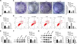
Silencing circ-RERE can inhibit autophagy and immune escape in AML cells. RT-qPCR to confirm circ-RERE expression change (A), followed by observations of viability (B), proliferation (C), apoptosis (D), cytotoxicity of CD8+ T-cells (E), PD-L1 expression (F), LC3-II/LC3-I ratio and Beclin-1 and p62 expression (G). Data are expressed as mean ± standard deviation (n = 3). * p < 0.05, ** p < 0.01, *** p < 0.001, **** p < 0.0001.
circ-RERE is a molecular sponge for miR-128-3p
Bioinformatic analysis using the starbase database predicted a binding site between circ-RERE and miR-128-3p (Fig. 3A). Luciferase reporter assays showed significantly decreased activity upon co-transfection of Wild-Type circ-RERE (WT-circ-RERE) with miR-128-3p mimics (Fig. 3B). Notably, miR-128-3p expression was downregulated in peripheral blood samples from AML patients and showed an inverse correlation with circ-RERE levels (Fig. 3C‒D). Furthermore, miR-128-3p expression was upregulated in Kasumi-1 cells transfected with sh-circ-RERE, while its expression was suppressed in oe-circ-RERE-transfected cells (Fig. 3E).

circ-RERE is a molecular sponge for miR-128-3p Predicted binding sites of circ-RERE and miR-128-3p on the starbase (A). Luciferase activity of WT/MUT-circ-RERE after miR-128-3p intervention (B). RT-qPCR detection of miR-128-3p expression in peripheral blood of AML patients (C); Correlation analysis of circ-RERE and miR-128-3p expression in AML peripheral blood (D); RT-qPCR detection of miR-128-3p expression after intervening circ-RERE expression (E). Data are expressed as mean ± standard deviation (n = 3). * p < 0.05, ** p < 0.01, *** p < 0.001, **** p < 0.0001.
miR-128-3p can block autophagy and immune escape in AML cells
To investigate miR-128-3p's function in AML cells, Kasumi-1 cells were transfected with either miR-128-3p mimic or inhibitor, with transfection efficiency verified by RT-qPCR (Fig. 4A). Functional assays demonstrated that miR-128-3p overexpression significantly suppressed cell viability and proliferation, while enhancing apoptosis and CD8+ T-cell-mediated cytotoxicity. Furthermore, it reduced PD-L1 expression and inhibited cellular autophagy (Fig. 4B‒G). Conversely, miR-128-3p knockdown produced opposite effects in Kasumi-1 cells (Fig. 4B‒G).

miR-128-3p can inhibit autophagy and immune escape in AML cells. RT-qPCR to confirm miR-128-3p expression change (A), followed by observations of viability (B), proliferation (C), apoptosis (D), cytotoxicity of CD8+ T-cells (E), PD-L1 expression (F); LC3-II/LC3-I ratio and Beclin-1 and p62 expression (G). Data are expressed as mean ± standard deviation (n = 3). * p < 0.05, ** p < 0.01, *** p < 0.001, **** p < 0.0001.
miR-128-3p targets the regulation of ZEB1 expression
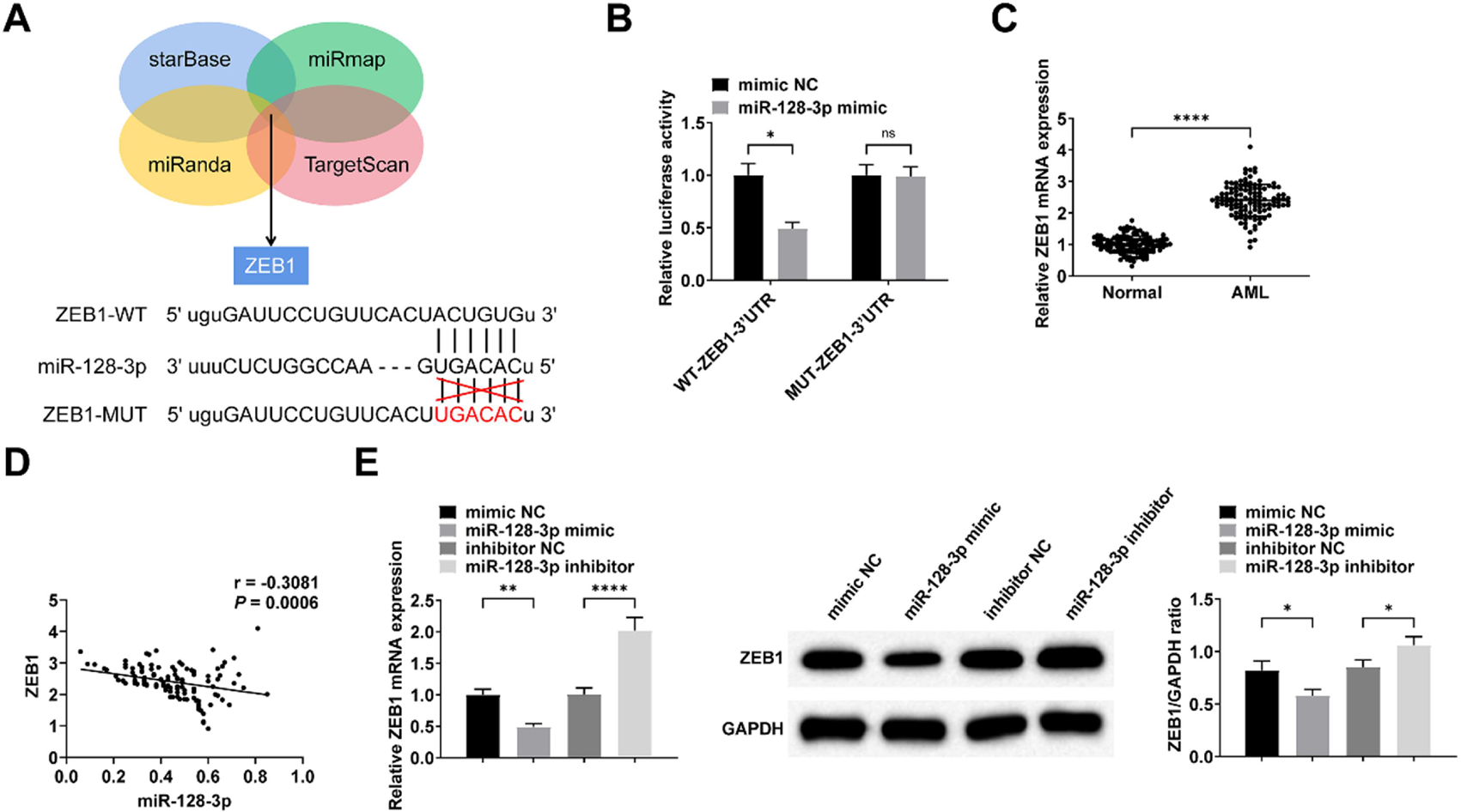
Bioinformatic analysis using multiple prediction tools (starBase, miRmap, miRanda, and TargetScan) identified ZEB1 as a potential downstream target of miR-128-3p (Fig. 5A). This targeting relationship was experimentally confirmed by dual-luciferase reporter assays (Fig. 5B). Consistent with these findings, ZEB1 expression was significantly upregulated in peripheral blood samples from AML patients and showed an inverse correlation with miR-128-3p levels (Fig. 5C‒D). Furthermore, transfection with miR-128-3p mimic or inhibitor resulted in corresponding downregulation or upregulation of ZEB1 expression in Kasumi-1 cells, respectively (Fig. 5E).

miR-128-3p targets regulation of ZEB1 expression. Predicted binding sites of ZEB1 and miR-128-3p (A). Luciferase activity of WT/MUT-ZEB1 after miR-128-3p intervention (B). RT-qPCR detection of ZEB1 expression in peripheral blood of AML patients (C); Inverse correlation between miR-128-3p and ZEB1 expression in AML patients (D); RT-qPCR and Western Blot detection of miR-128-3p expression after intervening miR-128-3p expression (E). Data are expressed as mean ± standard deviation (n = 3). * p < 0.05, ** p < 0.01, *** p < 0.001, **** p < 0.0001.
Up-regulation of ZEB1 reverses the effect of down-regulation of circ-RERE on AML cells
The functional role of the circ-RERE/miR-128-3p/ZEB1 axis was investigated through co-transfection of Kasumi-1 cells with sh-circ-RERE and pcDNA-ZEB1, with successful transfection confirmed by RT-qPCR and Western blot analysis (Fig. 6A). Functional assays demonstrated that pcDNA-ZEB1 effectively reversed the sh-circ-RERE-induced effects, including: (1) Decreased cell viability and proliferation, (2) Increased apoptosis and CD8+ T-cell cytotoxicity, and (3) Reduced PD-L1 expression and autophagy activity (Fig. 6B‒G). Furthermore, bioinformatic prediction using the JASPAR database identified multiple ZEB1 binding sites in the PD-L1 promoter region (Supplementary Fig. 1A‒B), which was experimentally validated by ChIP-qPCR showing direct ZEB1 binding to the PD-L1 promoter (Supplementary Fig. 1C).

ZEB1 involves circ-RERE-mediated regulation of autophagy and immune escape in AML cells. RT-qPCR and Western Blot to confirm ZEB1 expression change (A), followed by observations of viability (B), proliferation (C), apoptosis (D), cytotoxicity of CD8+ T-cells (E), PD-L1 expression (F); LC3-II/LC3-I ratio and Beclin-1 and p62 expression (G). Data are expressed as mean ± standard deviation (n = 3). * p < 0.05, ** p < 0.01, *** p < 0.001, **** p < 0.0001.
Circ-RERE knockdown suppresses tumor growth in vivo
Kasumi-1 cells transfected with either sh-NC or sh-circ-RERE were subcutaneously injected into immunocompetent BALB/c mice. The results demonstrated that circ-RERE knockdown significantly reduced both tumor volume and weight (Fig. 7A‒C). Immunohistochemical analysis further revealed enhanced CD8+ T-cell infiltration in tumors with circ-RERE silencing (Fig. 7D).

circ-RERE knockdown suppresses tumor growth in vivo. Representative images of xenograft tumors (A); Tumor volume measurements (B); Tumor weight analysis (C); CD8+ T-cell infiltration by immunohistochemical staining (D). Data are expressed as mean ± standard deviation (n = 5). * p < 0.05, ** p < 0.01, *** p < 0.001, **** p < 0.0001.
Discussion
CircRNAs are the focus of increasing research interest due to their characteristics20 and circRNA dysregulation in AML is functionally important and can influence disease onset and progression.21-24 This study investigated and finally discovered that circ-RERE was up-regulated in peripheral blood samples of AML patients and AML cells. Importantly, circ-RERE exerts oncogenic effects by regulating post-transcriptional translation.
The tumor microenvironment allows tumor cells to infiltrate lymphatics and blood vessels, evade immunity, and resist to antigen-specific T cells, thereby achieving distant metastasis and growth.25 A key immune checkpoint receptor, PD-1, principally expresses on some activated cells, called T, B, Dendritic (DC), Natural Killer (NK), and Treg. PD-1 is also associated with increased Treg-cell proliferation and enhanced immunosuppressive function.26 After the recognition of tumor antigens by peripheral T-Cell Receptors (TCRs), PD-L1 will bind PD-1 and activate downstream-related signaling pathways, blocking TCR signaling via feedback inhibition, and downregulate the expression of specific antiapoptotic protein molecules and pro-inflammatory factors, ultimately inhibiting T-cell survival, proliferation, and immune function.27 The blockade of PD-1 and PD-L1 has been determined to be of great benefit in the immunotherapy of AML.28 The present study provided evidence that circ-RERE deficiency reduced PD-L1 expression in AML cells and stimulated CD8+ T-cell cytotoxicity, suggesting that circ-RERE-regulated immune escape is mediated by preventing CD8+ T-cells from normal function.
For rapid proliferation and turnover, cancer cells generate high autophagic flux. Recent reports suggest that reduced autophagy gene expression may stimulate AML proliferation29 and that autophagic flux is associated with time to disease remission in AML.30 Autophagy refers to a process of continuous self-renewal and active proliferation that is required for normal hematopoiesis.31,32 As echoed here, silencing circ-RERE could inhibit autophagy gene expression in AML cells to inhibit AML cell proliferation and promote apoptosis.
Mechanistically, it is generally believed that circRNAs can compete with miRNAs to affect the functions of miRNAs.33 Consistent with the theory, miR-128-3p was sponged by circ-RERE here. MiR-128-3p acts as an oncogenic miRNA,34 and can even regulate breast cancer progression.35 Notably, miR-128-3p hampered the viability, proliferation, and autophagy of AML cells, whereas it drove apoptosis. This study further verified that miR-128-3p represses immune escape in AML cells by blocking PD-L1.
ZEB1 was originally identified as a lens-specific binding protein for the δ1-crystal enhancer and is a key transcription factor during the epithelial-mesenchymal transition process.36 ZEB1 high expression is associated with tumor onset and progression, metastasis, and treatment resistance of various malignancies.37-39 As a transcription factor, ZEB1 can activate or repress the transcription of target genes by recruiting different cofactors.40 Interestingly, ZEB1 can activate PD-L1 transcription by binding to the promoter of PD-L1 and promote tumor immune escape.41 ZEB1 was increased in AML and ZEB1 was negatively mediated by miR-128-3p targeting. Importantly, ZEB1 overexpression could reduce the influence of circ-RERE downregulation on PD-L1 expression. In addition, it was found that up-regulation of ZEB1 could reverse the inhibitory effect of down-regulation of circ-RERE on the proliferation, autophagy and immune escape of AML cells and the promotion of apoptosis.
This study has several limitations. First, the clinical samples were obtained from a single center, which may introduce selection bias, requiring validation in larger multicenter cohorts. Second, miR-128-3p may regulate additional target genes in AML, necessitating transcriptomic analyses to elucidate its complete molecular network. Furthermore, in the tumor immune microenvironment, targeting PD-L1 alone may be insufficient to fully restore T-cell function due to compensatory regulation by other checkpoints such as CTLA-4. Therefore, developing combination therapies with PD-L1 inhibitors and other immune checkpoint blockers holds significant clinical relevance.
Conclusion
Circ-RERE level is elevated in AML, and it can modify miR-128-3p to promote ZEB1 expression, thereby promoting autophagy and immune escape in AML cells. This study explores the underlying mechanism of circ-RERE function in AML cells, providing a potential target for AML therapy.
Ethics approval and consent to participate
This study was approved by the ethical committees of Yinzhou People's Hospital (n° 201902CN16). Written informed consent was obtained from all patients.
-
Funding
Not applicable.
Data availability statement
Data is available from the corresponding author on request.
Acknowledgments
Not applicable.
Supplementary materials
Supplementary material associated with this article can be found, in the online version, at doi:10.1016/j.clinsp.2025.100850.
Supplementary PDF
References
- 1 Short N.J., Konopleva M., Kadia T.M., Borthakur G., Ravandi F., DiNardo C.D., et al. Advances in the treatment of acute myeloid leukemia: new drugs and new challenges. Cancer Discov. 2020;10(4):506-25.
- 2 Bullinger L., Döhner K., Döhner H. Genomics of acute myeloid leukemia diagnosis and pathways. J Clin Oncol. 2017;35(9):934-46.
- 3 Benard B., Gentles A.J., Köhnke T., Majeti R., Thomas D. Data mining for mutation-specific targets in acute myeloid leukemia. Leukemia. 2019;33(4):826-43.
- 4 Thakral D., Gupta R., Khan A. Leukemic stem cell signatures in acute myeloid leukemia- targeting the Guardians with novel approaches. Stem Cell Rev Rep. 2022;18(5):1756-73.
- 5 Major C.K., Kantarjian H., Sasaki K., Borthakur G., Kadia T., Pemmaraju N., et al. Survivorship in AML ‒ a landmark analysis on the outcomes of acute myelogenous leukemia patients after maintaining complete remission for at least 3 years. Leuk Lymphoma. 2020;61(13):3120-7.
- 6 Kaito Y., Hirano M., Futami M., Nojima M., Tamura H., Tojo A., et al. CD155 and CD112 as possible therapeutic targets of FLT3 inhibitors for acute myeloid leukemia. Oncol Lett. 2022;23(2):51.
- 7 Yan Y., Upadhyaya R., Zhang V.W., Berg T. Epigenetic maintenance strategies after allogeneic stem cell transplantation in acute myeloid leukemia. Exp Hematol. 2022;109:1-10.e1.
- 8 Sibley C.R., Blazquez L., Ule J. Lessons from non-canonical splicing. Nat Rev Genet. 2016;17(7):407-21.
- 9 Salzman J., Chen R.E., Olsen M.N., Wang P.L., Brown P.O. Cell-type specific features of circular RNA expression. PLoS Genet. 2013;9(9):e1003777.
- 10 Jamal M., Song T., Chen B., Faisal M., Hong Z., Xie T., et al. Recent progress on circular RNA research in acute myeloid leukemia. Front Oncol. 2019;9:1108.
-
11 Ping L., Jian-Jun C., Chu-Shu L., Guang-Hua L., Ming Z. High circ_100053 predicts a poor outcome for chronic myeloid leukemia and is involved in imatinib resistance. Oncol Res. 2019. doi: 10.3727/096504018X15412701483326. Online ahead of print.
» https://doi.org/10.3727/096504018X15412701483326. - 12 Liu W., Cheng F. Circular RNA circCRKL inhibits the proliferation of acute myeloid leukemia cells via the miR-196a-5p/miR-196b-5p/p27 axis. Bioengineered. 2021;12(1):7704-13.
- 13 Zhou F., Wang D., Wei W., Chen H., Shi H., Zhou N., et al. Comprehensive profiling of circular RNA expressions reveals potential diagnostic and prognostic biomarkers in multiple myeloma. BMC Cancer. 2020;20(1):40.
- 14 Gao Y.L., Zhang M.Y., Xu B., Han L.J., Lan S.F., Chen J., et al. Circular RNA expression profiles reveal that hsa_circ_0018289 is up-regulated in cervical cancer and promotes the tumorigenesis. Oncotarget. 2017;8(49):86625-33.
- 15 Balatti V., Pekarky Y., Croce C.M. Role of microRNA in chronic lymphocytic leukemia onset and progression. J Hematol Oncol. 2015;8:12.
- 16 Ma D., Qin Y., Huang C., Chen Y., Han Z., Zhou X., et al. Circular RNA ABCB10 promotes non-small cell lung cancer progression by increasing E2F5 expression through sponging miR-584-5p Cell Cycle. 2020;19(13):1611-20.
- 17 Mets E., Van Peer G., Van der Meulen J., Boice M., Taghon T., Goossens S., et al. MicroRNA-128-3p is a novel oncomiR targeting PHF6 in T-cell acute lymphoblastic leukemia. Haematologica. 2014;99(8):1326-33.
- 18 Li L., Feng Y., Hu S., Du Y., Xu X., Zhang M., et al. ZEB1 serves as an oncogene in acute myeloid leukaemia via regulating the PTEN/PI3K/AKT signalling pathway by combining with P53. J Cell Mol Med. 2021;25(11):5295-304.
- 19 Rong D., Sun G., Zheng Z., Liu L., Chen X., Wu F., Gu Y., Dai Y., Zhong W., Hao X., Zhang C., Pan X., Tang J., Tang W., Wang X. MGP promotes CD8+ T cell exhaustion by activating the NF-κb pathway leading to liver metastasis of colorectal cancer. Int J Biol Sci. 2022;18(6):2345-61.
- 20 Issah M.A., Wu D., Zhang F., Zheng W., Liu Y., Fu H., et al. Epigenetic modifications in acute myeloid leukemia: the emerging role of circular RNAs (Review). Int J Oncol. 2021;59(6):107.
- 21 Singh V., Uddin M.H., Zonder J.A., Azmi A.S., Balasubramanian S.K. Circular RNAs in acute myeloid leukemia. Mol Cancer. 2021;20(1):149.
- 22 Shen Y., Jia Y., Zhang R., Chen H., Feng Y., Li F., et al. Using circ-ANAPC7 as a novel type of biomarker in the monitoring of acute myeloid leukemia. Acta Haematol. 2022;145(2):176-83.
- 23 Shang J., Chen W.M., Liu S., Wang Z.H., Wei T.N., Chen Z.Z., et al. CircPAN3 contributes to drug resistance in acute myeloid leukemia through regulation of autophagy. Leuk Res. 2019;85:106198.
- 24 Wang X., Jin P., Zhang Y., Wang K. CircSPI1 acts as an oncogene in acute myeloid leukemia through antagonizing SPI1 and interacting with microRNAs. Cell Death Dis. 2021;12(4):297.
- 25 Tettamanti S., Pievani A., Biondi A., Dotti G., Serafini M. Catch me if you can: how AML and its niche escape immunotherapy. Leukemia. 2022;36(1):13-22.
- 26 Gutic B., Bozanovic T., Mandic A., Dugalic S., Todorovic J., Stanisavljevic D., et al. Programmed cell death-1 and its ligands: current knowledge and possibilities in immunotherapy. Clinics (Sao Paulo). 2023;78:100177.
- 27 Brown J.A., Dorfman D.M., Ma F.R., Sullivan E.L., Munoz O., Wood C.R., et al. Blockade of programmed death-1 ligands on dendritic cells enhances T cell activation and cytokine production. J Immunol. 2003;170(3):1257-66.
- 28 Wang F., Yang L., Xiao M., Zhang Z., Shen J., Anuchapreeda S., et al. PD-L1 regulates cell proliferation and apoptosis in acute myeloid leukemia by activating PI3K-AKT signaling pathway. Sci Rep. 2022;12(1):11444.
- 29 Watson A.S., Riffelmacher T., Stranks A., Williams O., De Boer J., Cain K., et al. Autophagy limits proliferation and glycolytic metabolism in acute myeloid leukemia. Cell Death Discov. 2015;1:15008.
- 30 Piya S., Kornblau S.M., Ruvolo V.R., Mu H., Ruvolo P.P., McQueen T., et al. Atg7 suppression enhances chemotherapeutic agent sensitivity and overcomes stroma-mediated chemoresistance in acute myeloid leukemia. Blood. 2016;128(9):1260-9.
- 31 Mortensen M., Soilleux E.J., Djordjevic G., Tripp R., Lutteropp M., Sadighi-Akha E., et al. The autophagy protein Atg7 is essential for hematopoietic stem cell maintenance. J Exp Med. 2011;208(3):455-67.
- 32 Chen X., Clark J., Wunderlich M., Fan C., Davis A., Chen S., et al. Autophagy is dispensable for Kmt2a/Mll-Mllt3/Af9 AML maintenance and anti-leukemic effect of chloroquine. Autophagy. 2017;13(5):955-66.
- 33 Meng S., Zhou H., Feng Z., Xu Z., Tang Y., Li P., et al. CircRNA: functions and properties of a novel potential biomarker for cancer. Mol Cancer. 2017;16(1):94.
- 34 Shi Y., Yan F., Wang F., Pan L. MiR-128-3p suppresses tumor proliferation and metastasis via targeting CDC6 in hepatocellular carcinoma cells. Tissue Cell. 2021;72:101534.
- 35 Zhao J., Li D., Fang L. MiR-128-3p suppresses breast cancer cellular progression via targeting LIMK1. Biomed Pharmacother. 2019;115:108947.
- 36 Wu H.T., Zhong H.T., Li G.W., Shen J.X., Ye Q.Q., Zhang M.L., et al. Oncogenic functions of the EMT-related transcription factor ZEB1 in breast cancer. J Transl Med. 2020;18(1):51.
- 37 Fratini L., Jaeger M., de Farias C.B., Brunetto A.T., Brunetto A.L., Shaw L., et al. Oncogenic functions of ZEB1 in pediatric solid cancers: interplays with microRNAs and long noncoding RNAs. Mol Cell Biochem. 2021;476(11):4107-16.
- 38 Han X., Long Y., Duan X., Liu Z., Hu X., Zhou J., et al. ZEB1 induces ROS generation through directly promoting MCT4 transcription to facilitate breast cancer. Exp Cell Res. 2022;412(2):113044.
- 39 Xu H., Zhao J., Li J., Zhu Z., Cui Z., Liu R., et al. Cancer associated fibroblast-derived CCL5 promotes hepatocellular carcinoma metastasis through activating HIF1α/ZEB1 axis. Cell Death Dis. 2022;13(5):478.
- 40 Noman M.Z., Janji B., Abdou A., Hasmim M., Terry S., Tan T.Z., et al. The immune checkpoint ligand PD-L1 is upregulated in EMT-activated human breast cancer cells by a mechanism involving ZEB-1 and miR-200. Oncoimmunology. 2017;6(1):e1263412.
- 41 Zhao L., Liu Y., Zhang J., Liu Y., Qi Q. LncRNA SNHG14/miR-5590-3p/ZEB1 positive feedback loop promoted diffuse large B cell lymphoma progression and immune evasion through regulating PD-1/PD-L1 checkpoint. Cell Death Dis. 2019;10(10):731.
Edited by
-
Edited by
José Maria Soares Junior