 Karyotype and fluorescent banding in the interspecific F1 hybrid Passiflora 'Lamepiana', confirmed by GISH
Abstract
Passiflora 'Lamepiana' is the F1 hybrid from the cross between P. vitifolia and P. hatschbachii with ornamental characteristics. Based on cytogenetics techniques, this study aimed to confirm interspecific hybridization and characterize the karyotype of the hybrids and their parents using cytogenetic techniques. For all genotypes the predominance of metacentric chromosomes with two satellite pairs and a diploids karyotypes (2n = 18) were observed. It was possible to determine the occurrence of two satellite pairs in both the parents of the hybrids. The use of simultaneous probes of the F1 parents prepared according to GISH technique allowed to identify two chromosome lots each one inherited from both parents, confirming the hybrid character of the plants. Besides confirming hybridization, GISH did not demonstrate the existence of visible chromosomal alterations but rather cytogenetic stability in the hybrids.
Keywords:
Passionflower; karyomorphology; CMA 3 /DAPI banding; molecular cytogenetics
INTRODUCTION
Among the genera in the Passifloraceae family, Passiflora is the most significant because it encompasses a wide variety of species (Cervi and Imig 2013) and broad geographical distribution in neotropics (Ulmer and MacDougal 2004). Colorful flowers with different shapes can be found into the large number of species within this genus, which have aroused growing interest in the ornamental sector (Abreu et al. 2009), envisioning the use of passionflowers to decorate both indoor and outdoor environments.
Passiflora vitifolia Kunt (2n = 18; Storey 1950) is among the most beautiful and widespread passionflowers; it is found from Nicaragua to Venezuela and Peru and grows at altitudes of 200-1100 m (Vanderplank 2000, Ulmer and MacDougal 2004). Its very beautiful flowers reach an extraordinary diameter of 12-17 cm; the outer rank of the upright, short corona is red, while the two inner series are usually white (Ulmer and MacDougal 2004). Passiflora vitifolia belongs to supersection Coccinea, which was assigned to subgenus Passiflora; this supersection contains almost exclusively red-flowering species (Vanderplank 2000, Ulmer and MacDougal 2004). This species is very robust, can be cultivated in a protected, sunny location outdoors (Ulmer and MacDougal 2004), and has been used to produce interesting hybrids (Vanderplank 2000, Ulmer and MacDougal 2004, Santos et al. 2023).
Passiflora hatschbachii Cervi (2n = 18; Melo et al. 2014) is endemic to Brazil, occurring in Minas Gerais and possibly in Bahia, restricted to the Atlantic Forest biome; it was evaluated by CNCFlora (2016) as EN (endangered), according to União Internacional para a Conservação da Natureza (IUCN) Red List of Threatened Species criteria (Imig et al. 2018). Its flowers are delicate, white, small, with a diameter of 4.8-8.5 cm; it has a corona of filaments with two series, an outer filiform series and the following filiform to capillary series (Imig et al. 2018). It is a heliophyte species with robust branches.
As a favorable characteristic for the ornamental market, Passiflora ‘Lamepiana’ (2n = 18; Santos et al. 2023) is a F1 hybrid from P. vitifolia vs. P. hatschbachii which matches interesting floral characteristics such as intensity in flowering; petals and sepals red intense, and the potential for great acceptance in the ornamental plant market, being a promising genotype for the composition of gardens (Santos et al. 2023). P. ‘Lamepiana’ is a fertile plant, with regular meiosis (Souza et al. 2023), viable pollen and receptive stigma (Santos et al. 2023).
The use of Passiflora for ornamentation is still incipient; however, some crosses have yielded promising ornamental hybrids, such as the hybrid BRS Estrela-do-Cerrado (P. coccinea vs. P. setacea) (Faleiro et al. 2007a), in addition to hybrid cultivars such as P. ‘Aninha’, P. ‘Priscilla’, P. ‘Alva’ (Santos et al. 2012), P. ‘Bella’, P. ‘Gabriela’ (Belo et al. 2018), P. ‘Lamepiana’ (Santos et al. 2023).
Cytogenetic studies on Passiflora report the diploid characteristics of these species, with the occurrence of individuals 2n = 12, 2n = 18, 2n = 22, and 2n = 24 (Melo et al. 2001). In Passiflora, heterochromatic regions rich in GC colocalize with secondary constrictions and can be differentiated with CMA3 (chromomycin A3) and DAPI (4',6-diamidino-2-phenylindole) fluorochromes, demonstrating regions where constitutive heterochromatin occurs (Melo et al. 2014). GISH (Genomic In Situ Hybridization) enables chromosomal comparison among different species, different individuals, and both inter- and intraspecific hybrids. In addition, the application of GISH has allowed reliable confirmation of paternity in hybrids (Melo et al. 2017, Silva et al. 2018), whether they are natural or artificial (Silva and Souza 2013). The visualization of chromosomal recombination in backcrossed hybrids is also an important karyotypic analysis enabled by GISH (Melo et al. 2015).
Thus, the aim of this study was to perform karyomorphological characterization and apply CMA3/DAPI banding to identify GC-rich regions associated with satellites, and to confirm the hybridization of Passiflora F1 hybrids using GISH.
MATERIALS AND METHODS
Vegetal material and interspecific hybridization
The parent species P. vitifolia Kunt. (accession 481) and P. hatschbachii Cervi (accession 446), along with four F1 hybrids whose progeny was named HD26 (HD26-102, 104, 143, and 146) were assessed. More the one hybrid was obtained from crossing, however, no great dissimilarity for floral and vegetative characteristics occurred, leading to consider all as a part of the same group, referred as P. ‘Lamepiana’ (Santos et al. 2023). The parent plants were maintained in protected cultivation in the Passiflora Work Collection at the State University of Santa Cruz, Ilhéus, Bahia (lat 14° 39″ S, long 39° 10″ W, alt 78 m asl). From October 2015 to January 2016, artificial crosses were performed P. vitifolia x P. hatschbachii) during the early hours of the day, (between 7:00 and 8:00 a.m) in the anthesis flowers (Souza et al. 2023). Passiflora vitifolia was used as pollen recipient and P. hatschbachii was used as donor, respectively. Emasculation pre-anthesis and the protection with a paper bag in the previous day was conducted for the maternal parent (P. vitifolia). When conducting pollination besides collecting in bulk PG from the paternal parent (P. hatschbachii), curved stigmas were pollinated manually and protected again using paper bags. After artificial crossing, nylon nets were used to protect the fruits until maturation.
Preparing the slides
Roots from cuttings were collected and pre-treated by applying antimitotic 8-hydroxyquinoline solution at 0.002 M for 1 hour at room temperature (RT) and then for another 21 hours at 8-10 ºC. They were washed with distilled water for 5 minutes, fixed in Carnoy I solution (ethanol-glacial acetic acid [3:1, v/v]; Johansen 1940) for 3 hours at RT, and stored at -20 ºC. The roots were washed twice in distilled water for 5 minutes each and then incubated in 50 μL of an enzyme solution containing 2% cellulase and 20% pectinase for 1 hour and 20 minutes at 37 ºC. Before macerating, distilled water was used to wash the collected roots, which then was macerated using needles in 15 μL of 45% glacial acetic acid on a slide covered with a coverslip (18 × 18 mm), and gently pressed with filter paper to spread the chromosomes. Arter freezing with liquid nitrogen, the coverslips were removed and the slides were air-dried. Until being used, the slides were remained stored at -20 °C.
Giemsa staining and karyomorphological analysis
The slides were stained according to Guerra and Sousa (2002), with modifications. The material was stained with 4% Giemsa for 40 minutes, rinsed with distilled water, and air-dried. The slide was mounted using a 20 × 20 mm coverslip with Neomount medium. Karyomorphological data were obtained from five metaphases of each genotype. Absolute values were obtained for the lengths of the short arm (SA), long arm (LA), and satellites (SAT), with the satellite lengths added to their corresponding chromosomal arms. The arm ratio (r = LA/SA), the total chromosomal length for each chromosome (CLC = LA + SA + SAT), and the AAC (average of all chromosomes) were calculated. The statistical analysis was conducted using the average chromosome values from five metaphases per genotype. The data were analyzed using ANOVA, and the mean total chromosome lengths between genotypes were compared by Tukey test (p ≤ 0.05). The Sisvar software version 5.0 (Ferreira 2003) was used.
CMA3 and DAPI fluorochrome staining
Slides aged for a minimum of three days underwent CMA3/DAPI double staining according to Guerra and Souza (2002). 15 μL of CMA3 (0.25 mg-1) was applied to each slide and covered with glass coverslip. The slides were stored for 1 hour in a humid chamber at 37 °C. The coverslips were removed using a jet of distilled water and dried with an air pump. Then, 15 μL of DAPI (0.5 mg-1) was applied to each slide. The material was covered with a glass coverslip and stored in a dark box for 30 minutes. The slides were removed using a jet of distilled water and dried with an air pump. The slides were assembled by applying 15 μL of a 1:1 v/v glycerol/Macllvaine solution and covered with a 20 × 20 mm coverslip. The slides were aged in the dark for three days prior to microscopic analysis.
DNA extraction and fragmentation for GISH
Genomic DNA extraction from P. vitifolia and P. hatschbachii was performed according to the protocol established by Doyle and Doyle (1990), with some modifications (Melo et al. 2015). Young leaves, collected in duplicate, were macerated in liquid nitrogen and placed in 2% CTAB extraction buffer [1.5 M NaCl; 20 mM EDTA; 100 mM Tris-HCl; 0.2% β-mercaptoethanol]. The nucleic acids were isolated using a chloroform:isoamyl alcohol solution (24:1), and the DNA was resuspended in TE buffer [10 mM Tris (Promega), 1 mM EDTA]. The genomic DNA concentration was estimated by comparing it to 100 ng of lambda DNA on a 1% agarose gel stained with SYBR Safe. Approximately 30 μg of parental genomic DNA in a final volume of 1000 μL was fragmented using a sonicator set to 40% amplitude, alternating 2 seconds on and 2 seconds off for 5 minutes (Jauhar and Peterson 2006). The size of the fragments was verified on a 2% agarose gel using a 100 bp ladder as a reference marker. Fragmented DNA was purified using 10% of the final volume of 3 M sodium acetate and two times the final volume of ethanol. The material was kept at -20 ºC overnight and then centrifuged for 10 min at 14,000 rpm to remove the supernatant, followed by drying in TA for at least 1 hour. To resuspend the pellet, 120 μL of ultra-pure water was added to the tube. Subsequently, the sample was stored at ± 8 ºC and the following day, it was stored at -20 ºC until probe preparation.
Preparation of probes for GISH
The genomic DNA of P. vitifolia was labeled with digoxigenin-11-dUTP, while that of P. hatschbachii was labeled with biotin-16-dUTP. The probes were labeled by Nick Translation in the final concentration of 1 μg of cleaved DNA as per the manufacturer's protocol. The samples underwent 90 minutes of amplification in a programmed thermocycler. To purify the probes, 2 μL of sodium acetate (10% of the total reaction volume) and 40 μL of 100% ethanol (double the total reaction volume) were added to each sample. The material was stored overnight at -20 °C and then centrifuged the next day for 10 minutes at 13,000 rpm. The supernatant was discarded, and the pellet was dried at RT for at least 1 hour. The pellet was resuspended by adding 30 μL of ultra-pure water to the tube. The material was kept in the refrigerator at ±8 °C for 24 hours and subsequently stored at -20 °C until use.
Application of GISH
The slides were treated according to Schwarzacher and Heslop-Harrison (2000), with modifications (Melo et al. 2015). Slides were selected and dried in an oven at 37 °C for 1 hour. 50 µl of RNase solution was applied to each slide; the material was covered with a plastic coverslip and incubated for 1 hour at 37 °C in a pre-heated humid chamber. After two washes in 2×SSC (0.3 M sodium chloride; 0.03 M sodium citrate) at RT for 5 minutes each, with shaking (all washing steps mentioned below were performed on a shaker platform at 120 rpm (Biomixer, Mos-1)), 50 µl of 10 mM HCl (hydrochloric acid) was added to each slide, and the material was covered with a plastic coverslip and left to rest for 5 minutes. After removing excess HCl, 50 µl of pepsin solution (10 mg-1 pepsin; 10 mM HCl [1:100 v/v]) was added to each slide and covered with a plastic coverslip. The slides were incubated in a humid chamber for 20 minutes at 37 °C and washed twice in 2×SSC at RT for 5 minutes each. Subsequently, the slides were immersed in 4% paraformaldehyde at RT for 10 minutes and washed twice in 2×SSC for 5 minutes each. The slides were sequentially dehydrated with 70% ethanol and 96% ethanol for 5 minutes each and dried at RT for at least 30 minutes. After drying, the hybridization mixture was applied to the slides. For each slide, the hybridization mix consisted of 7.5 µl of 100% formamide, 3.0 µl of 50% dextran, 1.5 µl of 20×SSC, 0.2 µl of SDS (sodium dodecyl sulfate), and 1.4 µl of each probe. The hybridization mix was heated to 75 °C for 10 minutes in a thermocycler before being applied to the slides, and then immediately cooled on ice for 5 minutes. The cytological material containing the hybridization mixture was denatured in a thermal cycler with a slide adapter at 75 °C for 10 min and incubated overnight at 37 °C in a humid chamber. After hybridization, the slides were immersed in 2×SSC at RT for 5 minutes to remove the coverslips. Post-hybridization washes were performed as follows: two washes in 2×SSC at 42 °C for 5 minutes each, two washes in 0.1×SSC at 42 °C for 5 minutes each, and two additional washes in 2×SSC at 42 °C for 5 minutes each. Subsequently, the slides were washed once in 4×SSC/0.2% Tween 20 at RT for 5 minutes and treated with 50 μl of 5% bovine serum albumin (BSA). For detection, 20 µl of solution containing 0.7 μl of avidin-FITC, 0.7 μL of anti-digoxigenin-Rhodamine, and 18.6 μL of 5% BSA was applied to each slide. The slides containing the detection antibodies were incubated in a humid chamber for 1 hour at 37 °C. Three 5-minute washes were performed using 4×SSC/0.2% Tween 20 to remove excess antibodies. Finally, the slides were quickly immersed in 2×SSC and counterstained with DAPI/Vectashield. The slides were covered with 20 × 20 mm glass coverslips and stored in the dark at ± 8-10 °C for later analysis.
Photo documentation
The metaphases were photographed using an epifluorescence microscope equipped with an Olympus DP25 5M digital camera and DP2-BSW software. The CMA3/DAPI-stained metaphases were photographed: for DAPI visualization, the U-MWU filter (330-385 nm excitation/400 nm dichroic cut-off emission/>420 nm) was used, and for CMA3 visualization, the U-MWB filter (450-480 nm excitation/500 nm dichroic cut-off emission/>515 nm) was used. For GISH, the hybridizations detected with avidin-FITC were observed using the U-MWB filter (450-480 nm excitation/500 nm dichroic cut-off emission/>515 nm) and the hybridizations detected with anti-digoxigenin-rhodamine were visualized using the U-MWG filter (510-550 nm excitation/570 nm dichroic cut-off emission/>590 nm). The CMA3/DAPI and FITC/rhodamine overlays and plates were prepared using Photoshop SC5 software.
RESULTS AND DISCUSSION
The diploid chromosome number 2n = 18 was observed in both parent species, P. vitifolia and P. hatschbachii, as well as in the interspecific hybrids HD26-102, HD26-104, HD26-143, and HD26-146 (Figure 1), like other interspecific Passiflora (Santos et al. 2012, Souza et al. 2020). The chromosomal morphology of the parents and the four hybrids analyzed was predominantly metacentric, 2n = 18m (Table 1). The CLC of P. vitifolia ranged from 2.08 to 4.12 µm and that of P. hatschbachii from 1.78 to 3.46 µm. In hybrids, CLC from chromosomes 1 to 18 (homeologous) ranged from 1.67 µm to 4.14 µm for HD26-102, 1.58 µm to 3.92 µm for HD26-104, 1.55 µm to 3.41 µm for HD26-143, and 1.55 µm to 3.55 µm for HD26-146. ANOVA showed a significant difference for CLC among genotypes by the F test (p ≤ 0.05), with a mean square of 0.32 and a coefficient of variation of 12.63%. The maternal parent (P. vitifolia) and the hybrid HD26-143 showed the most significant differences for CLC, with the other four individuals showing intermediate chromosomal averages between these two genotypes, which had the highest and lowest averages, respectively. Tukey's test grouped the genotypes into three clusters based on AAC values: P. vitifolia, 3.02a; P. hatschbachii, 2.44ab; HD26-146, 2.49ab; HD26-104, 2.51ab; HD26-102, 2.75ab, and HD26-143, 2.30b.

Mitotic metaphases of parents and interspecific F1 hybrids (HD26) of Passiflora (2n = 18). (A) P. vitifolia; (B) P. hatschbachii; (C) HD26-102; (D) HD26-104; (E) HD26-143; (F) HD26-146. Scale Bar = 10 µm.
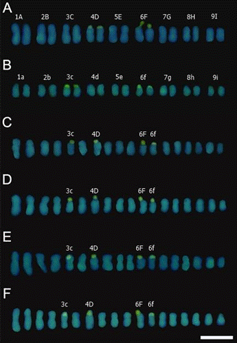
Satellites were identified in the genitors and hybrids (Figure 2). In P. vitifolia and P. hatschbachii, satellites were identified in chromosome pairs 4 and 6 in the former, and in pairs 3 and 6 in the latter (Figures 3A and 3B). In each hybrid, four chromosomes with CMA+ bands were observed in the chromosomes 5, 7, 11 and 12 (Figures 3C - 3F). The chromosomes 5 and 12 came from de male parent while chromosomes 7 and 11 came from the female parent. Chromosome pairing cannot occur in hybrids, as the chromosomes of the progeny are homeologous and the chromosomes of the parental species have very similar sizes, making it difficult to distinguish the chromosomes from each parent in the hybrid.
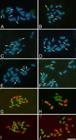
CMA3/DAPI banding and Genomic In Situ Hybridization (GISH) in mitotic metaphases of parents and interspecific F1 hybrids (HD26) of Passiflora (2n = 18). (A-F) CMA+/DAPI- Banding. (A) P. vitifolia (B) P. hatschbachii, (C) HD26-102, (D) HD26-104, (E) HD26-143, (F) HD26-146. The arrows point to CMA3+ blocks. (G-J) GISH. (G) HD26-102, (H) HD26-104, (I) HD26-143, (J) HD26-146. Scale bar = 10 μm.
Chromosomal alterations are not expected in F1 hybrids because of hybridization per se. The F1 hybrid is generated from the male and female gametes of the parents, and the gametes show recombination due to the meiosis that occurred during their production, in the parents. Therefore, the F1 hybrid has different characteristics in relation to the parents due to these recombined gametes that generated it. The hybrid karyotype differs from the karyotypes of the parents considering that the F1 hybrid has one set of chromosomes from the maternal genitor and one from the paternal genitor, and not necessarily due to possible chromosomal alterations. This is expected for diploid species. However, the occurrence of polyploidization in the ancient parent and hybridization events are believed to be the major drivers of plant diversification and speciation (Berankova et al. 2024). These mechanisms culminate in the amplification of chromosomal sets within genomes - a phenomenon termed "genome upsizing" (Wendel 2000) - which is succeeded by post-polyploid diploidization processes. These processes are believed to be linked to substantial DNA loss, referred to as "genome downsizing," as well as structural chromosomal alterations (Mandáková et al. 2017) Differences in CLC can be observed among various diploid Passiflora species (Souza et al. 2003). In hybrid chromosomes, CLC tends to vary too, showing intermediate values between the two species involved in the cross, as observed in hybrids from the P. edulis vs. P. setacea cross (Melo et al. 2016).
The comparison of karyotype length between P. hatschbachii and P. vitifolia showed that the latter species has larger chromosomes, and hybrids resulting from this cross have chromosomes of intermediate length between the two species. In the analyzed hybrids, differences were observed not only in quantitative aspects identified by conventional staining but also in the satellite positions relative to the parents. Differences in satellite position between chromosomes may originate from translocation due to structural rearrangements in backcrossed hybrids (Melo et al. 2017); however, such chromosomal changes were not observed with GISH.
Chromosomal morphology in Passiflora species is a conserved trait (Vieira et al. 2004), with most species showing a higher occurrence of metacentric chromosomes and few submetacentric chromosomes (Viana and Souza 2012, Amorim et al. 2014, Melo et al. 2014). In this study, the chromosomes of the hybrids were classified as metacentric, with a few classified as submetacentric, a trait inherited from the parents.
Using CMA3/DAPI banding, we observed CMA3+/DAPI- blocks confined to satellite regions/secondary constrictions (Figures 2 and 3), characteristics that were able to visualize with conventional staining. Four CMA3+/DAPI- blocks were observed in P. vitifolia, four in P. hatschbachii, and four in each hybrid.
Double staining with CMA3 and DAPI fluorochromes is crucial for identifying the number of satellites associated with GC-rich heterochromatin regions in Passiflora. In interspecific hybrids, the use of these fluorochromes to identify satellites enables reliable confirmation of hybridization, as these regions serve as important cytological markers (Melo et al. 2017). In the hybrids examined in this study, the CMA3/DAPI banding consistently enabled the observation of two pairs of satellites in both parents and hybrids. The occurrence of hybrids with different numbers of satellites may lead to chromosomal rearrangements if these individuals cross (Silva et al. 2018). The analysis of satellite numbers and their chromosomal positions in hybrids and their parental species is a crucial aspect of cytogenetic studies. In Passiflora species, these characteristics provide valuable chromosomal markers that facilitate comparisons among individuals (Silva eta al. 2018). This approach underscores the significance of satellite number and position as informative traits in distinguishing and comparing Passiflora genotypes.
The simultaneous use of maternal and paternal probes allowed for the distinct visualization of P. vitifolia and P. hatschbachii genomes in each of the four analyzed F1 hybrids. In each hybrid, nine chromosomes of maternal origin P. vitifolia (red) and nine chromosomes of paternal origin P. hatschbachii (green) were observed (Figure 3). Molecular cytogenetics has enabled rapid and accurate confirmation of hybrids. In Passiflora, the application of GISH has aided in the identification of interspecific hybrids and backcrosses (Melo et al. 2017).

Karyograms of parents and interspecific F1 hybrids (HD26) of Passiflora (2n = 18). (A) P. vitifolia with chromosomes pairs numbered 1 to 9 and A to I; (B) P. hatschbachii with chromosomes pairs numbered 1 to 9 and a to i; (C-F) hybrids with parental CMA+/DAPI- chromosomes; (C) HD26-102; (D) HD26-104; (E) HD26-143, and (F) HD26-146. Scale bar = 10 μm.
In this investigation, the application of probe-probe hybridization facilitated the identification of two distinct chromosomal sets within the hybrid plants' genomes - one originating from P. vitifolia and the other from P. hatschbachii. In addition to being efficient and accurate in confirming hybridization, GISH also allows visualization of recombinations in backcrossed hybrids, thus aiding in chromosomal understanding during pairing (Melo et al. 2016). Although no translocations or inversions were observed in the HD26 hybrids, the possibility of chromosomal changes cannot be dismissed, as some chromosomes in the hybrids were smaller than those in the parents. Some meiotic irregularities observed in interspecific Passiflora hybrids (Santos et al. 2012, Souza et al. 2020) may justify the existence of deletions.
CONCLUSION
The studies confirmed the interspecific hybridization of P. ‘Lamepiana’, a new ornamental hybrid. Karyotypic analysis revealed that the hybrids maintain the diploid chromosome number (2n = 18) and exhibit predominantly metacentric chromosomes, with intermediate chromosome length compared to the parental species. CMA³⁺/DAPI⁻ banding demonstrated that the hybrids have the same number of CMA³⁺ bands as the parents, enabling the identification of these marker chromosomes in the hybrids. Additionally, GISH technique allowed the distinction of chromosomal sets inherited from each parental species, confirming the hybrid origin of the plants.
ACKNOWLEDGEMENTS
We thank UESC, CNPq (Conselho Nacional de Desenvolvimento Científico e Tecnológico) and CAPES (Coordenação de Aperfeiçoamento de Pessoal de Nível Superior) for their financial support to the research; FAPESB (Fundação de Amparo à Pesquisa do Estado da Bahia) for the scholarships awarded to the first author; and CNPq for the scholarship awarded to the second author.
Data Availability
The datasets generated and analyzed during the current research are available from the corresponding author upon reasonable request.
REFERENCES
- Abreu PP, Souza MM, Santos EA, Pires MV, Pires MM, Almeida AAF2009 Passion flower hybrids and their use in the ornamental plant market: perspectives for sustainable development with emphasis on Brazil. Euphytica 166:307-315
- Amorim JS, Souza MM, Viana AJ, Corrêa RX, Araújo IS, Ahnert D2014 Cytogenetic, molecular and morphological characterization of Passiflora capsularis L. and Passiflora rubra L. Plant Systematic and Evolution 300:1147-1162
- Belo GO, Souza MM, Silva GS, Lavinscky MP2018 Hybrids of Passiflora: P. gardneri versus P. gibertii, confirmation of paternity, morphological and cytogenetic characterization. Euphytica 214:1-14
- Berankova D, Cizkova J, Majzlikova G, Dolezalova A, Mduma H, Brown A, Swennen R, Hribova E2024 Striking variation in chromosome structure within Musa acuminata subspecies, diploid cultivars, and F1 diploid hybrids. Frontiers in Plant Science 15:1387055
- Cervi AC, Imig DC2013 A new species of Passiflora (Passifloraceae) from Mato Grosso do Sul, Brazil. Phytotaxa 103:46-50
-
CNCFlora - Centro Nacional de Conservação da Flora2016 Passifloraceae. In Lista vermelha da flora brasileira versão 1. Available at <Available at http://cncflora.jbrj.gov.br/portal/pt-br/profile/Passiflora saccoi >. Accessed on September 13, 2016.
» http://cncflora.jbrj.gov.br/portal/pt-br/profile/Passiflora saccoi - Doyle JJ, Doyle JL1990 Isolation of plant DNA from fresh tissue. Focus 12:13-15
- Faleiro FG, Junqueira NTV, Junqueira KP, Braga MF, Borges RS, Peixoto JR, Andrade GA, Santos EC, Silva DGP2007 BRS Estrela do Cerrado: Híbrido de Passiflora para uso como planta ornamental. Revista Brasileira de Horticultura Ornamental 13 (Suplemento): 334.
- Ferreira DF2003 Programa Sisvar. Software 5.0. UFLA, Lavras.
- Guerra M, Sousa MJ2002 Como observar cromossomos: um guia de técnicas em citogenética vegetal, animal e humana. FUNPEC, Ribeirão Preto, 131p
- Imig DC, Milward-de-Azevedo Milward-de-Azevedo, Cervi AC2018 Passifloraceae sensu stricto from Minas Gerais, Brazil. Rodriguésia 69:1701-1735
- Jauhar PP, Peterson TS2006 Cytological analyses of hybrids and derivatives of hybrids between durum wheat and Thinopyrum bessarabicum, using multicolour fluorescent GISH. Plant Breeding 125:19-26
- Johansen DA1940 Plant microtechnique. Mc Graw Hill, New York, 523p
- Mandáková T, Pouch M, Harmanova K, Zhan SH, Mayrose I, Lysak MA2017 Multispeed genome diploidization and diversification after an ancient allopolyploidization. Molecular Ecology 22:6445-6462
- Melo CAF, Souza MM, Viana AP, Santos EA, Souza VO, Corrêa RX2016 Morphological characterization and genetic parameter estimation in backcrossed progenies of Passiflora L. for ornamental use. Science Horticulturae 212:91-103
- Melo CAF, Silva GS, Souza MM2015 Establishment of the genomic in situ hybridization (GISH) technique for analysis in interspecific hybrids of Passiflora. Genetics and Molecular Research 14:2176-2188
- Melo CAF, Souza MM, Abreu PP, Viana AJC2014 Karyomorphology and GC-rich heterochromatin pattern in Passiflora (Passifloraceae) wild species from Decaloba and Passiflora subgenera. Flora 209:620-631
- Melo CAF, Souza MM, Silva GS2017 Karyotype analysis by FISH and GISH techniques on artificial backcrossed interspecific hybrids involving Passiflora sublanceolata (Killip) MacDougal (Passifloraceae). Euphytica 213:161-177
- Melo NF, Cervi AC, Guerra M2001 Karyology and cytotaxonomy of the genus Passiflora L. (Passifloraceae). Plant, Systematics and Evolution 226:69-84
- Santos AP, Souza MM, Silva GS, Souza VO2023 Passiflora 'Lamepiana': a new F1 hybrid of Passiflora L. with potential for introduction in the ornamental plant market. Brazilian Journal of Botany 46:893-906
- Santos EA, Souza MM, Abreu PP, Araújo IS, Viana AP, Almeida AAF, Freitas JCO2012 Confirmation and characterization of interspecific hybrids of Passiflora L. (Passifloraceae) for ornamental use. Euphytica 184:389-399
- Schwarzacher T, Haslop-Harrison P2000 Practical in situ hybridization. Bios Scientific Publishers, Oxford, 203p
- Silva GS, Souza MM2013 Genomic in situ hybridization in plants. Genetics and Molecular Research 12:2953-2965
- Silva GS, Souza MM, Melo CAF, Urdampilleta JD, Forni-Martins ER2018 Identification and characterization of karyotype in Passiflora hybrids using FISH and GISH. BMC Genetics 19:26-36
- Souza AC, Souza MM, Viana AP, Silva GS, Lavinscky MP, Souza VO, Freitas JCO2023 Meiosis and fertility in F1 interspecific hybrids (Passiflora vitifolia vs. Passiflora hatschbachii). Euphytica 219:2
- Souza MM, Pereira TNS, Viana AP, Pereira MG, Bernacci LC, Sudré CP, Silva LC2003 Meiotic irregularities and pollen viability in Passiflora edmundoi Sacoo (Passifloraceae). Caryologia 56:161-169
- Souza VO, Souza MM, Viana AP, Silva GS, Melo CAF, Freitas JCO, Souza AC2020 Morphological and cytogenetic characterization of new ornamental Passiflora hybrids (P. 'Vivis' and P. 'Jhovi'. Crop Breeding and Applied Biotechnology 20: e-276920313.
- Storey WB1950 Chromosome numbers of some species of Passiflora occurring in Hawaii. Pacific Science 4:37-42
- Ulmer T, MacDougal JM2004 Passionflowers of the world. Timber Press, Portland, 430p
- Vanderplank J2000 Passion flowers. The MIT Press, Massachusetts, 224p
- Viana AJC, Souza MM2012 Comparative cytogenetics between the species Passiflora edulis and Passiflora cacaoensis. Plant Biology 14:820-827
- Vieira MLC, Barbosa LV, Mayeda LY2004 Citogenética dos maracujazeiros (Passiflora spp.). In Lima AA and Cunha MAP (eds) Maracujá: produção e qualidade na passicultura. EMBRAPA, Cruz das Almas, p. 45-65
- Wendel FJ2000 Genome evolution in polyploids. Plant Molecular Biology 42:225-249