 In vitro propagation and root anatomical features of Phragmipedium kovachii J.T. Atwood, Dalström & Ric.Fernández
ABSTRACT
Phragmipedium kovachii is a renowned orchid for its striking flowers and ecological significance. This study aimed to develop an efficient protocol for its micropropagation and conservation. Shoots measuring 0.7-1.0 cm were used as explants and cultured on half-strength Murashige and Skoog (MS) medium with various combinations of 6-benzylaminopurine (6-BAP) and indole-3-acetic acid (IAA), with or without 100 mL L-¹ coconut water. The highest protocorm-like bodies (PLBs) formation rate (96.7%) and the maximum PLBs per explant (12.58 ± 0.33) were obtained on medium containing 2.0 mg L-¹ 6-BAP, 0.6 mg L-¹ IAA, and coconut water. During the regeneration phase, PLBs regenerated on media supplemented with either 0.5 mg L-¹ 6-BAP and coconut water, as well as those on media containing 0.5 mg L-¹ meta-Topolin (mT) and banana homogenate, produced optimal plantlets for subsequent rooting stages. Rooting was enhanced using 1.0 mg L-¹ IAA with coconut water or 0.5 mg L-¹ indole-3-butyric acid (IBA) with banana homogenate. Plantlets grown on coconut water-supplemented media developed longer roots with a pentarch stele, well-differentiated sclerenchyma tissue, and numerous metaxylem vessels, exhibiting the highest survival rate. In contrast, those cultured on banana homogenate produced thicker roots with a polyarch stele. Acclimatization was successfully achieved using substrates consisting of equal proportions of bark, perlite, and peat, as well as a mixture of soil, sand and perlite (2:1:1), ensuring high ex vitro survival rates (87 and 80%, respectively). This study provides an effective protocol for the ex situ conservation and large-scale production of P. kovachii plants.
Index terms:
Orchidaceae; growth regulators; organic additives; plant anatomy
RESUMO
Phragmipedium kovachii é uma orquídea renomada por suas flores impressionantes e importância ecológica. Este estudo teve como objetivo desenvolver um protocolo eficiente para sua micropropagação e conservação. Brotos de 0,7-1,0 cm foram usados como explantes e cultivados em meio Murashige e Skoog (MS) a meia concentração, suplementado com diferentes combinações de 6-benzilaminopurina (6-BAP) e ácido indol-3-acético (AIA), com ou sem 100 mL L-¹ de água de coco. A maior taxa de formação de corpos semelhantes a protocormos (PLBs) (96,7%) e maior número de PLBs por explante (12,58 ± 0,33) ocorreram no meio com 2,0 mg L-¹ de 6-BAP, 0,6 mg L-¹ de AIA e água de coco. Na regeneração, os PLBs cultivados em meio com 0,5 mg L-¹ de 6-BAP e água de coco ou com 0,5 mg L-¹ de meta-topolina (mT) e homogeneizado de banana produziram plântulas ideais para o enraizamento. O enraizamento foi estimulado em meio com 1,0 mg L-¹ de AIA e água de coco, ou 0,5 mg L-¹ de ácido indol-3-butírico (AIB) e homogeneizado de banana. Plântulas em meio com água de coco desenvolveram raízes mais longas, com estele pentarco, tecido esclerênquima diferenciado e muitos vasos de metaxilema, apresentando maior sobrevivência. As cultivadas em homogeneizado de banana produziram raízes mais espessas com estele poliárquico. A aclimatação foi bem-sucedida em substratos de casca, perlita e turfa, ou solo, areia e perlita (2:1:1), com taxas de sobrevivência ex vitro de 87% e 80%, respectivamente. Este protocolo é eficaz para a conservação ex situ e produção em larga escala de P. kovachii.
Termos para indexação:
Orchidaceae; reguladores de crescimento; aditivos orgânicos; anatomia vegetal
Introduction
The genus Phragmipedium Rolfe is one of the most important genera within the subfamily Cypripedoideae, which has received significant attention in recent studies of tropical species (Parizaca, Díaz-Morales, & Pupulin, 2019). Most of the species belonging to this genus are included in the CITES Appendix as they are all threatened with extinction (Perdue, Díaz-Morales, & Pupulin, 2021). This genus contains about 15 species that are distributed from southern Mexico and Guatemala to Bolivia and Brazil. Most of these species are terrestrial and lithophytic, growing in tropical regions in conditions of high-humidity within volcanic fissures and on the remains of river systems (Plants of the world online - POWO, 2025). Phragmipedium plants are grown as specimen plants and usually bloom in spring, and the plant can remain in bloom for more than 6 months with multiple flowers appearing along the stem. It can also be grown as indoor plants (Kehew, 1989; Schiff, 2018).
Phragmipedium kovachii J.T. Atwood, Dalström & Ric. Fernández is one of the most ornamental orchids discovered in the last century. First described in 2001, it was named after James Kovacs, who illegally transported the species from Peru to the United States. Native to Peru, it grows on southwest-facing limestone slopes in primary montane forests at 1900-2000 m (Atwood, Dalström, & Fernandez, 2002). This terrestrial species has 25-30 cm inflorescences and striking 10-20 cm flowers, with white to pink sepals, bright purple petals, and a fuchsia-red lip (Cribb, 2005; Zelenko, 2007). Due to habitat destruction and overcollection, it is listed under CITES Appendix I, restricting international trade to artificially propagated specimens from legal sources (Hinsley et al., 2018).
In the tissue culture of orchids, Protocorm-like bodies (PLBs) are described as rounded, spherical, clustered structures that resemble in structure and function the protocorms. In many orchid clonal micropropagation protocols, the production and regeneration of PLBs is an important aim due to their ability to differentiate and produce large numbers of plants in a short period of time. Cytokinin and auxin are among the most important PGRs used to stimulate the formation and proliferation of PLBs (Cardoso, Zanello, & Chen, 2020). They can be used individually or in combination, depending on the plant part used for PLBs induction and the studied species. Recently, several studies have been successfully stimulated PLBs formation from different explants by using cytokinins alone or in combinations with auxins in several orchid species and hybrids such as Rhynchostylis gigantea (Lindl.) Ridl., Bulbophyllum auricomum Lindl., Vanda cristata Wall. ex Lindl., and Mokara Sayan × Ascocenda Wangsa gold (Prasongsom, Thammasiri, & Chuenboonngarm, 2016; Gantait, Subrahmanyeswari, & Sinniah 2022; Pathak et al., 2022; Win et al., 2022).
Besides PGRs, the addition of organic nutrient sources such as coconut water, banana homogenate, and potato homogenate in culture media has also been found to enhance orchid growth, PLBs proliferation, and rooting in vitro (Tiwari et al., 2022). The use of these additives helps reduce costs and the need for synthetic ingredients. They are also low cost and environmentally friendly due to their high content of vitamins, minerals, and natural carbon sources that play an important role in metabolic processes within the plant cell (Rohmah & Taratima, 2022; Jolman et al., 2022). In several studies, the effectiveness of using cytokinins and coconut water together for the propagation of protocorms of epiphytic and terrestrial orchid species, such as Eulophia flava (Lindl.) Hook., as well as auxins and banana homogenate for the rooting of orchid plants, such as Paphiopedilum insigne (Wall. ex Lindl.) Pfitzer, has been proven (Poniewozik et al., 2021; Vasupen, Bundithya, & Potapohn 2023).
Clonal micropropagation of terrestrial orchids, such as Phragmipedium species, is more challenging than that of epiphytic orchids (Park et al., 2023). Given the limited information available on the clonal micropropagation of P. kovachii, as well as its difficulties in propagation and its status as a threatened species (Sánchez, 2021; Tiwari et al., 2024), this study aimed to develop an effective protocol for the induction of PLBs, plantlet regeneration, and rooting of P. kovachii, contributing to its ex situ conservation. For this purpose, the effect of different concentrations of auxins, cytokinins, and organic additives on the growth and development of regenerants at different stages of clonal micropropagation was assessed, and the anatomical features of the roots of P. kovachii were identified.
Material and Methods
Plant materials and in vitro culture conditions
The experimental materials used in this study were shoots of about 0.7-1.0 cm in length, which developed from protocorms obtained through asymbiotic seed germination. These protocorms were cultivated for eight months on half-strength Murashige and Skoog (1962) medium (1/2MS), supplemented with 0.5 mg L⁻¹ cefotaxime and 100 mL L⁻¹ coconut water. The aseptic culture was obtained from the Laboratory of Plant Cellular Biotechnology at the Central Botanical Garden of the National Academy of Sciences of Belarus. The medium used in all experiments consisted of 1/2MS supplemented with 20 g L-1 sucrose, 0.7 g L-1 activated charcoal, and 0.1 g L-1 myo-inositol. The medium was dispensed into baby-food jars, with 30 mL of culture medium per container. The sterilization of the culture medium and equipment used in the experiment was done by autoclaving in a WAC-60 autoclave (Daihan Scientific, South Korea) at 121°C and 15 lbs of pressure for 20 minutes. The pH of the medium before sterilization was 5.9 ± 0.2. The explants were cultured on nutrient medium in laminar boxes (Claire, Australia), according to the rules for working with sterile materials. All culture vessels were incubated in a growth room at 24 ± 2 °C, the photoperiod was 16/8 h (light/dark), and the light intensity was 46-60 μmol m-2 s-1.
Multiplication stage
For shoot initiation and PLBs proliferation, the aseptic materials were cultured on 1/2MS medium supplemented with various combinations of 6-BAP and IAA (Sigma, USA), in the presence or absence of 100 mL L⁻¹ coconut water. The percentage of PLBs formation and the number of shoots and PLBs were evaluated after 70-90 days of cultivation.
Regeneration of PLBs and plantlet formation
The PLBs formed at the previous stage were individually separated and subcultured on 1/2MS medium with the addition of different organic additives [banana homogenate (50 g L⁻¹, from pale yellow banana fruit), coconut water (100 mL L⁻¹)] and containing 0.5 mg L⁻¹ of various cytokinins (6-BAP, kinetin (Kin), mT). As a control, medium without organic additives was used. The number of adventitious shoots per explant, plantlet height, and the number of roots and leaves were recorded after 70-90 days of culture.
Rooting of plantlet
Plantlets regenerated from PLBs with root primordia and shoots formed during the multiplication stage were transferred to the same nutrient medium but with different organic additives (30 g L-1 potato homogenate, 50 g L−1 banana homogenate, 100 mL L-1 coconut water) and auxins IBA and IAA at different concentrations of 0.5 and 1.0 mg L-1. As a control, nutrient medium without auxins was used. After 70-90 days of cultivation, the rooting percentage, number of leaves, plantlet length, number of roots, and root length were recorded.
Acclimatization
After the rooting stage, the plant roots were rinsed under tap water to remove the agar, and each group of plants grown on a media supplemented with different organic additives was separately planted in substrates. Various substrates, including soil, perlite, sand (2:1:1); bark, peat (1:1); bark, peat, perlite (1:1:1); and bark, peat, perlite, sphagnum moss (1:1:1:1), were used. The survival rate was recorded after 90 days of cultivation.
Root anatomy
Roots were collected from each group of plants grown on ½MS medium supplemented with organic additives in vitro and fixed in 70% ethanol. To identify the peculiarities of the anatomical structure of the fixed roots, both permanent and temporary slides were prepared. Specimen sections, 80-100 µm thick, were cut using an MS-2 sliding microtome (Tochmedpribor, Kharkiv, Ukraine) with an OMT-2802E freezing stage (LLC ‘KB TEKHOM’, Yekaterinburg, Russia). The specimens for detecting lignification zones were stained with safranin and alcian blue (Prozina, 1960; O’Brien & McCully, 1981). The slides were examined using an Olympus CX41 light microscope (Olympus Corp., Tokyo, Japan), and photographs of the sections were taken with a Canon 7D Mark II digital camera (Canon Inc., Tokyo, Japan), attached to the microscope via an adapter. Morphometric measurements of the following parameters were carried out: root diameter (µm), velamen thickness (µm), exodermis (µm), cortex thickness (µm), endodermis thickness (µm), and stele diameter (µm) using the ImageJ program (NIH, MD).
Experimental design and statistical analysis
The investigation was conducted at the Laboratory of Plant Biotechnology of the Main Botanical Garden of the Russian Academy of Sciences Named After N.V. Tsitsin. Each experiment was conducted using a completely randomized design (CRD) and repeated three times, with 30 explants per treatment. Statistical analysis of the obtained data was performed carried out using Microsoft Office Excel 2019 and SPSS Statistics 26 software packages. Analysis of variance (ANOVA) was performed to examine the differences between groups of variables, and if significant, Duncan’s multiple range test at P ≤ 0.05 was used for post hoc analysis. The tables and graphs present the mean values ± their standard deviation (SD).
Results and Discussion
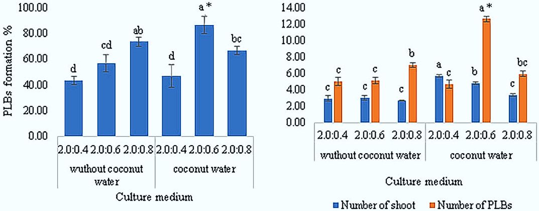
In our study, we started with a small number of plant materials, so the shoots were cultivated on ½MS medium supplemented with a combination of 6-BAP and IAA to produce a large number of regenerants. The results indicated that a significantly larger number of shoots formed on the medium supplemented with 2.0 mg L-1 6-BAP, varying concentrations of IAA, and coconut water, compared to the medium without coconut water. A significant increase in the number of shoots (5.67 ± 0.17 per explant) was observed on the medium containing 2.0 mg L-1 6-BAP, 0.4 mg L-1 IAA, and 100 mL L-1 coconut water, when compared to the other treatments. This can be attributed to the addition of coconut water, which, in combination with cytokinins and auxins, promotes cell division during the development of plant tissue in vitro. The presence of natural hormones, such as zeatin and kinetin, in coconut water may contribute to this stimulatory effect (Win et al., 2022). Increasing the concentration of IAA in the nutrient medium led to a 17-20% reduction in shoot formation (Figure 1).
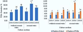
Effect of the combination of 6-BAP and IAA without/with coconut water on PLBs formation and shoot multiplication after 90 days of cultivation *Values are mean ± SD, and the letters “a”, “b”, and “c” denote groups by Duncan’s multiple range test at the 5% level.
In general, the percentage of PLBs formation was low, due to shoot aging, their ability to regenerate weakens (Tanaka et al., 1975). Regarding the induction of PLBs, the ½MS nutrient medium supplemented with 2.0 mg L-1 6-BAP, 0.6 mg L-1 IAA, and 100 mL L-1 coconut water produced the highest number of PLBs (12.58 ± 0.33 per explant) and PLBs formation rate (87%). These results are consistent with the results of other studies, which have shown that high concentrations of cytokinin, in combination with auxin, stimulate PLBs formation (Hossen et al., 2021). Conversely, it was noted that the percentage of PLBs formation increased by approximately 16% in the medium without coconut water as the concentration of IAA was raised (Figure 1). Additionally, in media containing coconut water, an increase in auxin concentration led to a color change in the formed PLBs from green to yellow and pale green (Figure 2).

Effect of the combination of 6-BAP and IAA without/with coconut water on PLBs formation and shoot multiplication after 90 days of cultivation. Abbreviations: blue arrows, old shoot; yellow arrows, new shoot; red arrows, PLBs. Scale bars: 1 cm.
This is consistent with the results of other studies (Cordova et al., 2024), as an imbalance of nutrients or essential growth-stimulating components may lead to the yellowing or necrosis of PLBs. Additionally, this may be due to the fact that high concentrations of auxins can be toxic, leading to suppression of cell growth and causing chromosomal damage (Mei et al., 2012).
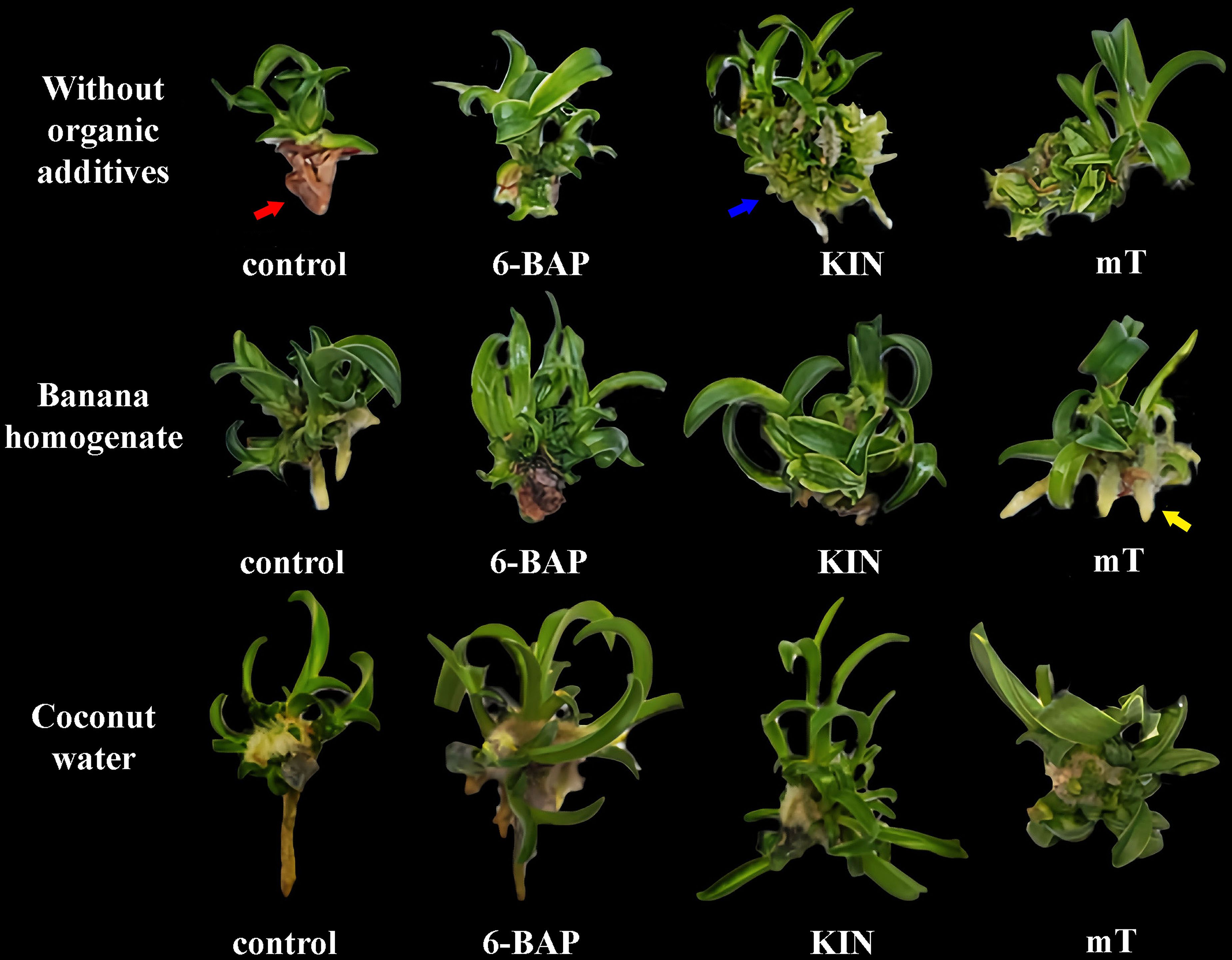
Modification of nutrient medium components, including PGRs and organic additives, is essential for different types of growth responses in explants (Kumara et al., 2022). The results showed a significant effect of organic additives, cytokinins, and their interaction on the regeneration of PLBs and plantlet formation (Figure 3).
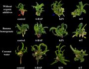
Plantlet regeneration of P. kovachii from PLBs cultured in ½MS medium supplemented with different combinations of organic additives and cytokinins. Abbreviations: blue arrows, PLBs proliferation; yellow arrows, root induction; red arrows, necrosis. Scale bars: 1 cm.
The use of cytokinins is essential for PLBs regeneration, but the frequency and course of regeneration vary depending on the type of cytokinin used (Pathak et al., 2022). Exogenous cytokinins contribute significantly to the induction of regenerative tissues through the indirect regeneration pathway in most ornamental plants (Zhu et al., 2024). The PLBs cultivated on ½MS medium supplemented with 0.5 mg L-1 6-BAP and 100 mL L-1 coconut water produced the highest average number of shoots (5.58 ± 0.22 per explant) compared to the other treatments. The shoot length and number of leaves were significantly affected by the presence of organic additives compared to the medium without organic additives, regardless of the cytokinin used (Table 1).
The number of leaves (3.50 ± 0.14 per explant) and shoot length (1.98 ± 0.06 cm) were also higher in the medium supplemented with 0.5 mg L-1 6-BAP and coconut water. This effect can be attributed to the biochemical composition of coconut water, which contains vitamins, natural plant hormones, and sugars such as sorbitol and mannose. These compounds contribute to regulating the biological pathway for cell wall formation and ion absorption. Additionally, gibberellins (GAs), which are present in coconut water, promote shoot elongation and regulates cambial activity in plant cells (Lazim et al., 2015; Qiao Er Wong et al., 2024). A significant effect of organic additives, cytokinins, and their interaction on rooting percentage was also observed (Table. 1). This trait is important for plants during the rooting stage. The highest rooting percentage (63%) was obtained on the nutrient medium supplemented with 0.5 mg L-1 mT and 50 g L-1 banana homogenate. The addition of mT enhances rooting by modifying the activities of antioxidant enzymes and promoting chloroplast differentiation compared to other cytokinins (Zaytseva et al., 2021). Furthermore, organic additives alone had a significant effect on root number. The highest number of roots was formed on the medium supplemented with banana homogenate, regardless of the cytokinin added (Table 1). This may be due to the high potassium content in bananas. A study by Xu et al. (2020) demonstrated that potassium ion supply stimulates and regulates ATPase activity in the plasma membrane, generating an acidic environment that facilitates cell wall loosening and hydrolase activation, thereby promoting root cell growth. However, the highest number of roots (2.33 ± 0.33 per explant) was recorded on the medium supplemented with 0.5 mg L-1 mT and 50 g L-1 banana homogenate. In addition, a high necrosis rate was observed on the media free of organic additives and supplemented only with cytokinin (Figure 3). The highest rate (57 %) was recorded in media without cytokinin and organic additives. Also, a higher percentage of new PLBs proliferation was observed on the media supplemented only with cytokinins compared to the media containing both organic additives and cytokinins (Figure 3). Such excessive proliferation is undesirable for conservation efforts aimed at maintaining genetic diversity (Bartel & Downing, 2024). The highest percentage of PLBs proliferation was recorded in media supplemented with 0.5 mg L-1 mT and kinetin (67% and 53.33%, respectively) compared to the other treatments.
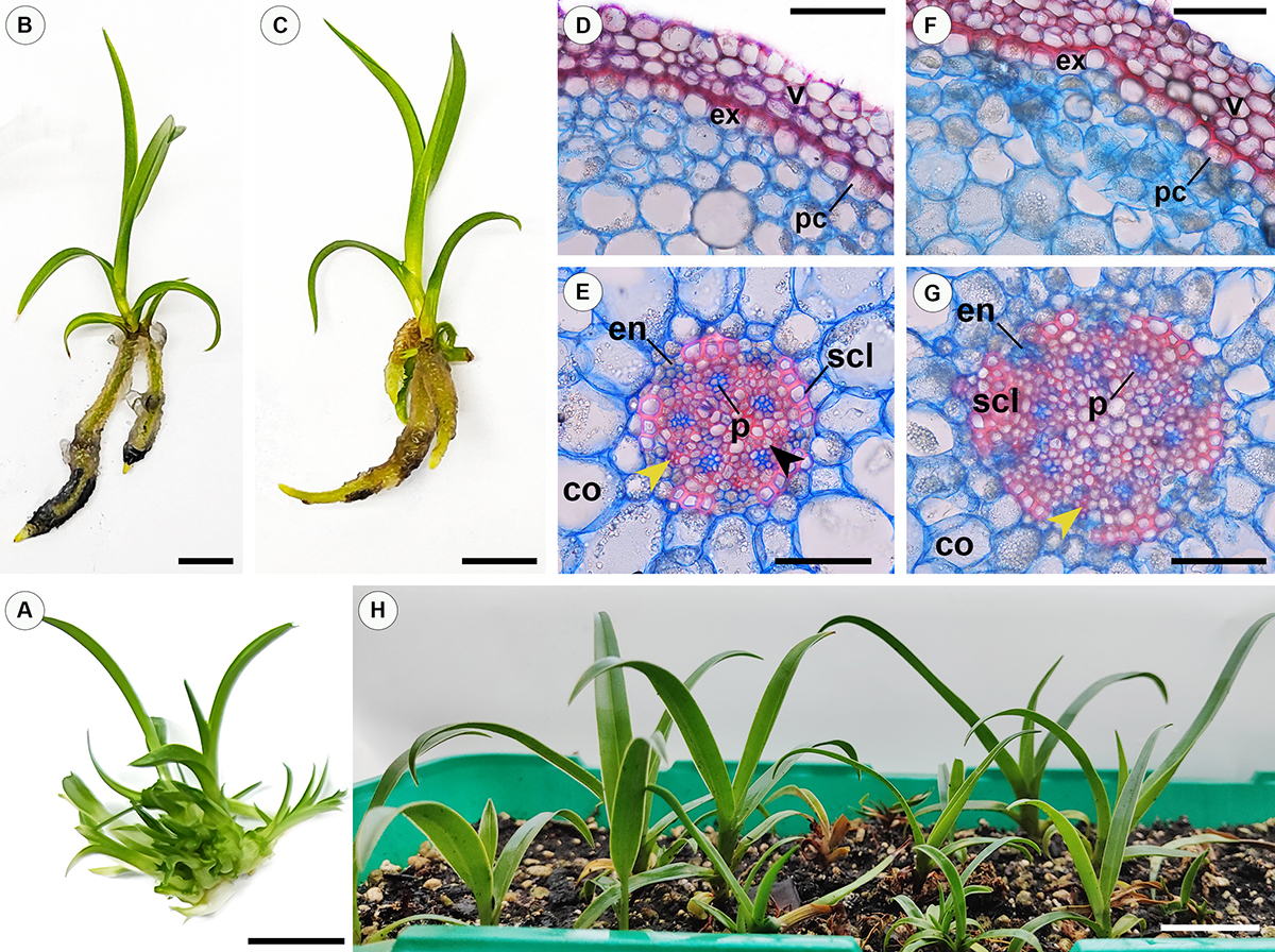
During the rooting stage, plantlets regenerated from PLBs, with developed root primordia, exhibited better growth and rooting efficiency. In contrast, shoots formed during the multiplication stage, when transferred to the rooting medium, developed adventitious shoots but did not form roots (Figure 4A), rendering them unsuitable for acclimatization.
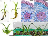
Rooting features of P. kovachii plantlets. А, multiple shoots derived from regenerates at the multiplication stage on rooting medium. B, plantlets cultivated on modified MS medium with coconut water and 1.0 mg L-1 IAA. C, plantlets cultivated on modified MS medium with banana homogenate and 0.5 mg L-1 IBA. D, cross-sections of roots grown on modified MS medium with coconut water and 1.0 mg L-1 IAA showing velamen and exodermis. E, cross-sections of roots grown on modified MS medium with coconut water and 1.0 mg L-1 IAA showing cortex and central cylinder. F, cross-sections of roots grown on modified MS medium with banana homogenate and 0.5 mg L-1 IBA showing velamen and exodermis. G, cross-sections of roots grown on modified MS medium with banana homogenate and 0.5 mg L-1 IBA showing cortex and central cylinder. H, acclimatized P. kovachii plantlets. Abbreviations: yellow arrows, protoxylem; black arrows, metaxylem; white arrows, starch grains. velamen (v) exodermis (ex) cortex (co), endodermis (en), passage cells (pc), phloem (p) embedded in sclerenchymatous tissue (scl). Scale bars: A-C = 1 cm, D - F= 10 µm, E - J= 50 µm, H= 5 cm.
This may be due to the high cytokinin concentration in the medium, which likely accumulated in the plant tissues, inhibiting root formation while promoting shoot proliferation. The absorption and metabolism of both exogenous and endogenous cytokinins regulate the cytokinin pool in plant tissues, thereby affecting in vitro growth and development (Li et al., 2022). Therefore, PLB regeneration by culturing them on media containing low concentrations of PGRs and organic additives is a more effective approach for obtaining plantlets suitable for rooting. This protocol has been successfully applied for the propagation of various orchid species, such as Trichopilia suavis and Cattleya gaskelliana (Hussien, Molkanova, & Mitrofanova, 2023; Hussien et al., 2024). Statistical analysis revealed a significant influence of organic additives on rooting percentage. The highest rooting rate of regenerated plantlets derived from PLBs with root primordia was observed in media supplemented with coconut water and banana homogenate, compared to media containing potato homogenate, regardless of the type or concentration of auxin (Table 2).
The plantlets grown on 1/2MS nutrient medium supplemented with 0.5 mg L-1 IBA and 50 g L-1 banana homogenate developed the highest number of roots (4.33± 0.19 per plantlet) (Figure 4C). This effect can be attributed to the role of IBA in enhancing the translocation of carbohydrates, plant growth regulators, and nutrients to the root formation zone (Khandaker et al., 2022). Several studies have demonstrated that as the concentration of auxins in the rooting medium increases, the ability to stimulate root growth decreases (Tien et al., 2020; Yoon et al., 2021). However, both the type and concentration of auxins significantly influenced root development, with higher auxin concentrations generally reducing root number (Table 2). An exception was noted in media containing coconut water, where higher auxin concentrations led to a non-significant increase in root number. Furthermore, plantlets grown on media containing coconut water exhibited longer root development. The highest root length (4.27 ± 0.20 cm) was achieved on a medium supplemented with 1.0 mg L-1 IAA and 100 mL L-1 coconut water, highlighting the significant effect of both auxin type and concentration on root elongation (Figure 4B).
Statistical analysis of vegetative growth parameters demonstrated a significant effect of organic additives. The longest plantlets (5.20 ± 0.07 cm) and the highest number of leaves (4.34± 0.19 per plantlet) were observed in regenerants cultured on media supplemented with 1.0 mg L-1 IAA and 100 mL L-1 coconut water. This could be due to the fact that coconut water contains IAA and GAs, which enhance cell division, elongation, and both stem and root growth (Aishwarya, Seenivasan, & Naik, 2022).
With the established differences in the quantitative characteristics of the regenerant roots when cultivated on nutrient media containing different organic additives, a study of their anatomical structure was conducted. The roots of P. kovachii plantlets grown in vitro were observed to consist of a multiseriate velamen, exodermis, cortex, endodermis, and stele. This anatomical structure plays a crucial role in adaptation to the natural habitat, as P. kovachii is a terrestrial species that grows in volcanic rock crevices (Stern, 2014). The number of velamen cell layers varied depending on the organic additives present in the culture medium. Roots grown on a medium containing potato homogenate and coconut water developed 2-3 layers of velamen cells, while those grown on a medium with banana homogenate formed velamen consisting of 3-4 layers of oval and polygonal cells, in cross section (Figure 4 D-F). Additionally, the velamen layer in roots grown on the medium with banana homogenate was thicker (18.05±0.39 μm) compared to those grown on the potato homogenate and coconut water media (13.94±0.75μm and 11.89±0.84 μm, respectively) (Table 3). The exodermis, located between the velamen and cortex, consisted of a single layer of U-thickened cells (Figure 4D-F). Passage cells within the exodermis, characterized by their thinner walls compared to adjacent exodermal cells, contained starch grains. It should be noted that the thickness of these layers has accounted for approximately 3 % of the root diameter, regardless of the organic additives in the culture medium. Perhaps this is due to the growth of roots under in vitro conditions with high humidity.
The endodermis consisted of a single layer of O-thickened cells, including thin-walled passage cells. This layer constituted approximately 2% of the root diameter. The outer exodermis and the inner endodermis provide mechanical support to the roots due to the deposition of lignin and suberin on their walls. Both layers are characterized by the presence of the Casparian strip in their cell walls, forming a barrier between cells (Martins et al., 2020). The Casparian strip contributes to the selectivity of solute uptake and serves as an apoplastic barrier, preventing the flow of ions from the vascular cylinder into the cortex (Santos & Silva, 2023).
The cortex was composed of 7-8 layers of thin-walled parenchymatous cells containing starch grains with intercellular spaces. The availability of starch granules in the cortex area has also been observed, which is unique and probably helps in the storage function (Muthukumar & Shenbagam, 2018). In transverse sections, the outer cortical cells appeared small and oval, whereas the cells in the middle and inner layers, next to the central cylinder, were larger and more elongated. This characteristic was clearly observed in roots formed on½MS medium supplemented with coconut water. The cortex occupied the largest part of the roots grown on medium with coconut water (73%), followed by those grown on potato homogenate (67%) and banana homogenate (64%) (Table 3).
The pericycle was multilayered, and the vascular bundles were embedded within a matrix of sclerenchymatous cells. This characteristic was particularly prominent in roots grown on a medium containing coconut water. Under in vitro conditions, roots grown on a medium supplemented with banana homogenate had a larger diameter compared to those grown on media containing coconut water or potato homogenate (Table 3). Roots grown on a medium containing banana homogenate exhibited a more developed stele with a polyarch structure (Figure 4G), constituting approximately 18% of the root cross-section and displaying a significant number of protoxylem and phloem poles. This increase in stele diameter is likely due to the enlargement of the phloem and protoxylem poles (Rai, Bhutia, & Moktan, 2024). In contrast, roots grown on a coconut water medium had a smaller diameter and comprising about 14% of the root cross-section, but were characterized by the presence of metaxylem vessels, as well as sclerenchyma cells (Figure 4E). The formation of secondary tissues, such as metaxylem and sclerenchyma tissue in the stele, leads to mechanical support and contributes to the axial transfer of water and nutrients to the vegetative parts, which explains the higher plant length and the increased number of leaves on the medium supplemented with coconut water compared to plants growing on media supplemented with other organic additives (Lynch et al., 2021).
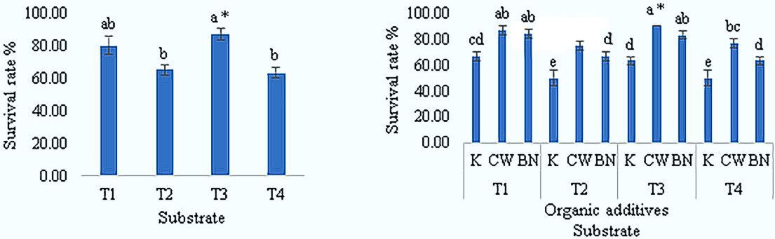
The problems of the acclimatization process have not been considered in the published works on the micropropagation of P. kovachii (Sánchez, 2021). In this study, we have studied the process of acclimatization to ex vitro conditions on different substrates. The substrate in which plants grow plays an important role because it mainly affects the quality of plants (|De Faria et al., 2018). According to the statistical analysis, there was a significant effect of the substrates and the type of organic additives in the culture medium on which the plantlets were grown at the rooting stage on the acclimatization process. After 90 days, the plantlets rooted on medium supplemented with 100 mL L-1 coconut water showed a higher survival rate (Figure 4H). This can also be explained by the formation of metaxylem vessels, which give the plants the ability to adapt to ex vitro conditions, such as low humidity and others (Reeger et al., 2021). The highest survival rates were observed for plantlets grown on substrates composed of bark, peat moss, and perlite (1:1:1) and soil, sand and perlite (2:1:1) compared to the other substrates (Figure 5).
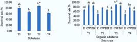
Effect of different substrates on the survival rate of P. kovachii plantlets under ex vitro conditions after 90 days. *Values are mean ± SD, the letters “a”, “b”, and “c” denote groups by Duncan’s multiple range test at the 5% level. Abbreviations: soil, perlite, sand (2:1:1) (T1); bark, peat (1:1) (T2); bark, peat, perlite (1:1:1) (T3); bark, peat, perlite, sphagnum moss (1:1:1:1) (T4); potato homogenate (K); coconut water (CW); banana homogenate (BN).
In general, it was noted that P. kovachii plants do not prefer standing water in the substrate. Therefore, it was noted that many plants died in a substrate containing sphagnum, which negatively affected the survival rate. In addition, P. kovachii can tolerate slight drought between waterings.
Conclusions
In this study, PLBs of P. kovachii were successfully induced from shoots on ½MS medium supplemented with 2.0 mg·L⁻¹ 6-BAP, 0.6 mg·L⁻¹ IAA, and coconut water. PLBs regeneration was achieved using either 6-BAP with coconut water or mT with banana homogenate. Plantlets with root primordia grown on 1.0 mg·L⁻¹ IAA and coconut water developed longer roots and showed higher survival rates. Acclimatization was successfully achieved using substrates composed of sand, soil, and perlite, or bark, peat moss, and perlite.
Acknowledgements
This research was funded by assignments № 122042700002-6 from the Ministry of Science and Higher Education of the Russian Federation.
References
- Aishwarya, P. P., Seenivasan, N., & Naik, D. S. (2022). Coconut water as a root hormone: Biological and chemical composition and applications. The Pharma Innovation Journal, 11(12):1678-1681.
- Atwood, J. T., Dalström, S., & Fernandez, R. (2002). Phragmipedium kovachii, a new species from Peru. Selbyana, 23:1-4.
- Bartel, L. G., & Downing, J. (2024). Comparison of in-vitro orchid media solutions for Prosthechea cochleata (L.) W.E. Higgins vars. diandra and triandra. Plant Cell, Tissue and Organ Culture (PCTOC), 158:27.
- Cardoso, J. C., Zanello, C. A., & Chen, J. T. (2020). An overview of orchid protocorm-like bodies: Mass propagation, biotechnology, molecular aspects, and breeding. International Journal of Molecular Sciences, 21(3):985.
- Cordova, E. M. et al. (2024). Induction of protocorm-like bodies (PLBs) and plant regeneration in Cymbidium chloranthum (Orchidaceae).BIO Web of Conferences. EDP Sciences, 131:01002.
- Cribb, P. J. (2005). Phragmipedium kovachiiCurtis’s Botanical Magazine, 22(1):8-11.
- De Faria, R. T. et al. (2018). Substrates for the cultivation of epiphytic orchids.Semina: Ciências Agrárias, 39(6):2851-2866.
- Gantait, S., Subrahmanyeswari, T., & Sinniah, U. R. (2022). Leaf-based induction of protocorm-like bodies, their encapsulation, storage and post-storage germination with genetic fidelity in Mokara Sayan× Ascocenda Wangsa gold. South African Journal of Botany, 150:893-902.
- Hinsley, A. et al. (2018). A review of the trade in orchids and its implications for conservation. Botanical Journal of the Linnean Society, 186(4):435-455.
- Hossen, M. M. et al. (2021). Effects of plant growth regulators on in vitro growth and development of orchid (Dendrobium sp.) from protocorm-like bodies (PLBs). Journal of the Bangladesh Agricultural University, 19(3):294-301.
- Hussien, M. et al. (2024). Plant growth regulators and organic additives on the proliferation of protocorm-like bodies and plantlet regeneration of Cattleya gaskelliana (N.E. Br.) B.S. Williams. Ciência e Agrotecnologia, 48:e018223.
- Hussien, M., Molkanova, O., & Mitrofanova, I. V. (2023). Micropropagation of Trichopilia suavis Lindl. & Paxton. Ornamental Horticulture, 29(3):365-374.
- Jolman, D. et al. (2022). The challenges of growing orchids from seeds for conservation: An assessment of asymbiotic techniques. Applications in Plant Sciences, 10(5):e11496.
-
Kehew, K. A. (1989). Growing paphiopedilums and phragmipediums in your home. American Orchid Society Bulletin (USA), 164p. Available in:https://secure.aos.org/media/Content-Images/PDFs/Phrag_home_culture.pdf
» https://secure.aos.org/media/Content-Images/PDFs/Phrag_home_culture.pdf - Khandaker, M. M. et al. (2022). Effects of indole-3-butyric acid (IBA) and rooting media on rooting and survival of air layered wax apple (Syzygium samarangense) CV Jambu Madu. Brazilian Journal of Biology, 82:e256277.
- Kumara, P. U. et al. (2022). Low-cost tissue culture technology for the orchid regeneration. Peruvian Journal of Agronomy, 6(2):147-158.
- Lazim, M. I. M. et al. (2015). Quantification of cytokinins in coconut water from different maturation stages of Malaysia’s coconut (Cocos nucifera L.) varieties. Journal of Food Processing & Technology, 6:515.
- Li, Y. Y. et al. (2022). Profiles of cytokinins metabolic genes and endogenous cytokinins dynamics during shoot multiplication in vitro of Phalaenopsis International Journal of Molecular Sciences, 23(7):3755.
- Lynch, J. P. et al. (2021). Root anatomy and soil resource capture. Plant and Soil, 466:21-63.
- Martins, J. P. R. et al. (2020). Anatomical and physiological changes of in vitro-propagated Vriesea imperialis (Bromeliaceae) in the function of sucrose and ventilated containers. Plant Biosystems - An International Journal Dealing with all Aspects of Plant Biology, 154(1):87-99.
- Mei, T. A. et al. (2012). Exquisite protocol of callus induction and protocorm-like bodies (PLBs) regeneration of Dendrobium ‘Sonia-28’. Australian Journal of Crop Science, 6(5):793-800.
- Murashige, T., & Skoog, F. (1962). A revised medium for rapid growth and bioassays with tobacco tissue culture. Physiologia Plantarum, 15(3):473-497.
- Muthukumar, T., & Shenbagam, M. (2018). Vegetative anatomy of the orchid Bulbophyllum sterile (Orchidaceae: Epidendroideae). Lankesteriana, 18(1):13-22.
- O’Brien, T. P., & McCully, M. E. (1981). The study of plant structure: Principles and selected methods Melbourne: Termarcarphi.
- Parizaca, A. D., Díaz-Morales, M., & Pupulin, F. (2019). A new species of Phragmipedium (Orchidaceae: Cypripedioideae) from Peru. Phytotaxa, 423(4):259-265.
- Park, H. B. et al. (2023). Asymbiotic seed germination and in vitro seedling development of the endangered orchid species Cypripedium guttatum Plants, 12(22):3788.
- Pathak, P. et al. (2022). Regeneration competence of an endangered orchid, Vanda cristata Wall. ex Lindl. using leaf explants: A study in vitro South African Journal of Botany, 151:1018-1024.
- Perdue, R. T. (2021). Who needs the dark web? Exploring the trade in critically endangered plants on eBay. American Journal of Criminal Justice, 46:1006-1017.
-
Plants of the world online - POWO. Phragmipedium Rolfe. Available in: Available in: <Available in: Available in: https://powo.science.kew.org/taxon/urn:lsid:ipni.org:names:312553-2/general-information >. Access in: January 8, 2025.
» https://powo.science.kew.org/taxon/urn:lsid:ipni.org:names:312553-2/general-information - Poniewozik, M., Szot, P., & Parzymies, M. (2021). Tissue culture multiplication of Paphiopedilum insigne depending on the medium type, growth regulators, and natural supplements. Acta Scientiarum Polonorum Hortorum Cultus, 20(4):125-134.
- Prasongsom, S., Thammasiri, K., & Chuenboonngarm, N. (2016). Efficient adventitious shoot regeneration from shoot tip culture of Rhynchostylis gigantea (Lindl.) Ridl.(amethyst-purple), a rare Thai orchid species.Walailak Journal of Science and Technology (WJST),13(9):757-767.
- Prozina, M. N. (1960). Botanical microtechniques Moscow: Vysschaya Schkola, 207 p.
- Qiao Er Wong, S. et al. (2024). Effects of coconut water and banana homogenate on shoot regeneration of Meyer lemon (Citrus× meyeri). Pertanika Journal of Tropical Agricultural Science, 47(1):147-158.
- Rai, P., Bhutia, K., & Moktan, S. (2024). Comparative study on root anatomy of six species of Himalayan Dendrobium Swartz. Flora, 310:152424.
- Reeger, J. E. et al. (2021). Targeted mutation of transcription factor genes alters metaxylem vessel size and number in rice roots. Plant Direct, 5(6):e00328.
- Rohmah, K. N., & Taratima, W. (2022). Effect of chitosan, coconut water and potato extract on protocorm growth and plantlet regeneration of Cymbidium aloifolium (L.) Sw. Current Applied Science and Technology, 22(2):1-10.
- Sánchez, M. E. R. (2021). Micropropagación de Phragmipedium kovachii, con fines de conservación genética. Revista Agrotecnológica Amazónica, 1(1):45-61.
- Santos, I. S. D., & Silva, M. J. D. (2023). Anatomy and histochemistry of the vegetative system of Brachystele guayanensis (Lindl.) Schltr. (Orchidaceae), a potential medicinal species. Plants, 12(14):2635.
- Schiff, J. L. (2018). Rare and exotic orchids: Their nature and cultural significance Springer International Publishing, 115p.
- Stern, W. L. (2014). Anatomy of the monocotyledons Volume X: Orchidaceae. Oxford: Oxford University Press, 257p.
- Tanaka, M., Hasegawa, A., & Goi, M. (1975). Studies on the clonal propagation of monopodial orchids by tissue culture I. Formation of protocorm-like bodies from leaf tissue in Phalaenopsis and Vanda Journal of the Japanese Society for Horticultural Science, 44(1):47-58.
- Tien, L. H. et al. (2020). Effect of auxins (IAA, IBA and NAA) on clonal propagation of Solanum procumbens stem cuttings. Plant Cell Biotechnology and Molecular Biology, 21(55-56):113-120.
-
Tiwari, P. et al. (2022). Biotechnological interventions in orchids: Recent updates, translational success, and commercial outcomes PREPRINT (Version 1). Available in: <https://doi.org/10.21203/rs.3.rs-1382018/v1>.
» https://doi.org/10.21203/rs.3.rs-1382018/v1 - Tiwari, P. et al. (2024). Advances in orchid biology: Biotechnological achievements, translational success, and commercial outcomes. Horticulturae, 10(2):152.
- Vasupen, E., Bundithya, W., & Potapohn, N. (2023). Effects of coconut water, benzylaminopurine, and naphthalene acetic acid on seed germination and rhizome food reserve: In vitro culture of Eulophia flava (Lindl.) Hook. f. Current Applied Science and Technology, 23(3):1-13.
- Win, T. A. et al. (2022). Plantlet regeneration and PLBs propagation of Bulbophyllum auricomum Lindl. Korean Journal of Plant Resources, 35(4):508-514.
- Xu, X. et al. (2020). Effects of potassium levels on plant growth, accumulation and distribution of carbon, and nitrate metabolism in apple dwarf rootstock seedlings. Frontiers in Plant Science, 11:904.
- Yoon, A. et al. (2021). Plant growth regulators and rooting substrates affect growth and development of Salix koriyanagi cuttings. Rhizosphere, 20:100437.
- Zaytseva, Y. G., Ambros, E.V., & Novikova, T. I. (2021). Meta-topolin: Advantages and disadvantages for in vitro propagation. In N. Ahmad., & M. Strnad,Meta-topolin: A growth regulator for plant biotechnology and agriculture Springer, Singapore, (pp.119-141).
- Zelenko, H. (2007). Not a single orchid... Lankesteriana International Journal on Orchidology, 7(1-2):164-166.
- Zhu, L. et al. (2024). Regeneration of ornamental plants: Current status and prospects. Ornamental Plant Research, 4:e022.
-
Editor de seção:
Renato Paiva