Clinical and bacteriological diagnosis of infectious keratoconjunctivitis in ruminants from herds in the state of Maranhão, Brazil
Abstract
Infectious keratoconjunctivitis, caused mainly by Moraxella spp., is a common disease in ruminants such as cattle, goats and sheep, manifested by ocular signs such as conjunctival hyperemia, epiphora and corneal opacity, which can lead to blindness. Transmission occurs by direct contact, aerosols, fomites and mechanical vectors such as flies. This study aimed to diagnose and provide data on the occurrence of infectious keratoconjunctivitis in herds of Maranhão. A total of 5814 animals (4348 goats, 1426 sheep and 40 cattle) were evaluated, of which 16 presented clinical signs of keratoconjunctivitis. The highest prevalence was observed in cattle (5 %, n=2), followed by sheep (0.56 %, n=8) and goats (0.13 %, n=6). Most of the affected goats were less than one year old (83.33 %), and among sheep, the Santa Ines breed was the most affected (62.5 %). Among cattle, both affected animals were females of the Holstein breed. The clinical signs observed were consistent between species, including corneal opacity, lacrimation and purulent discharge. Microbiological analysis confirmed Moraxella spp. in 7 samples (3 goats, 2 sheep, 2 cattle), confirming it as the probable cause. Five samples were contaminated and three showed no bacterial growth. The lack of biosecurity measures, such as vector control and quarantine, may have contributed to the spread of the disease. This study, the first of its kind in the region, highlights the urgent need for preventive measures and more effective diagnostic strategies in Maranhão, as well as the need for research to improve the clinical and epidemiological understanding of this disease.
Keywords
Moraxella spp; veterinary ophthalmology; goats; sheep; cattle
Resumo
A ceratoconjuntivite infecciosa, causada principalmente por Moraxella spp., é uma doença comum em ruminantes como bovinos, caprinos e ovinos, manifestada por sinais oculares como hiperemia conjuntival, lacrimejamento e opacidade corneana, que pode levar à cegueira. A transmissão ocorre por contato direto, aerossóis, fômites e vetores mecânicos como moscas. Este estudo teve como objetivo diagnosticar e fornecer dados sobre a ocorrência da ceratoconjuntivite infecciosa em rebanhos maranhenses. Foram avaliados 5.814 animais (4.348 caprinos, 1.426 ovinos e 40 bovinos), dos quais 16 apresentaram sinais clínicos de ceratoconjuntivite. A maior prevalência foi observada em bovinos (5 %, n=2), seguida de ovinos (0,56 %, n=8) e caprinos (0,13 %, n=6). A maioria dos caprinos afetados tinha menos de um ano (83,33 %), e nos ovinos, a raça Santa Inês foi a mais afetada (62,5 %). Nos bovinos, ambos os animais afetados eram fêmeas da raça Holandesa. Os sinais clínicos observados foram consistentes entre as espécies, incluindo opacidade corneana, lacrimejamento e secreção purulenta. A análise microbiológica identificou Moraxella spp. em 7 amostras (3 caprinos, 2 ovinos, 2 bovinos), confirmando-a como causadora provável. Cinco amostras foram contaminadas e três não apresentaram crescimento bacteriano. A falta de medidas de biossegurança, como controle de vetores e quarentena, pode ter contribuído para a propagação da doença. Este estudo, inédito na região, destaca a necessidade urgente de medidas preventivas e estratégias de diagnóstico mais eficazes no Maranhão além da necessidade de realizar pesquisas para melhorar a compreensão clínica e epidemiológica desta doença.
Palavras-chave
Moraxella spp.; oftalmologia veterinária; caprinos; ovinos; bovinos
1. Introduction
Infectious keratoconjunctivitis in ruminants, also known as 'Pink Eye' and New Forest Disease, is a seasonal, cosmopolitan disease that can affect cattle, regardless of breed, age, or sex (1). It is also known to affect goats and sheep (2). This disease is often caused by the gram- negative bacterium Moraxella spp., however, only microorganisms that express virulence factors such as pili, lipopolysaccharides, phospholipase B, and outer membrane proteins are capable of developing it, as these factors enable the bacteria to adhere to the cornea and produce necrosis through toxins, thereby overcoming host defenses (3). Other agents, such as Mycoplasma spp., Chlamydophila spp., are also significant contributors, particularly in small ruminants (4).
The most common clinical signs of this disease comprise acute inflammatory reaction of the conjunctival tissue, which is followed by scleral hyperemia, epiphora, blepharospasm and photophobia. In some cases, this disease leads to corneal opacity, ulceration and perfuration, which can progress to temporary or irreversible blindness (5). This contagious disease is transmitted by direct contact, aerosols and fomites contaminated with infected animals’ nasal or ocular secretion. Infectious keratoconjunctivitis can also be transmitted by mechanical vectors, a fact that makes this disease cases more frequent in the rainy season or in case of flies-population increase (6). Predisposing factors, such as dust, twigs, dry forage and wind, can harm animals' eye surface and leave them vulnerable to infection onset (7).
The diagnosis involves collecting evidence to categorize the disease, as most ocular conditions lack pathognomonic clinical signs, requires a combination of evidence, including history, clinical signs, and laboratory diagnostics. Definitive diagnosis relies on isolating and characterizing the causative agent through techniques such as bacterial culture, direct immunofluorescence, or serological testing (1). Little is known about its impacts on animal welfare; however, pain assessment studies have shown that it is a painful and irritating condition. Although it is not a fatal disease, it has significant economic impact, since animals’ vision loss leads to weight loss, reduced milk production, handling difficulties and treatment-related costs (8).
In Maranhão State, the lack of diagnostic resources and preventive measures in rural herds may exacerbate the disease's impact, making studies on its clinical and epidemiological aspects essential. This factor, combined with the lack of technical assistance to breeders and the lack of previous studies on the subject in the region, highlights the importance of the current research, which seeks to provide unprecedented data on the occurrence of the disease. Thus, the objectives of the present study were to clinically and bacteriologically diagnose infectious keratoconjunctivitis caused by Moraxella spp. in ruminants from herds in the State of Maranhão.
2. Material and methods
2.1 Ethical aspects of animal research
This study was approved the Ethics and Animal Experimentation Committee - CEEA of the Veterinary Medicine Course at State University of Maranhão (UEMA), according to protocol no. 14/2021.
2.2 Study local and contextualization
The study was conducted in 18 Maranhão State’s municipalities, including 87 properties, in partnership with the State Health-Defense Agency, in goat-, sheep- and cattle-breeding farms. Sampling was conveniently performed based on the number of animals presenting clinical signs of infectious keratoconjunctivitis in each assessed herd.
Clinical findings resulting from eye examination comprised the following changes: epiphora, blepharospasm, photophobia and, in some cases, corneal opacity, ulceration and perfuration. Only animals showing clinical signs consistent with infectious keratoconjunctivitis were included in the study; asymptomatic individuals were excluded from sampling. Clinical evaluations were performed by veterinarians using specific ocular examination instruments such as a focal light and a direct ophthalmoscope.
Conjunctival swabs were collected from affected animals using sterile swabs soaked in 0.9 % saline solution. Swabs were stored in transport media and promptly sent to the laboratory for microbiological processing.
2.3 Samples processing
Samples were seeded on plates added with MacConkey agar and sheep blood agar (5 %), incubated at 37°C under aerobic conditions, and subjected to readings, at 24 and 48 hours after incubation, in order to identify and isolate colonies. Colonies were phenotypically identified based on colonial morphology by taking into consideration the following aspects: shape, size, appearance, color and hemolysis. Gram staining reaction was conducted based on Newprov® manufacturer’s recommendations. Colonies were also assessed to biochemical tests (oxidase, catalase, indole, motility, citrate and carbohydrate fermentation) and growth on MacConkey agar (9).
2.4 Epidemiological data
Data were tabulated and organized in Microsoft Excel® spreadsheets. Statistical calculations of the study population necessary for this disease were performed by taking into consideration animals’ herd, sex, breed, age and the municipality they were bred in. Results concerning the frequency of animals presenting clinical signs suggesting infectious keratoconjunctivitis were calculated in Epi Info statistical software.
3. Results
In total, 5,814 animals were assessed: 4348 goats; 1426 sheep and 40 cattle. Sixteen of them presented clinical signs suggesting infectious keratoconjunctivitis: 0.13 % of them were goats (n=6), 0.56 % were sheep (n=8) and 5 % were cattle (n=2). Although cattle exhibited the highest prevalence among the evaluated species, goats and sheep accounted for a greater absolute number of cases due to their larger population in the study.
Goats affected by keratoconjunctivitis did not exhibit a defined breed pattern and were equally distributed between males and females. The majority of affected goats (83.33 %) were younger than one year old. Clinical signs observed in these animals comprised hyperemia, epiphora, corneal opacity, purulent secretion around the eyes and corneal ulcer (Figure 1).

Clinical signs suggesting infectious keratoconjunctivitis in goats. A - Corneal ulcer. B - Purulent secretion. C- Corneal opacity. D - Hyperemia and epiphora.
Among the sheep, most cases (62.5 %, n=5) occurred in Santa Inês breed, while 37.5 % (n=3) were Dorper sheep. All affected sheep were female, with 62.5 % (n=5) being older than four years. The observed clinical signs comprised corneal opacity, conjunctival hyperemia, epiphora and purulent discharge around the eyes (Figure 2).
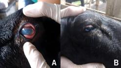
Clinical signs suggesting infectious keratoconjunctivitis in sheep. A - Corneal opacity and conjunctival hyperemia. B – Discharge around the eye.
In cattle, both affected animals were female Holstein cows aged 8 and 10 years, respectively. This limited sample size makes it challenging to assess breed or age predispositions for this species. Clinical signs observed in this group comprised corneal opacity and edema.There was higher incidence of affected females (81.25 %) among animals who tested positive for the investigated disease. Corneal opacity was the most common clinical sign of it; it accounted for 75 % of cases. On the other hand, 68.75 % of animals only had one eye affected in comparison to 31.25 %, who presented bilateral lesions.
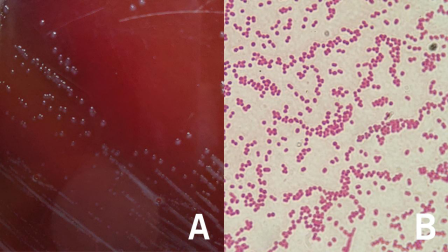
In microbiological analyses bacteria compatible with Moraxella spp. were isolated from 7 samples (3 from goats, 2 from sheep and 2 from cattle). The colonies presented typical characteristics of Moraxella spp., including coccoid-bacillary morphology, gray coloration, small size and beta-hemolysis on blood agar (Figure 3). Biochemical tests showed positive reactions for catalase and oxidase, and no growth on MacConkey agar. Five samples were contaminated, presenting Gram-positive and Gram-negative bacteria. Three samples showed no bacterial growth. Based on the clinical signs, epidemiology and microbiological results, Moraxella spp. is suggested as the causative agent in the cases investigated.

Moraxella sp. colonies based on sheep blood agar (5 %) (A) and Gram staining reaction under a 100x oil immersion lens (B).
None of the breeders who had animals presenting clinical signs of keratoconjunctivitis subjected newly acquired animals to quarantine. Additionally, they did not control vectors, specifically the species Musca domestica, as observed in Figure 4. The facilities mostly had inadequate structure, and the absence of vaccination against infectious keratoconjunctivitis was observed in all herds.

4. Discussion
While Infectious keratoconjunctivitis has been widely studied in other parts of Brazil, this is the first study conducted in Maranhão State, providing essential data on the occurrence of the disease in this region. Although the overall prevalence in this study was low, the identification suggests that the disease is present and may be underreported in the region. This underscores the importance of conducting further research to evaluate the disease's full impact and to implement better diagnostic and control strategies. However, the lack of more extensive data from the Maranhão region makes it difficult to draw definitive conclusions about species-specific risk factors.
Despite the lower prevalence of infectious keratoconjunctivitis in cattle, the rate is significantly higher compared to goats and sheep. This difference in prevalence may be related to species susceptibility, management, or environmental conditions. This observation may be relevant for management strategies, considering that small ruminants are raised in extensive to ultra-extensive conditions, which allows for greater exposure of animals to infectious agents present in the environment (10).
Infectious keratoconjunctivitis more often affects younger animals, due to their immunological vulnerability (11). Additionally, in this study, Santa Inês sheep showed a higher prevalence of the disease, which may suggest a breed predisposition or the influence of environmental and management factors specific to this breed. However, no literature was found to corroborate this fact.
The clinical signs found in all species resemble those described by Kalaiselvi et al. (9), who conducted a study focused on investigating infectious keratoconjunctivitis incidence associated Moraxella bovis in Ruminants. Detailed observation of clinical signs, especially in the early stages, is essential for the early diagnosis and management of the disease.
Microbiology results are consistent with those reported by Carmo et al. (12), who described infectious keratoconjunctivitis outbreak in calves. The identification of Moraxella sp. as an etiological agent reinforces the importance of precise and targeted laboratory diagnostics, which allow the correct identification of the pathogen and, consequently, the adoption of effective treatments.
A limitation of this study is the lack of molecular identification of Moraxella spp. strains. While phenotypic identification provides useful preliminary data, molecular methods such as PCR could have confirmed the identity of the pathogen at a species level. Furthermore, the focus solely on animals with clinical signs, excluded potential asymptomatic carriers, which could provide a more accurate estimate of the disease's prevalence and the role of carriers in maintaining the transmission cycle.
Contamination in some samples may have been caused by failures in the sample collection or handling process, or even the presence of opportunistic microorganisms that may be favoring or complicating the primary infection. Infectious keratoconjunctivitis is often described as a multietiological condition, in which Moraxella spp. may be the main agent, but not the only one involved (1). In addition, the absence of bacterial growth can be explained by several factors, such as variations in culture conditions, degradation of the bacterial genetic material during transport, or the presence of another microorganism that cannot be cultivated using conventional methods.
Although the identification of Moraxella spp. is suggestive, it is important to consider that the definitive diagnosis must integrate the observed clinical signs, such as conjunctivitis and ocular discharge, together with the epidemiological data. Based on these results, it can be concluded that Moraxella spp. plays an important role in ruminant eye infections, being a relevant factor to be considered in conjunctivitis outbreaks in Maranhão.
The absence of biosecurity measures, likely facilitated the spread of this disease on the farms. Flies (particularly Musca domestica) are known to act as mechanical vectors for Moraxella spp. (12). The failure to control these vectors may have contributed to the persistence and transmission of the disease. Furthermore, the lack of veterinary assistance and the absence of vaccination programs for keratoconjunctivitis in herds may have exacerbated the problem, allowing the disease to spread undetected.
Additionally, environmental factors, such as the humid climate and seasonal changes, may create favorable conditions for the proliferation of flies, particularly in the rainy season. These factors need to be considered in future studies to fully understand the epidemiology of infectious keratoconjunctivitis in Maranhão and to develop more effective prevention and control strategies.
Lack of research in this field underscores the importance of investigating both the clinical signs and epidemiology of this disease. It is worth emphasizing the need of taking prophylactic measures to help controlling this disease in herds, since breeders often lack the necessary knowledge to take animal health-related measures without effective technical assistance.
5. Conclusion
This study is the first to investigate the occurrence of infectious keratoconjunctivitis in ruminants in the state of Maranhão, although the prevalence was low, the findings highlight the need for more rigorous monitoring of the disease in the region, in addition to the implementation of preventive measures, such as vector control and vaccination, to reduce the economic impacts caused by the disease. The identification of Moraxella spp. reinforces the importance of accurate laboratory diagnostics for the definition of appropriate therapeutic strategies. Future research should explore the diversity of pathogens involved in infectious keratoconjunctivitis in ruminants, including investigation of asymptomatic carriers and molecular confirmation of Moraxella species present in the region, in order to provide a more solid basis for the implementation of effective animal health programs.
Data availability statement
The complete dataset supporting the results of this study is available upon request from the corresponding author.
-
Generative AI use statement
The authors did not use generative artificial intelligence tools or technologies in creating or editing any part of this manuscript.
References
-
1 Kneipp M. Defining and Diagnosing Infectious Bovine Keratoconjunctivitis. Veterinary Clinics of North America: Food Animal Practice [Internet]. 2021 [cited 2024 Dec 10];37(2):237–252. Available from: https://doi.org/10.1016/j.cvfa.2021.03.001
» https://doi.org/10.1016/j.cvfa.2021.03.001 -
2 Jayasekara PP, Jenkins C, Gerber PF, Olmo L, Xaikhue T, Theppangna W, Walkden-Brown SW. Case-control study to identify the causative agents of ophthalmia and conjunctivitis in goats in Savannakhet province of Lao PDR. Veterinary Microbiology [Internet]. 2024 [cited 2024 Dec 10];296:110195. Available from: https://doi.org/10.1016/j.vetmic.2024.110195
» https://doi.org/10.1016/j.vetmic.2024.110195 -
3 Bilbao L, Acquistapace S, Umpiérrez A, Smircich P, Alonzo P, Sotelo-Silveira JR, Zunino P. Genomic characterization of Moraxella bovis and Moraxella bovoculi Uruguayan strains isolated from calves with infectious bovine keratoconjunctivitis. Revista Argentina de Microbiologia [Internet]. 2024 [cited 2024 Dec 9];56(2):165–174. Available from: https://doi.org/10.1016/j.ram.2023.12.003
» https://doi.org/10.1016/j.ram.2023.12.003 -
4 Gulaydin A, Gulaydin O, Akgul MB, Sindak N, Yildirim O. Investigation of the presence of Chlamydia spp., Mycoplasma spp., and Moraxella ovis in infectious keratoconjunctivitis cases in sheep and goats in Siirt province and evaluation of clinical findings. Polish Journal of Veterinary Sciences [Internet]. 2024 [cited 2024 Dec 10];183–191. Available from: https://doi.org/10.24425/pjvs.2024.149348
» https://doi.org/10.24425/pjvs.2024.149348 -
5 Angelos, JA. Infectious Bovine Keratoconjunctivitis (Pinkeye). Veterinary Clinics of North America: Food Animal Practice [Internet]. 2015 [cited 2024 Dec 10];31(1):61–79. Available from: https://doi.org/10.1016/j.cvfa.2014.11.006
» https://doi.org/10.1016/j.cvfa.2014.11.006 -
6 Maier, G, Doan, B, O’Connor, AM. The Role of Environmental Factors in the Epidemiology of Infectious Bovine Keratoconjunctivitis. Veterinary Clinics of North America: Food Animal Practice [Internet]. 2021 [cited 2024 Dec 10];37(2):309–320. Available from: https://doi.org/10.1016/j.cvfa.2021.03.006
» https://doi.org/10.1016/j.cvfa.2021.03.006 -
7 Angelos JA, Elizalde P, Griebel P. Bovine Immune Responses to Moraxella bovis and Moraxella bovoculi Following Vaccination and Natural or Experimental Infections. Veterinary Clinics of North America: Food Animal Practice [Internet]. 2021 [cited 2024 Dec 10];37(2):253–266. Available from: https://doi.org/10.1016/j.cvfa.2021.03.002
» https://doi.org/10.1016/j.cvfa.2021.03.002 -
8 Hadi NS, Jaber NN, Sayhood MH, Mansour FT. Isolation and genetic detection of Moraxella bovis from bovine keratoconjunctivitis in Basrah city. Iraqi Journal of Agricultural Science [Internet]. 2021 [cited 2024 Dec 10];52(4):925– 931. Available from: https://doi.org/10.36103/ijas.v52i4.1401
» https://doi.org/10.36103/ijas.v52i4.1401 -
9 Kalaiselvi G, Balakrishnan G, Raman A, Jayanthi N, Ramya R, Soundararajan C. Molecular Confirmation of Moraxella bovis Associated Infectious Keratoconjunctivitis in Ruminants. Indian Journal of Animal Research [Internet]. 2024 [cited 2024 Dec 13]; Available from: https://doi.org/10.18805/IJAR.B-5349
» https://doi.org/10.18805/IJAR.B-5349 -
10 Soares DM, Alves MS, Barros TM, Policarpo WA, Rocha TB, Campos NRCL, Pereira H de M. Isolamento e Identificação Molecular de Corynebacterium pseudotuberculosis em Caprinos e Ovinos com Linfadenite. Revista Ciência Animal [Internet]. 2024 Apr 12 [cited 2024 Dec 13];34(1):71–80. Available from: https://revistas.uece.br/index.php/cienciaanimal/article/view/12861
» https://revistas.uece.br/index.php/cienciaanimal/article/view/12861 -
11 El-Khabaz KAS, Zaitoun AMA, Elseleny MGA. Clinical study on Moraxella-associated infectious keratoconjunctivitis (IKC) of small ruminants. Benha Veterinary Medical Journal [Internet]. 2021 [cited 2024 Dec 13];40:1–4. Available from: https://doi.org/10.21608/bvmj.2021.80682.1436
» https://doi.org/10.21608/bvmj.2021.80682.1436 -
12 Carmo P, Vargas AC, Rissi DR, Oliveira-Filho JC, Pierezan F, Lucena RB, Leivas Leite FL, Barros CSL. Surto de ceraconjuntivite infecciosa bovina e hemoncose causando mortalidade em bezerros. Pesquisa Veterinária Brasileira [Internet]. 2011 [cited 2024 Dec 13];31(5):374–378. Available from: https://doi.org/10.1590/S0100-736X2011000500002
» https://doi.org/10.1590/S0100-736X2011000500002 -
13 Chaves NST, Lima AMV, Amaral AVC. Surto de ceratoconjuntivite infecciosa em ovinos causada por Moraxella spp. no Estado de Goiás, Brasil. Ciência Animal Brasileira [Internet]. 2008 [cited 2024 Dec 13];9(1):256–261. Available from: https://revistas.ufg.br/vet/article/view/3695
» https://revistas.ufg.br/vet/article/view/3695
Edited by
-
Editor:
Luiz Augusto B. Brito