Fiberglass posts reduced fractures in endodontically treated teeth restored with veneers: a case-control study
Abstract
This study aimed to evaluate the effects of fiberglass posts on fracture behavior in endodontically treated teeth (ETT) restored with composite veneers. Adult patients who had endodontically treated anterior teeth restored with composite veneers, with or without fiberglass posts, were divided into the case (with fractured teeth) and control (without fractured teeth) groups. All fracture patterns were sorted by a 6-point scoring system: 0, no fracture; 1, veneer buccal fracture; 2, incisal edge fracture; 3, coronal middle-third fracture; 4, coronal cervical fracture; 5, coronal and root fracture; and 6, root fracture. The odds ratio was calculated concerning the outcome (fracture) and exposed factor (post presence). Fracture patterns were analyzed by the Mann–Whitney and Fisher exact tests, with a significance level of 95%. Of the 89 ETT restored with composite veneers (31 with posts; 58 without posts), 30 were fractured. The odds ratio revealed a reduction in fracture risk to 34% compared with ETT without posts. Teeth without posts fractured more frequently, showing more complex fracture patterns. In conclusion, fiberglass posts decreased the risk of fractures in ETT restored with composite veneers.
Composite Resins; Dental Veneers; Case-Control Studies
Introduction
Dental materials and restorative techniques have quickly advanced owing to the esthetic demands of dental treatment, particularly in the anterior oral region. Esthetic procedures include dental bleaching methods, ceramic crowns, ceramic veneers, and direct composite veneers.1-5
Direct composite veneers require preparing the buccal face of the tooth, usually involving the enamel and dentin. In non-vital endodontically treated teeth (ETT), significant loss of coronal tissue may compromise the retention, integrity, and mechanical strength of the remaining tooth. Therefore, preparations can accentuate the reduction in fracture strength.6,7 To restore the extensively damaged teeth, prefabricated fiber posts cemented into the root canal can improve the lost retention and stress distribution throughout the tooth, increasing the mechanical behavior of the weakened tooth.8-10
Endodontic posts can be made of fiberglass, fiber-carbon, ceramic, metallic cores, and even resin-reinforced polyethylene fiber11 in different sizes (diameter) and shapes.12-14 Although metallic cores demonstrate the highest fracture strength,15 they are frequently associated with tooth catastrophic fracture patterns, whereas fiber posts induce less problematic and restorable fracture patterns, usually in the coronal portion of the tooth.9,16 Although laboratory data indicate that fiber posts results better than metallic cores, this advantage is not observe clinically. Studies have shown similar clinical performance for both posts.17,18
Tooth fractures occur daily in the dental clinic for various reasons regardless of endodontic posts, including coronal fractures in the case of composite direct veneers.7 The influence of endodontic posts on the fracture strength of ETT restored with composite veneers remains unclear. A study showed that the presence of posts did not affect fracture resistance.19 Conversely, a meta-analysis study revealed the positive influence of endodontic posts on the fracture strength of ETT restored with veneers.9 These findings are poorly broached in scientific literature, particularly concerning their prevalence and clinical outcomes.
Regarding the lack of clinical evidence and the disagreement in the literature about the use of posts in direct composite veneers, this case-control study aimed to evaluate whether the presence of fiberglass posts in ETT restored with composite veneers can reduce the risk of fractures of restorations and ETT. The null hypothesis was that the presence of fiberglass posts did not affect the number of fractures and/or fracture patterns in the ETT restored with direct resin composite veneers.
Methods
Study design
This observational retrospective case-control study was conducted to evaluate the relationship between intracanal glass fiber post-placement and fracture behavior in ETT restored with composite veneers.20
Sample selection
This study was evaluated and approved by the local ethical committee (protocol #17606 – 11/11/2010).
The required sample size was calculated by Epi Info for Windows (sample size and power), considering a significance level of 5%, power of 80%, calculated association as odds ratio of 5, and 25% of exposure to exposition factor (fiberglass posts) in the control (no fracture). Thus, a sample size of 30 was estimated considering the control and case groups.
Patients aged 18–65 years of both sexes, with anterior teeth (incisors and canines) either upper or lower restored with direct composite resin veneers, endodontically treated, with or without cemented intracanal fiberglass posts were selected from undergraduate and post-graduate clinics of a dental school. Evaluations were performed after their regular assistance appointment in the maintenance program visit or urgent dental care between 2010 and 2012. The teeth were divided into cases (ETT restored with composite veneers, with or without posts, which were fractured, regardless of the fracture pattern) and control (teeth in the same description of the cases, but without fractures). The groups and criteria are shown in Table 1.
Evaluation criteria
The use of direct veneers, built of the light-cured resin composite, regardless of the size of the inorganic particles, dentin-bonding agent, and time in service, were evaluated. This study included intracanal prefabricated glass fiber posts cemented with resin-luting cement for teeth with posts. Conversely, teeth with cast metal posts and noncomposite veneers were excluded. All teeth selected had at least 50% of the coronal structure with ferrule and may have had previous class III, IV, or V restorations. The endodontic access should have been made in the lingual surface of the crown, just coronal to the cingulum. With the bur at right angles to the long axis of the tooth, the cavity was extended to remove the entire pulp chamber roof cervico-incisally and mesiodistally. The teeth with an opening extended > 2 mm from the incisal edge were not included.
In this study, the endodontic treatment should be wholly infra bony and extended until 2 mm from the apical apex. The endodontic post should fill 2/3 of the root length or at least of the same length as the tooth crown.
A single examiner with Kappa calibration (K = 1) performed the clinical evaluation of veneers through visual and probe examinations in a dental office of the school of dentistry. The clinical evaluation considered the location and size commitment of the fractures. The experimental conditions of the teeth concerning the fractures were classified according to scores (Table 2).
Location and size commitment scores (patterns) of veneers/tooth fractures (modified from D’Arcangelo et al, 2010)
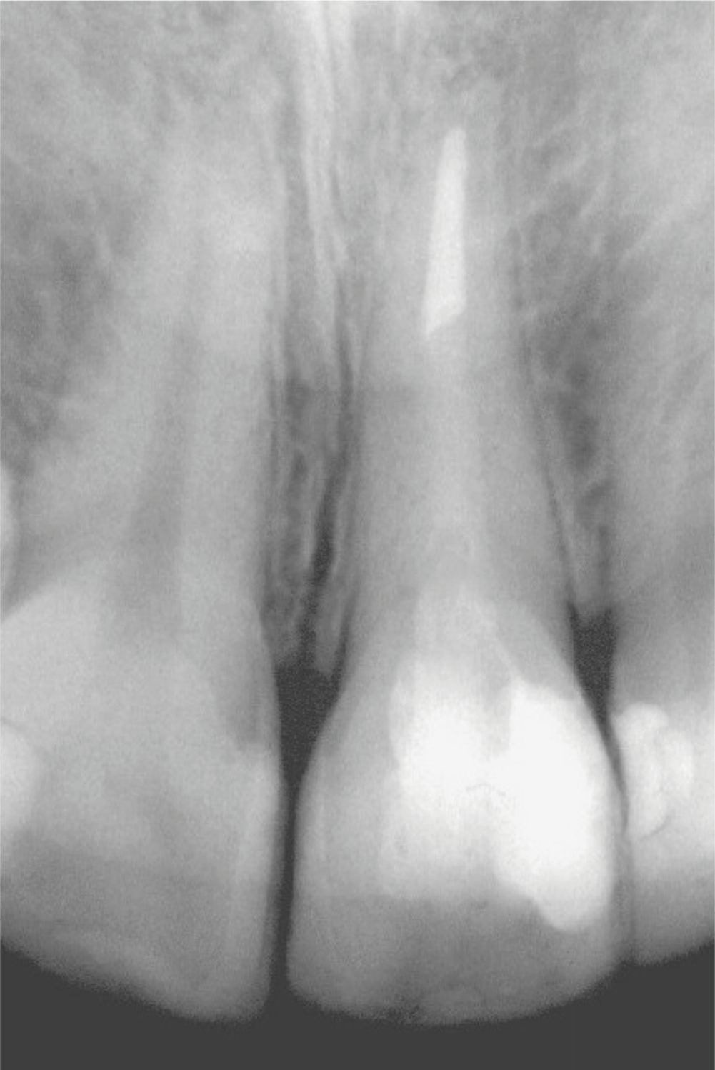
The presence of the endodontic was checked through patients’ clinical records and confirmed by radiographic inspection (Figure 1).

Radiographic image used to confirm the presence and condition of the endodontic treatment and fiberglass post.
Statistics
Data analyzed were the odds ratio of the outcome (fracture) to the exposition factor (with presence). Furthermore, fracture patterns were analyzed by the Mann–Whitney and Fisher exact test, with the significance level set at 95%.
Results
The study included a total of 52 patients (mean age, 43.4 years old). Of the 89 ETT restored with composite veneers were evaluated, 31 had prefabricated fiberglass posts and 58 had no posts. Of the 89 teeth, 30 presented with fractures (Table 3). The time in service of the ETT restored with veneers varies from 3 to 132 (mean, 49) months.
An odds ratio of < 1.0 denotes that the presence of fiberglass posts reduced the fracture risk of ETT restored with composite veneers. Odds ratio analysis showed a 34% of risk to fractures with post compared with the teeth without posts (0.34 times the outcome chance).
The relationship between the restored teeth with or without posts and the outcome (fracture) was analyzed by Fisher’s exact test (Table 3). This test showed significant differences in fracture events (p = 0.0295), which were more frequent in restored teeth without posts.
The different fracture patterns (Figure 2) in the ETT restored with composite veneers with or without posts were analyzed by the Mann–Whitney test (Table 4).

Representative images of the most prevalent failure patterns: a) pattern 1, the chip fracture is pointed by the arrow; b) pattern 3, fracture in the coronal and middle third portions of the crown; and c) pattern 4, fracture involving the coronal and cervical regions.
Discussion
The results revealed a positive influence of the fiberglass post, reducing the risk of fracture to ETT restored with composite veneers, which corroborates with previous studies8, 9. Therefore, the null hypothesis, i.e., the presence of the fiberglass post would not influence the number of fractures and/or fracture patterns in the restored teeth, was rejected.
Despite the observed reduction of the fracture risk, the presence of fiberglass posts has always been associated with retention, not mechanical strength. Some factors, such as tooth structure remains, adhesive luting procedures, and fiber post features (fiberglass and carbon fiber posts), must be carefully evaluated. The ETT usually have a more extensive dental structure loss because of the pulp assessment, restorations, and associated carious lesions. These situations often need complex rehabilitation to avoid higher failure rates.7 In addition, the tooth weakens the remaining structure.19,21 In the anterior teeth, the veneer preparation also reduces stiffness6. Thus, to compensate for the loss of mechanical properties, a post with Young’s modulus closer to that of the dentin and adhesive cementation cemented adhesively in the root dentin walls was developed.22.
In the fracture pattern analysis, a few fractures had patterns 1 and 2, which are probably little or not influenced by the post. The preparation form and tissue loss may have significantly influenced these two types of failure. Both fractures occurred in the veneer buccal fracture (chipping) and incisal edge of the restoration in areas where the dental tissue was replaced by the direct restorative composite, away from the region where the posts are seated. Chipping fractures are associated with the properties of the composite, type of filler particles, silanization, degree of conversion,23 applied force angle, and distance from the edge,24 which may be associated with occlusal interferences.
The fracture patterns associated with the coronal middle (pattern 3) and cervical (pattern 4) thirds were found only in ETT restored without fiberglass posts. These results are consistent with the stress distribution of different dental rehabilitation types seen in a previously published finite-element analysis. The highest stress concentration in sound teeth occurred in the cervical region, close to the cement–enamel density, which can increase the risk of tooth fracture in the case of fatigue, or an increased pressure level. The same study demonstrated that when less rigid coronal restorations associated with fiberglass posts are used, the tension in this region is dissipated, which may reduce the fracture risk, as was found in the present study.25 Given the preparation for resin veneers evaluated in this study, all teeth had at least 50% of the coronal structure with a ferrule. However, previous class III and IV restorations may have contributed to patterns 3 and 4 for teeth without an endodontic post. The more favorable stress distribution induced by the post and adhesive cementation may have influenced these results if patterns 3 and 4 did not occur in teeth with endodontic posts. Some studies have shown that the fiber posts had reduced stress concentration at the root compared with other post systems.13,26,27
The unique coronal and root fracture (pattern 5) was recorded in one tooth restored with a fiberglass post. As a retrospective study, standardizing all factors involved is very tricky. Despite this, for inclusion in the study, the tooth examined presented the same clinical and radiographic parameters as the other elements included in the research, with a filling of 2/3 of the length of the canal and a minimum length equal to the size of the dental crown. This catastrophic failure was probably the result of areas weakened by preparations that could not be detected on radiographs. With greater stress distribution along the post region, some root regions may present stress concentration points in the cement core close to the interface with the dentin when compared with the tooth without posts25. Thus, stress, even at low values, concentrating in a fragile area increases the fracture risk. Given this behavior and the fractures similar to the one described in this sample, the idea that fiberglass reinforces the remaining tooth structure cannot be proven so far.
Despite the clear reduction in the fracture risk in ETT restored with fiberglass posts, this result is an effect of the reinforcement of the remaining structure by fiberglass post because several factors are involved in this treatment. Differences between the post materials and cementation, root canal geometry, angulation and magnitude of the oblique force, extension and material of restoration, and conditions of remaining tissues contribute to the results.13,21,22,25-29 This difficulty is noticeable based on the results of previous studies. Although in vitro studies have indicated that fiberglass posts improve the fracture resistance of the ETT due to better stress distribution,9,13,25-27 these results are not as clear in clinical studies. A randomized controlled trial revealed similar clinical performances of metal and fiberglass posts after 9 years of follow-up,18 which were confirmed by a systematic and meta-analysis that evaluated controlled trials and prospective clinical trials.30 The factors involved may have generated this similarity in the result. The authors highlight the low number of studies with > 5 years of follow-up as a limitation. Even in these in vitro studies, which allow for better standardization, many of the analyzed studies still have high or medium levels of bias,9 making precise analysis and, consequently, obtaining a clearer answer more difficult. These aforementioned points reinforce the already observed need for clinical and even laboratory studies to describe their methods more precisely.
Based on the present findings, within the limitations of this study, intracanal prefabricated glass fiber posts can be used to restore ETT associated with composite veneers to reduce the risk of severe fractures. To certify these findings, more longitudinal and controlled clinical trials are needed, particularly with extended follow-up periods.
Conclusions
The results of this study imply that the presence of post decreased the risk of fractures in ETT restored with composite veneers. Intracanal glass fiber posts could prevent complex fractures in the coronal middle third (pattern 3) and coronal cervical (pattern 4) regions in veneered teeth when fractures occur.
References
-
1 Gomes GH, Corbellini AO, Rotta WG, Martos J, Boeira GF. Interdisciplinary esthetic approach in clinical dental rehabilitation. J Conserv Dent. 2021;24(5):519-23. https://doi.org/10.4103/jcd.jcd_441_21
» https://doi.org/10.4103/jcd.jcd_441_21 -
2 Nicholas LS, Yew Christopher QE, Fei Frank LK. Conservative esthetic management of brown enamel fluorosis using combination therapy: A clinical report. J Conserv Dent. 2023;26(3):349-54. https://doi.org/10.4103/jcd.jcd_632_20
» https://doi.org/10.4103/jcd.jcd_632_20 -
3 Poggio CE, Bonfiglioli R, Dosoli R. A patient presentation: planning and executing a difficult case in a full digital workflow. J Esthet Restor Dent. 2021 Jan;33(1):135-42. https://doi.org/10.1111/jerd.12715
» https://doi.org/10.1111/jerd.12715 -
4 Silva JS, Herkrath A, Pontes DG, Queiroz AC, Medina PO, Herkrath FJ, et al. Multidisciplinary Management of Intrusive Luxation: Four-year Clinical Follow-up. Oper Dent. 2022 Nov;47(6):603-11. https://doi.org/10.2341/21-060-S
» https://doi.org/10.2341/21-060-S -
5 Vieira A, Emerenciano NG, Moda MD, Silva Ú, Fagundes TC, Danelon M, et al. Treatment of Molar-incisor Hypomineralization: A Case Report of 11-year Clinical Follow-up. Oper Dent. 2023 Mar;48(2):121b-129. https://doi.org/10.2341/21-150-S
» https://doi.org/10.2341/21-150-S -
6 Hussain SK, McDonald A, Moles DR. In vitro study investigating the mass of tooth structure removed following endodontic and restorative procedures. J Prosthet Dent. 2007 Oct;98(4):260-9. https://doi.org/10.1016/S0022-3913 (07)60110-3
» https://doi.org/10.1016/S0022-3913 (07)60110-3 -
7 Meijering AC, Creugers NH, Roeters FJ, Mulder J. Survival of three types of veneer restorations in a clinical trial: a 2.5-year interim evaluation. J Dent. 1998 Sep;26(7):563-8. https://doi.org/10.1016/S0300-5712 (97)00032-8
» https://doi.org/10.1016/S0300-5712 (97)00032-8 -
8 D'Arcangelo C, De Angelis F, Vadini M, D'Amario M, Caputi S. Fracture resistance and deflection of pulpless anterior teeth restored with composite or porcelain veneers. J Endod. 2010 Jan;36(1):153-6. https://doi.org/10.1016/j.joen.2009.09.036
» https://doi.org/10.1016/j.joen.2009.09.036 -
9 Jurema AL, Filgueiras AT, Santos KA, Bresciani E, Caneppele TM. Effect of intraradicular fiber post on the fracture resistance of endodontically treated and restored anterior teeth: A systematic review and meta-analysis. J Prosthet Dent. 2022 Jul;128(1):13-24. https://doi.org/10.1016/j.prosdent.2020.12.013
» https://doi.org/10.1016/j.prosdent.2020.12.013 -
10 Salameh Z, Sorrentino R, Ounsi HF, Sadig W, Atiyeh F, Ferrari M. The effect of different full-coverage crown systems on fracture resistance and failure pattern of endodontically treated maxillary incisors restored with and without glass fiber posts. J Endod. 2008 Jul;34(7):842-6. https://doi.org/10.1016/j.joen.2008.03.025
» https://doi.org/10.1016/j.joen.2008.03.025 - 11 Piovesan EM, Demarco FF, Cenci MS, Pereira-Cenci T. Survival rates of endodontically treated teeth restored with fiber-reinforced custom posts and cores: a 97-month study. Int J Prosthodont. 2007;20(6):633-9.
-
12 Ausiello P, Gloria A, Maietta S, Watts DC, Martorelli M. Stress Distributions for Hybrid Composite Endodontic Post Designs with and without a Ferrule: FEA Study. Polymers (Basel). 2020 Aug;12(8):1836. https://doi.org/10.3390/polym12081836
» https://doi.org/10.3390/polym12081836 -
13 Ince Yusufoglu S, Saricam E, Ozdogan MS. Finite element analysis of stress distribution in root canals when using a variety of post systems instrumented with different rotary systems. Ann Biomed Eng. 2023 Jul;51(7):1436-48. https://doi.org/10.1007/s10439-023-03145-w
» https://doi.org/10.1007/s10439-023-03145-w - 14 Vitale MC, Chiesa M, Coltellaro F, Bignardi C, Celozzi M, Poggio C. FEM analysis of different dental root canal-post systems in young permanent teeth. Eur J Paediatr Dent. 2008 Sep;9(3):111-7.
-
15 Al-Wahadni AM, Hamdan S, Al-Omiri M, Hammad MM, Hatamleh MM. Fracture resistance of teeth restored with different post systems: in vitro study. Oral Surg Oral Med Oral Pathol Oral Radiol Endod. 2008 Aug;106(2):e77-83. https://doi.org/10.1016/j.tripleo.2008.03.038
» https://doi.org/10.1016/j.tripleo.2008.03.038 -
16 Martelli H Jr, Pellizzer EP, Rosa BT, Lopes MB, Gonini A Jr. Fracture resistance of structurally compromised root filled bovine teeth restored with accessory glass fibre posts. Int Endod J. 2008 Aug;41(8):685-92. https://doi.org/10.1111/j.1365-2591.2008.01424.x
» https://doi.org/10.1111/j.1365-2591.2008.01424.x - 17 Naumann M, Sterzenbac G, Alexandra F, Dietrich T. Randomized controlled clinical pilot trial of titanium vs. glass fiber prefabricated posts: preliminary results after up to 3 years. Int J Prosthodont. 2007;20(5):499-503.
-
18 Sarkis-Onofre R, Amaral Pinheiro H, Poletto-Neto V, Bergoli CD, Cenci MS, Pereira-Cenci T. Randomized controlled trial comparing glass fiber posts and cast metal posts. J Dent. 2020 May;96:103334. https://doi.org/10.1016/j.jdent.2020.103334
» https://doi.org/10.1016/j.jdent.2020.103334 -
19 Baratieri LN, Andrada MA, Arcari GM, Ritter AV. Influence of post placement in the fracture resistance of endodontically treated incisors veneered with direct composite. J Prosthet Dent. 2000 Aug;84(2):180-4. https://doi.org/10.1067/mpr.2000.108415
» https://doi.org/10.1067/mpr.2000.108415 -
20 Schulz KF, Grimes DA. Case-control studies: research in reverse. Lancet. 2002 Feb;359(9304):431-4. https://doi.org/10.1016/S0140-6736 (02)07605-5
» https://doi.org/10.1016/S0140-6736 (02)07605-5 -
21 Coelho-De-Souza FH, Camacho GB, Demarco FF, Powers JM. Fracture resistance and gap formation of MOD restorations: influence of restorative technique, bevel preparation and water storage. Oper Dent. 2008;33(1):37-43. https://doi.org/10.2341/07-27
» https://doi.org/10.2341/07-27 -
22 Trushkowsky RD. Esthetic and functional consideration in restoring endodontically treated teeth. Dent Clin North Am. 2011 Apr; 55(2):403-10, x. https://doi.org/10.1016/j.cden.2011.01.009
» https://doi.org/10.1016/j.cden.2011.01.009 -
23 Zadeh PN, Stawarczyk B, Hampe R, Liebermann A, Mayinger F. Edge chipping resistance of veneering composite resins. J Mech Behav Biomed Mater. 2021 Apr;116:104349. https://doi.org/10.1016/j.jmbbm.2021.104349
» https://doi.org/10.1016/j.jmbbm.2021.104349 - 24 Quinn GD, Quinn GD. Fractography of ceramics and glasses. Washington, DC: National Institute of Standards and Technology; 2007.
-
25 Zarone F, Sorrentino R, Apicella D, Valentino B, Ferrari M, Aversa R, et al. Evaluation of the biomechanical behavior of maxillary central incisors restored by means of endocrowns compared to a natural tooth: a 3D static linear finite elements analysis. Dent Mater. 2006 Nov;22(11):1035-44. https://doi.org/10.1016/j.dental.2005.11.034
» https://doi.org/10.1016/j.dental.2005.11.034 -
26 Badami V, Ketineni H, Pb S, Akarapu S, Mittapalli SP, Khan A. Comparative evaluation of different post materials on stress distribution in endodontically treated teeth using the finite element analysis method: a systematic review. Cureus. 2022 Sep;14(9):e29753. https://doi.org/10.7759/cureus.29753
» https://doi.org/10.7759/cureus.29753 -
27 Kharboutly NA, Allaf M, Kanout S. Three-dimensional finite element study of endodontically treated maxillary central incisors restored using different post and crown materials. Cureus. 2023 Jan;15(1):e33778. https://doi.org/10.7759/cureus.33778
» https://doi.org/10.7759/cureus.33778 -
28 Carvalho MA, Lazari PC, Gresnigt M, Del Bel Cury AA, Magne P. Current options concerning the endodontically-treated teeth restoration with the adhesive approach. Braz Oral Res. 2018 Oct;32 suppl 1:e74. https://doi.org/10.1590/1807-3107bor-2018.vol32.0074
» https://doi.org/10.1590/1807-3107bor-2018.vol32.0074 -
29 Skupien JA, Cenci MS, Opdam NJ, Kreulen CM, Huysmans MC, Pereira-Cenci T. Crown vs. composite for post-retained restorations: a randomized clinical trial. J Dent. 2016 May;48:34-9. https://doi.org/10.1016/j.jdent.2016.03.007
» https://doi.org/10.1016/j.jdent.2016.03.007 -
30 Martins MD, Junqueira RB, de Carvalho RF, Lacerda MF, Faé DS, Lemos CA. Is a fiber post better than a metal post for the restoration of endodontically treated teeth? A systematic review and meta-analysis. J Dent. 2021 Sep;112:103750. https://doi.org/10.1016/j.jdent.2021.103750
» https://doi.org/10.1016/j.jdent.2021.103750