 Meloxicam Inhibited the Glycation Phenomenon via Scavenging Di-Carbonyl Moieties
Abstract
Senescence or aging is often associated with onset of morbidities, which have been attributed to the harmful phenomenon of glycation. To date, no drug is available to combat this deleterious process. The drug repurposing is a robust and cost-effective approach to identify potential candidate molecules for drug discovery programs. In present study, Meloxicam (MEL) was evaluated for its capacity to be repurposed against glycation. Using the Fructose-BSA model, the anti-glycation effect was evaluated by measuring intrinsic fluorescence of Advanced Glycation End Products (AGEs). The fructoseamine load and free lysine availability was estimated using NBT and TNBSA assays respectively. The BSA secondary structure was assessed by Thioflavin-T, Congo red and Circular Dichroism tests. Finally, lysine blockade and carbonyl entrapment was evaluated as a possible mode of anti-glycation action. Our data showed that MEL (0.5, 1, and 2mM) has significantly reduced the AGEs formation (IC50 = 0.25mM). The load of fructosamine adducts along with free lysine availability was found to be reduced. The secondary structure of BSA was preserved. Regarding mode of action, MEL did not block lysine residues, which was also supported by computational data. However, it was found to entrap carbonyl intermediates. In conclusion, the present study demonstrate that MEL possess anti-glycation potential, which can be attributed to entrapment of carbonyl intermediates. Hence, MEL, a clinically used NSAIDs, present itself as promising candidate to be repurposed against deleterious phenomenon of glycation.
Keywords:
Glycation; Meloxicam; Carbonyl entrapping; Fructose; BSA
INTRODUCTION
Aging and associated morbidities have been linked to various pathological mechanisms (Campisi et al., 2019). One such phenomenon is termed as glycation, which states that aging co-relates with load of advanced glycation end products (AGEs) in the body (Anguizola et al., 2013). Literature suggests that AGEs contribute to the progressive decline in cellular functions thereby leading to the loss of bodily functions over time (Chaudhuri et al., 2018). Glycation (also known as Maillard reaction) is a non-enzymatic interaction between carbohydrates (carbonyl groups) and proteins, which are rich in lysine and arginine residues (Srikanth et al., 2011). The reaction initially results in the formation of reversible Schiff bases, which later progress to more stable Amadori products. Subsequent irreversible dehydration, oxidation, polymerization, and cross-linking reactions lead to the formation of AGEs (Srikanth et al., 2011).
Glycation occurs ubiquitously in cells but is heightened in various pathological conditions, particularly hyperglycemia (Biedrzycki et al., 2023). AGEs load have been implicated in cognitive impairment, diabetic complications, renal, and hepatic insufficiencies. Furthermore, AGEs upon binding to its receptor (RAGE) triggers set the inflammatory processes collectively termed glycative stress (Rungratanawanich et al., 2021). Among the various di-carbonyl compounds, the Methylglyoxal (MGO) is the predominant precursor of AGEs (Chaudhuri et al., 2018). Accumulation of MGO occurs in conditions of hyperglycemia, impaired glucose metabolism, and oxidative stress (Allaman et al., 2015). This MGO reacts with proteins, DNA, and other biomolecules, resulting in the formation of AGEs (Schalkwijk, Stehouwer, 2020). Consequently, MGO accumulation contributes aging, diabetic complications (Yeh et al., 2017) and cognitive impairments (Shen et al., 2020).
Numerous efforts were made to develop the inhibitors of glycation. Unfortunately, to date, no drug reached the market. A notable example is Aminoguanidine (AG), which demonstrated some success in scavenging dicarbonyl intermediates (Rowan et al., 2018). However, its clinical utility was hindered by occurrence of adverse events. Under such circumstances, the drug repurposing presents a robust and economical approach for identifying candidate molecules for anti-glycation drug discovery programs (Pushpakom et al., 2019).
The concept of structure-activity relationship plays a crucial role in the preliminary identification of lead molecules for a drug discovery program. In this regard, the sulfur-containing compounds have been reported to possess anti-glycation properties (Younus, Anwar, 2016) via modulation of pathways involved in the formation of AGEs (Perrone et al., 2020). Considering this, MEL was picked from the arsenal of clinically used drugs for present study. It belongs to the class of “oxicams”, which contain a Thiazole ring. It is a five-membered aromatic heterocyclic compound containing nitrogen and sulfur atoms (Barnette et al., 2020), along with a cyclic tertiary sulfonamide moiety (De Monte et al., 2015). These chemical attributes are believed to endow anti-glycation potential to MEL. Keeping this into account, the current study aimed to investigate the anti-glycation potential of MEL (Figure 1), which is currently a clinically used NSAID (Luger et al., 1996).

MATERIAL AND METHODS
Chemicals
The chemicals used in the study are as follows: Acetic acid, formic acid, sodium azide, sodium carbonate, sodium dihydrogen phosphate, sodium hydrogen phosphate, β-mercapethanol, 2, 4, 6-trinitrobenzenesulfonic acid (TNBSA), fructose, nitroblue tetrazolium (NBT), O-phenyldiamine, and phosphate buffer saline were obtained from Sigma-Aldrich. AG and N-α-acetyl-L-lysine were obtained from Chemcruz. Bovine serum albumin (BSA) originated from Imumed®. Congo Red (CR) and sodium bicarbonate were acquired from Bio Basic Inc., (Canada). Dimethyl sulfoxide (DMSO) was obtained from Merck. Ethanol and methanol were provided by Serva (Germany). Meloxicam was a gift from Hilton Pharma (Pakistan). 2-MethylQuinoxaline (2-MQ) and O-phthaldialdehyde 98% (OPA) were procured from Alfa Aesar. Sodium dodecyl sulfate (SDS) was obtained from Kanto Chemical (Japan). Sodium thiopental came from Abbott Laboratories, Pakistan. Thioflavin T (ThT) was sourced from Santa-Cruz Biotechnology.
It is noteworthy that this study is part of a larger project aimed at testing multiple drugs for their potential to be repurposed against glycation. A portion of this work, including data on the vehicle and positive control (Ahmed et al., 2024), has been previously published and is also utilized in this manuscript.
AGEs Inhibition Assay
The experiment was performed as described earlier with some minor adaptations (Khan et al., 2017). The reaction mixture was prepared by mixing bovine serum albumin (BSA, 10 mg/ml) with fructose (100 mM) in a phosphate buffer (0.2 M, pH 7.4) supplemented with sodium azide (0.1%). AG (5 mM) and MEL (0.5, 1, and 2 mM) was added positive control and test group vials. The mixtures were subjected to heating (60°C) for 24 hours followed by assessment of fluorescence intensity (excitation and emission lambda of 360/40 and 460/40 nm respectively) using spectrofluorometer (JASCO, Japan).
Fructosamine Adduct Assay
The quantification of Amadori (fructosamine) adducts present in aforementioned reaction mixtures were estimated using NBT assay (Arfat et al., 2014). Samples (100 µl each) from native BSA, glycated BSA, AG and MEL were separately mixed with sodium carbonate buffer (100 mM, pH 10.35, 1000 µl) containing NBT (0.25 mM). After incubation for 2 hours in darkness, the absorbance (525 nm) was determined using a spectrophotometer (JASCO, Japan).
TNBSA Assay
Quantification of available free amino groups was determined via TNBSA assay (2,4,6-trinitrobenzenesulfonic acid) as described earliers (Xie, Chen, 2013). The TNBSA solution was prepared by combining 0.01% w/v of TNBSA in sodium bicarbonate buffer (0.1 M, pH 8.5). Briefly, the protein (15 µg) from each sample was mixed with sodium bicarbonate buffer (250 µl). After incubation for 2 hours, the absorbance (335 nm) was measures using spectrophotometer (JASCO, Japan).
Protein Conformation Assays
Thioflavin-T Test
The assay will be performed as described earlier with slight modification (Pang et al., 2020). A stock solution of Thioflavin-T (ThT) was prepared in dark by diluting 100 µM of ThT in 5 ml of phosphate buffer (pH 7.3). Subsequently, 20 µM (160 µl) of ThT stock solution was combined with 1 µM (40 µl) of each protein sample and incubated for one hour. Following incubation, fluorescence intensity were measured (excitation and emission lambda of 460 nm and 485 nm) using spectrofluorometer (JASCO, Japan).
Congo Red Assay
Congo red (CR) assay was performed as reported earlier (Almeida, Brito, 2020). A stock solution of 12.5 µM dye was prepared in Tris buffer (pH 7.3). Briefly, 40 µl CR stock (0.5 µM) was mixed with 1.6 mg (160 µl) of protein samples. After keeping samples for 20 minutes at room temperature, the absorbance (530 nm) was taken using spectrophotometer (JASCO, Japan).
Circular Dichroism
The secondary structure of BSA in samples was evaluated using CD as described earlier (Schalkwijk, Stehouwer, 2020). Using spectropolarimeter (JASCO, Japan), the protein samples were scanned (4x) within the Far-UV amide region (190–250 nm) and the UV region (250–400 nm) at ambient temperature. Later, the CD spectral analysis was performed using the Dichro Web server thereby enriching the precision and depth of the structural evaluation (Shahidi, Ambigaipalan, 2015).
Mechanistic Assays
Lysine Blockade Assay
The ability of MEL to block of reactive lysine was assessed through the OPA assay (Goodno et al., 1981). The OPA reagent was formulated by combining specific components in an amber-colored reagent bottle. For a 100 ml reagent, the constituents included o-phthalaldehyde (80 mg) dissolved in pure ethanol (2 ml). To this mixture, 50 ml of 0.1 M sodium tetraborate alkaline buffer (pH: 10), 5 ml of 20% (w/w) sodium dodecyl sulfate (SDS), and 200 µl of β-mercaptoethanol were added. All reagents were thoroughly mixed and stored in an airtight dark bottle. The test involved mixing 25 µg of BSA (50 µl) with freshly prepared OPA reagent (3 ml) and allowed to stay at room temperature for 20 minutes. Subsequently, the fluorescence (excitation at 360 nm and emission at 460 nm, respectively) was measured using a spectrofluorometer (JASCO, Japan).
Carbonyl Entrapping Assay
The ability of MEL to entrap carbonyl intermediated was assessed by method reported earlier (Mesías et al., 2013). The stock solution (0.4 mg/ml) of Methylglyoxal (MGO) was prepared in sodium phosphate buffer (0.1 M, pH 7.4). The derivatizing agent (o-phenylenediamine, OPD) was dissolved in pure methanol (10.8 mg/ ml). The 5-Methylquinoxaline (5-MQ) served as an internal standard. The 2-Methylquinoxaline (2-MQ) was dissolved in 50% methanol at the strength of 1 mg/ ml. AG was used as a positive control. An aliquot of MGO was combined with PBS (850 µl) and 5-MQ (50 µl). Test compounds (0.5, 1, and 2 mM) were prepared. After thorough mixing, samples were heated 60°C for 24 hours. Post-incubation, 200 µl of OPD solution was added to each vial, vortexed for five seconds, and incubated for 30 minutes in the dark to complete the derivatization reaction. The quantification (AUC) 2-MQ (derivatization product) obtained in each sample was done using High-performance liquid chromatography (HPLC). The setup consisted of Shimadzu Prominence HPLC system equipped with a pump (LC-20A), autosampler (SIL-20A), diode array UV detector (SPD-M20A), and a communicating bus module (CBM-20A) connecting hardware to the LC solutions software. Chromatography separation utilized a Hibar® 250, 4-6 LiChrospher® RP 18e (5 µM) column preceded by a guard column (nucleosil 100-5 C18). The mobile phase consisted of 50% methanol and 5% glacial acetic acid. Prior to injection (10 µl), each sample was filtered using a syringe filter (0.22 µm). A flow rate of 0.5 ml/min was maintained.
Computational Study
The 3D crystal structure of BSA (PBB ID: 4F5S) was retrieved by protein data bank. The 2D of MEL was downloaded using PUBCHEM as SDF files and was transformed to 3D format by PYMOL 3.11. Finally, the molecular docking was performed using Auto dock vina 1.1.2 (Trott, Olson, 2010). Both the prepared Receptor and ligands were transformed to pdbqt format respectively for the docking. The active sites were predicted by Prank web database and the grid was set as x = 100, y = 100 and z = 100 along with the coordinate sizes x= 4.368, y = 16.991 and z = 106.819, respectively. Once docking was complete, the most stable confirmation of ligand-protein interaction was used for the analysis of docking results with the help of Biovia Discovery tool 2021 client.
Statistical Analysis
The data is presented as mean ± standard error of the mean (n=3). The statistical differences among means were done by One-way ANOVA, followed by LSD (Least Significant Difference) using IBM SPSS 21.0 software. A minimum level of significance level was set at p<0.05.
RESULTS
AGE Inhibition Assay
The fluorescence intensity exhibited a significant (p<0.005) increase in gBSA group as compared to nBSA (Figure 2). Treatment with AG (5mM) and MEL (0.5, 1 and 2mM) demonstrated a decrease (IC50 ~ 0.25mM) in fluorescence as compared to gBSA.

The figure shows the formation of advance glycation end products (as intrinsic fluorescence) in the presence of AG and MEL. The gBSA group exhibit enhanced fluorescence as compared to nBSA. The AG and MEL has significantly reduced the formation of AGEs. The hash (###) represents the significant (p<0.005) difference as compared to nBSA while asterisks [* (p<0.05), ** (p<0.01) and *** (p<0.005)] represents the statistical comparison with the gBSA. All values are expressed as mean ± SEM of intrinsic AGEs fluorescence intensity (n=3).
Estimation of Fructosamine Adducts
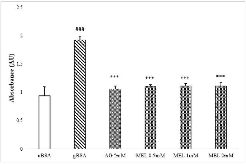
Glycation, a multi-step phenomenon, initiates with the formation of fructosamine adducts. The absorbance was found to be significantly (p<0.005) elevated in gBSA group as compared to nBSA (Figure 3). Our data further demonstrates a significantly diminished load of these adducts in reaction mixtures treated with MEL (0.5, 1, and 2 mM) in comparison to the glycated control (Figure 3).

The figure shows the mean ± SEM of Fructosamine adducts absorbance in the presence of AG and MEL (n=3). The gBSA group exhibit enhanced formation of fructosamine adducts as compared to nBSA. The AG and MEL (0.5 mM) has significantly reduced the formation of these adducts as compared to the gBSA. The hash (###) represents the significant (p<0.005) difference as compared to nBSA while asterisks [* (p<0.05), ** (p<0.01) and *** (p<0.005)] represents the statistical comparison with the gBSA.
TNBS Assay
The gBSA shows significant (p<0.005) decrease in absorbance as compared to nBSA (Figure 4). The presence of AG (5mM) and MEL (0.5, 1 and 2 mM) demonstrated a significant increase in absorbance as compared to gBSA.

The figure shows mean ± SEM of absorbance (indicative of free lysine availability) in the presence of AG and MEL. The hash (###) indicates a significant (p<0.005) difference compared to nBSA, while asterisks [* (p<0.05), ** (p<0.01), and *** (p<0.005)] represent statistical comparisons with gBSA. All values are presented as mean ± SEM of TNBS assay absorbance (n=3).
Protein Conformation Assays
Thioflavin T Assay
In comparison to nBSA, the gBSA exhibited significantly (p<0.005) enhanced fluorescence intensity (Figure 5a). Treatment with AG (5mM) significantly decreased fluorescence as compared to gBSA. The MEL (0.5, 1 and 2mM) demonstrated a decrease in fluorescence too as compared to gBSA but it was found to be increasing with dose i.e. the highest tested dose caused minimal significant different (p<0.05).

The figure illustrates the mean ± SEM of ThT fluorescence intensity (n=3). The gBSA shows significantly increased fluorescence as compared to nBSA. However, the AG and MEL treatments caused significant reduction in ThT fluorescence. It is of note that lowest tested dose of MEL (0.5 mM) caused maximal reduction in ThT fluorescence. The hash (###) denotes a significant (p<0.005) difference compared to nBSA, while asterisks [* (p<0.05), ** (p<0.01), and *** (p<0.005)] indicate statistical comparisons with gBSA.
Congo Red Assay
The absorbance was found to be significantly (p<0.005) increased in gBSA group as compared to nBSA (Figure 5b). Treatment with AG (5mM) and MEL (0.5, 1 and 2 mM) demonstrated a significant (p<0.005) decrease in absorbance as compared to gBSA.

The figure depicts the mean ± SEM (n=3) of Congo red absorbance under various treatments. A significant increase in gBSA group absorbance was noted as compared to nBSA. The AG (5mM) as well as MEL (0.5, 1, and 2 mM) treatments caused significant decrease in the absorbance as compared to gBSA. The hash sign (###) denotes a statistical difference (p < 0.005) compared to nBSA, while asterisks (***) indicate a statistical difference (p < 0.005) compared to gBSA.
Circular Dichorism
The nBSA showed the characteristic spectra with two lambda max at 208 and 222 nm, while the spectra of gBSA exhibited single lambda max at 218 nm (Figure 5c). The spectra’s of AG and MEL (0.5, 1 and 2 mM) are similar to that of nBSA.

The figure illustrates the CD analysis of BSA subjected to glycation in the presence and absence of Meloxicam or AG. The native BSA (nBSA) showed its distinctive spectra, characterized by two lambda max at 208 and 222 nm. In contrast, the glycated BSA (gBSA) displayed a solitary lambda maximum at 218 nm. Exposure to AG and MEL yielded BSA spectra similar to that of nBSA.
Mechanistic Assays
Lysine Blockade Assay
Our data indicate that none of the treatment groups caused a significant change in fluorescence intensity (Figure 6a). This suggests that neither AG nor MEL possess the ability to interfere with the initial fructose-BSA (lysine) interaction.

The figure exhibit mean ± SEM of fluorescence intensity in the presence of various treatments (n=3). The OPA reacts with free primary amines, such as lysine, to produce fluorescent derivative. If a compound blocks lysine residues, it reduce free amines on BSA surface. Presumably, the OPA fluorescence reduced. Our data showed that none of the treatment i.e. AG (5mM) and MEL (0.5, 1 or 2mM) caused significant changes in fluorescence as compared to control thereby suggesting lack of lysine blockade by the test agents.
Computational Study
Molecular docking studies indicated that MEL interacted with various BSA residues i.e. GLY, HIS, ILE, LYS, PHE, TRP, and VAL within chain A (pink), possessing binding energies of around -7.6 kcal/mol (Figure 6b).

The figure depicts the interaction of MEL with BSA (PBB ID: 4F5S) using computational tool (Autodock vina). The interacting residues were found to be GLY, HIS, ILE, LYS, PHE, TRP, and VAL.
Carbonyl Entrapping Assay
The AG (5mM) caused 76% inhibition in the AUC (area under the curve) of 2-MQ peak. The MEL treatment showed the inhibition of 98%, 94% and 92% at the tested doses of 0.5, 1 and 2 mM respectively. The representative HPLC chromatograms depicting the distinctive profiles of various treatment groups are shown in Figure 6c.

The figure illustrates representative chromatograms obtained from the carbonyl entrapping assay conducted in the presence of AG and MEL. The 5-MQ acts as an internal standard, while 2-MQ is produced as a result of OPD-mediated derivatization reaction of MGO. In case, the MGO is entrapped by test agent, it is not available for derivatization to 2-MQ thereby resulting in its diminished peak, as observed following treatments with AG (5mM) and MEL (0.5, 1 and 2 mM).
DISCUSSION
Glycation has been reported to play a pivotal role in morbidities linked with aging and diabetes mellitus. Currently, no treatment option is available to counteract this harmful phenomenon. Repurposing offers a robust and cost-effective approach to introduce a new molecule into the drug discovery process. Considering this into account, the present study investigates the anti-glycation potential of MEL, a clinically used NSAID.
The concepts of medicinal chemistry play a crucial role in the identification of lead molecule(s) for a drug discovery program. In this regard, the sulfur-containing compounds have been reported to possess anti-glycation properties (Younus, Anwar, 2016) through modulation of various pathways involved in the formation of AGEs (Perrone et al., 2020). Keeping this in view, the MEL was chosen from existing clinically used drugs for this study. It belongs to the class of “oxicams”. It cotains five-membered aromatic heterocyclic thiazole ring with nitrogen and sulfur atoms (Barnette et al., 2020) along with a cyclic tertiary sulfonamide moiety (De Monte et al., 2015). Taken together, these chemical attributes makes MEL a likely molecule to possess anti-glycation potential. Our experimental approach adopts a well-established and validated model of BSA-fructose (Biedrzycki et al., 2023). Bovine serum albumin (BSA) stands as the pre-eminent protein for in vitro investigations, owing to its substantial homology with human albumin, elevated purity, and the stability inherent in its prosthetic groups (Raut, Khullar, 2023). Our preliminary data shows that MEL (0.5, 1, and 2 mM), in similarity with standard AG, has the ability to inhibit the formation of AGEs (Figure 2). Hence, our SAR based selection was found to be effective (Spasov et al., 2021).
Glycation, a multi-step phenomenon, initiates with the formation of fructosamine adducts. Our data (NBT assay) demonstrates a significantly diminished load of these adducts in reaction mixtures treated with MEL (0.5, 1, and 2 mM) in comparison to the glycated control (Figure 3). The graph shows that the lowest tested dose of MEL was most effective in reducing this burden; the notion worthy of further investigations (Younus, Anwar, 2016).
Moreover, the progression of glycation involves the interaction of fructose with the lysine amino acid within BSA (Arasteh et al., 2014). Lysine residue 524 of BSA (equivalent to 525 in human serum albumin) is considered to be the primary participant in the glycation process through interaction with fructose (Rabbani, Ahn, 2019). To assess the availability of free lysine in BSA, the TNBSA assay was conducted. Our results indicate a significant reduction in lysine residues in glycated samples compared to the control (Figure 4). This suggests the engagement of these residues in mediating the reaction of glycation. Remarkably, MEL-treated samples exhibits an augmented availability of free lysine relative to glycated samples. This reinforces the proposition that meloxicam possesses the capability to intervene in the glycation process.
Bovine serum albumin (BSA), a 66 kDa protein, is primarily characterized by its α-helical structure (Babcock, Brancaleon, 2013). Glycation is believed to alter its secondary structures to ordered amyloid-like β-sheets (Zaman et al., 2019). In order to assess these conformational changes, the ThT and Congo red dyes were used (Biancalana, Koide, 2010). The ThT dye, known for its specificity in binding with amyloid-like aggregates, revealed elevated fluorescence intensity in AGE samples (Figure 5a). However, the presence of MEL and AG resulted in decreased fluorescence suggesting lower load of amyloid-like aggregates. In similar lines, the CR data also revealed higher load of protein aggregates in gBSA as compared to nBSA, which was reduced by treatment with MEL and AG (Figure 5b). Hence, it can be deduced that MEL has the ability to reduces amyloid-like aggregates formation in glycation prone environment (Awasthi, Saraswathi, 2015).
Circular Dichorism stands as a reliable methodology for the discernment of protein secondary structures. The native BSA produce a discernible positive band at 192 nm and two negative bands around 208 and 222 nm (Ranjbar, Gill, 2009), a characteristic spectra suggestive of an alpha-helical rich structure. Our data also showed similar spectra in nBSA samples (Figure 5c). Moreover, in the glycated BSA (gBSA) samples, a notable shift towards alterations in protein conformation from alpha helical to beta sheet was evidenced by positive bands around 195 nm and a distinct negative band at 218 nm. It is noteworthy that our CD spectra analysis revealed that AG and Meloxicam treated samples exhibited spectral profiles similar to that of nBSA. This underscores their capacity to uphold the alpha helical integrity of BSA within an environment susceptible to glycation.
Lysine blockade and carbonyl trapping represent two crucial pharmacological targets for anti-glycation compounds (Chen et al., 2018). Therefore, both mechanisms were assessed to elucidate the potential mode of action of MEL. Our data from the lysine blockade assay (OPA assay) revealed an absence of such activity following AG treatment (Figure 6a). This finding aligns with existing literature, as AG is a well established carbonyl entrapper (Khan et al., 2020). This validates our methodology as well. It is noteworthy that MEL also did not exhibit any lysine-blocking activity at any of the tested doses. The lysine blockade activity was further evaluated using a computational approach. Molecular docking studies indicated that MEL interacted with various BSA residues i.e. GLY, HIS, ILE, LYS, PHE, TRP, and VAL within chain A (Figure 6b). A review of the literature revealed that fructose binds to specific catalytic residues in BSA, particularly LYS-524, to initiate the glycation reaction (Mou et al., 2022; Qin et al., 2021). Our computational data demonstrate that MEL failed to interact with this catalytic residues of BSA (Figure 6b). This finding corroborates the absence of lysine blockade as a potential mechanism for the anti-glycation action of MEL.
The methylglyoxal (MGO) trapping assay was also employed to explore the potential anti-glycation mechanism of MEL (Ahmed, Thornalley, 2005). Dicarbonyl intermediates, specifically methylglyoxal (MGO) and glyoxal (Campisi et al., 2019), are pivotal contributors to the glycation process (Schalkwijk, Stehouwer, 2020). Entrapping these deleterious compounds represents a significant pharmacological target for impeding glycation. In our study, AG demonstrated a significant reduction (76%) in the AUC of 2-MQ peak (Figure 6c). This is suggestive of the entrapment of MGO. This result further validates our experimental conditions, as AG is a well-known carbonyl entrapping agent (Goh, Cooper, 2008). It is of note that MEL (0.5, 1, and 2 mM) was also found to significantly entrap MGO (≥ 90%). Literature revealed that scavengers of reactive oxygen species also exhibit the ability to neutralize reactive carbonyl species (RCS), thereby inhibiting the glycation process (Jomova et al., 2023, Singh et al., 2014). In this context, MEL has been previously reported to exhibit antioxidant activity (Pawlukianiec et al., 2020, Samra et al., 2023), which further substantiates its carbonyl trapping activity observed in the current study. The carbonyl moieties generated during the glycation reaction offer a promising pharmacological target. Compared to established anti-glycation agents like AG, MEL presents the advantage of a well-characterized safety profile and dual anti-inflammatory and anti-glycation activity. However, further comparative studies are warranted to fully elucidate its efficacy and clinical relevance in glycation-associated complications. Here, it is further elaborated that the absence of lysine blockade by MEL suggests a more targeted mode of action thereby potentially minimizing probable interference with normal protein function. This distinction may be relevant, when considering safety and specificity in therapeutic applications.
The present study deduced that MEL has the ability to inhibit the phenomenon of glycation, which can most likely be attributed to scavenging of di-carbonyl compounds. Hence, MEL, a clinically used NSAID, presents itself as a promising candidate to be repurposed as anti-glycation agent.
ACKNOWLEDGEMENTS:
Declared None.
Abbreviations:
- AGEs Advanced Glycation End Products
- AG Aminoguanidine
- BSA Bovine Serum Albumin
- CD Circular Dichroism
- gBSA Glycated BSA
- MEL Meloxicam
- nBSA Native BSA
- NBT Nitroblue Tetrazolium
- TNBSA 2,4,6-Trinitrobenzene Sulfonic Acid
-
FUNDING:
None.
DATA AVAILABILITY STATEMENT
Use of data not disclosed
REFERENCES
- Ahmed H, Fayyaz TB, Khatian N, Usman S, Nisar U, Abid M, et al. Phloroglucinol inhibited glycation via entrapping carbonyl intermediates. PLoS One. 2024;19:e0307708.
- Ahmed N, Thornalley PJ. Peptide mapping of human serum albumin modified minimally by methylglyoxal in vitro and in vivo. Ann N Y Acad Sci. 2005;1043:260-6.
- Allaman I, Bélanger M, Magistretti PJ. Methylglyoxal, the dark side of glycolysis. Front Neurosci. 2015;9:23.
- Almeida ZL, Brito RM. Structure and aggregation mechanisms in amyloids. Molecules. 2020;25:1195.
- Anguizola J, Matsuda R, Barnaby OS, Hoy K, Wa C, Debolt E, et al. Glycation of human serum albumin. Clin Chim Acta. 2013;425:64-76.
- Arasteh A, Farahi S, Habibi-rezaei M, Moosavi-movahedi AA. Glycated albumin: an overview of the in vitro models of an in vivo potential disease marker. J Diabetes Metab Disord. 2014;13:49.
- Arfat MY, Ashraf JM, Arif Z, Alam K. Fine characterization of glucosylated human IgG by biochemical and biophysical methods. Int J Biol Macromol. 2014;69:408-15.
- Awasthi S, Saraswathi N. Silybin, a flavonolignan from milk thistle seeds, restrains the early and advanced glycation end product modification of albumin. RSC Adv. 2015;5:87660-5.
- Babcock JJ, Brancaleon L. Bovine serum albumin oligomers in the E-and B-forms at low protein concentration and ionic strength. Int J Biol Macromol. 2013;53:42-53.
- Barnette DA, Schleiff MA, Osborn LR, Flynn N, Matlock M, Swamidass SJ, et al. Dual mechanisms suppress meloxicam bioactivation relative to sudoxicam. Toxicology. 2020;440:152478.
- Biancalana M, Koide S. Molecular mechanism of Thioflavin-T binding to amyloid fibrils. Biochim Biophys Acta Proteins Proteomics. 2010;1804:1405-12.
- Biedrzycki G, Wolszczak-Biedrzycka B, Dorf J, Michalak D, Zendzian-Piotrowska M, Zalewska A, et al. Antioxidant and anti-glycation potential of H2 receptor antagonists—in vitro studies and a systematic literature review. Pharmaceuticals. 2023;16:1273.
- Campisi J, Kapahi P, Lithgow GJ, Melov S, Newman JC, Verdin E. From discoveries in ageing research to therapeutics for healthy ageing. Nature. 2019;571:183-92.
- Chaudhuri J, Bains Y, Guha S, Kahn A, Hall D, Bose N, et al. The role of advanced glycation end products in aging and metabolic diseases: bridging association and causality. Cell Metab. 2018;28:337-52.
- Chen JH, Lin X, Bu C, Zhang X. Role of advanced glycation end products in mobility and considerations in possible dietary and nutritional intervention strategies. Nutr Metab (Lond). 2018;15:72.
- De Monte C, Carradori S, Gentili A, Mollica A, Trisciuoglio D, Supuran CT. Dual cyclooxygenase and carbonic anhydrase inhibition by nonsteroidal anti-inflammatory drugs for the treatment of cancer. Curr Med Chem. 2015;22:2812-8.
- Goh SY, Cooper ME. The role of advanced glycation end products in progression and complications of diabetes. J Clin Endocrinol Metab. 2008;93:1143-52.
- Goodno CC, Swaisgood HE, Catignani GL. A fluorimetric assay for available lysine in proteins. Anal Biochem. 1981;115:203-11.
- Jomova K, Raptova R, Alomar SY, Alwasel SH, Nepovimova E, Kuca K, et al. Reactive oxygen species, toxicity, oxidative stress, and antioxidants: Chronic diseases and aging. Arch Toxicol. 2023;97:2499-574.
- Khan M, Liu H, Wang J, Sun B. Inhibitory effect of phenolic compounds and plant extracts on the formation of advance glycation end products: A comprehensive review. Food Res Int. 2020;130:108933.
- Khan SA, Haider A, Mahmood W, Roome T, Abbas G. Gamma-linolenic acid ameliorated glycation-induced memory impairment in rats. Pharm Biol. 2017;55:1817-23.
- Luger P, Daneck K, Engel W, Trummlitz G, Wagner K. Structure and physicochemical properties of meloxicam, a new NSAID. Eur J Pharm Sci. 1996;4:175-87.
- Mesías M, Navarro M, Gökmen V, Morales FJ. Antiglycative effect of fruit and vegetable seed extracts: inhibition of AGE formation and carbonyl-trapping abilities. J Sci Food Agric. 2013;93:2037-44.
- Mou L, Hu P, Cao X, Chen Y, Xu Y, He T, et al. Comparison of bovine serum albumin glycation by ribose and fructose in vitro and in vivo. Biochim Biophys Acta Mol Basis Dis. 2022;1868:166283.
- Pang B, Bian X, Xing J, Liu S, Liu Z, Song F. Effects of lithospermic acid on hIAPP aggregation and amyloid-induced cytotoxicity by multiple analytical methods. Biochim Biophys Acta Proteins Proteomics. 2020;1868:140283.
- Pawlukianiec C, Gryciuk ME, Mil KM, Zendzian-Piotrowska M, Zalewska A, Maciejczyk M. A new insight into meloxicam: assessment of antioxidant and anti-glycating activity in in vitro studies. Pharmaceuticals. 2020;13:240.
- Perrone A, Giovino A, Benny J, Martinelli F. Advanced glycation end products (AGEs): biochemistry, signaling, analytical methods, and epigenetic effects. Oxid Med Cell Longev. 2020;2020:9069120.
- Pushpakom S, Iorio F, Eyers PA, Escott KJ, Hopper S, Wells A, et al. Drug repurposing: progress, challenges and recommendations. Nat Rev Drug Discov. 2019;18:41-58.
- Qin C, Li Y, Zhang Y, Liu L, Wu Z, Weng P. Insights into oat polyphenols constituent against advanced glycation end products mechanism by spectroscopy and molecular interaction. Food Biosci. 2021;43:101313.
- Rabbani G, Ahn SN. Structure, enzymatic activities, glycation and therapeutic potential of human serum albumin: A natural cargo. Int J Biol Macromol. 2019;123:979-90.
- Ranjbar B, Gill P. Circular dichroism techniques: biomolecular and nanostructural analyses—a review. Chem Biol Drug Des. 2009;74:101-20.
- Raut SK, Khullar M. Oxidative stress in metabolic diseases: Current scenario and therapeutic relevance. Mol Cell Biochem. 2023;478:185-96.
- Rowan S, Bejarano E, Taylor A. Mechanistic targeting of advanced glycation end-products in age-related diseases. Biochim Biophys Acta Mol Basis Dis. 2018;1864:3631-43.
- Rungratanawanich W, Qu Y, Wang X, Essa MM, Song BJ. Advanced glycation end products (AGEs) and other adducts in aging-related diseases and alcohol-mediated tissue injury. Exp Mol Med. 2021;53:168-88.
- Samra MM, Hafeez H, Azam M, Imran M, Basra MAR. Bi(III) complexes of piroxicam and meloxicam: Synthesis, characterization, antioxidant, anti-inflammatory and DNA cleavage studies. J Mol Struct. 2023;1272:134234.
- Schalkwijk C, Stehouwer C. Methylglyoxal, a highly reactive dicarbonyl compound, in diabetes, its vascular complications, and other age-related diseases. Physiol Rev. 2020;100:407-61.
- Shahidi F, Ambigaipalan P. Phenolics and polyphenolics in foods, beverages and spices: Antioxidant activity and health effects–A review. J Funct Foods. 2015;18:820-97.
- Shen CY, Lu CH, Wu CH, Li KJ, Kuo YM, Hsieh SC, et al. The development of maillard reaction, and advanced glycation end product (AGE)-receptor for AGE (RAGE) signaling inhibitors as novel therapeutic strategies for patients with AGE-related diseases. Molecules. 2020;25:5591.
- Singh VP, Bali A, Singh N, Jaggi AS. Advanced glycation end products and diabetic complications. Korean J Physiol Pharmacol. 2014;18:1-14.
- Spasov A, Ozerov A, Vassiliev P, Kosolapov V, Gurova N, Kucheryavenko A, et al. Synthesis and multifaceted pharmacological activity of novel quinazoline NHE-1 inhibitors. Sci Rep. 2021;11:24380.
- Srikanth V, Maczurek A, Phan T, Steele M, Westcott B, Juskiw D, et al. Advanced glycation endproducts and their receptor RAGE in Alzheimer’s disease. Neurobiol Aging. 2011;32:763-77.
- Trott O, Olson AJ. AutoDock Vina: improving the speed and accuracy of docking with a new scoring function, efficient optimization, and multithreading. J Comput Chem. 2010;31:455-61.
- Xie Y, Chen X. Structures required of polyphenols for inhibiting advanced glycation end products formation. Curr Drug Metab. 2013;14:414-31.
- Yeh WJ, Hsia SM, Lee WH, Wu CH. Polyphenols with antiglycation activity and mechanisms of action: A review of recent findings. J Food Drug Anal. 2017;25:84-92.
- Younus H, Anwar S. Prevention of non-enzymatic glycosylation (glycation): Implication in the treatment of diabetic complication. Int J Health Sci (Qassim). 2016;10:261-8.
- Zaman M, Khan AN, Zakariya SM, Khan RH. Protein misfolding, aggregation and mechanism of amyloid cytotoxicity: An overview and therapeutic strategies to inhibit aggregation. Int J Biol Macromol. 2019;134:1022-37.
Edited by
-
Associated Editor:
Carlota Rangel Yagui