 Enhancement of irinotecan oral bioavailability through quality by design approach in nanobubble formulation development
Abstract
Irinotecan (IRO), a potential drug approved by the USFDA used in colorectal cancer, hinders its therapeutic applications due to low solubility and very poor bioavailability. The current research aims to use Dextran sulphate (DEX) nanobubbles (NBs) as a delivery system for IRO to improve the bioavailability and enhance its beneficial effect. Using emulsification technique we formulated IRO-loaded dextran sulphate NBs and optimized utilizing a Box Behnken design with input of process and formulation parameters. The characterization of NBs checked for particle size, drug loading, entrapment efficiency, Fourier transform infrared spectroscopy (FTIR), differential scanning calorimeter (DSC), X-ray diffraction studies, and also stability studies, in vitro and in-vivo studies on rats. The optimized NBs presented 89.8 ± 9.5 nm of particle size, -18.6 ± 1.2 mV of zeta potential, and 0.284 ± 0.108 of polydispersity index. NBs showed 72.86 ± 1.54% of entrapment efficiency and 33.57 ± 2.10% of drug loading, while in vitro study showed that ultrasound enhanced the release of drugs by 99%, compared to plain drug, which was only 35%. Characterization studies confirmed the absence of drug-polymer interaction. In conclusion, the findings of this study suggested that this type of formulation might be an alternative for cancer treatment.
Keywords:
Box-Behnken design; Colorectal cancer; Dextran sulphate; Nanobubbles; Irinotecan.
INTRODUCTION
Colorectal cancer (CRC) is a leading cause of cancerrelated deaths and third most common cancer globally with around 400,000 new cases reported each year (Duan et al., 2022). It is estimated that 50-60% of individuals diagnosed with CRC will develop colorectal hepatic metastases (CRLM) one of the most prevalent distant metastases (Bhaskaran et al., 2022). The metastatic process involves cancer cells escaping from the primary tumour, surviving in the circulation, seeding at distant sites, and growing, facilitated by communication between tumour cells and immune cells through the secretion of cytokines, growth factors, and proteases that remodel the tumour microenvironment. While surgical removal of metastatic lesions provides the longest survival time, only 10-15% of patients are eligible for this treatment. Additionally, approximately half of patients who undergo liver metastases resection will experience a recurrence of metastatic lesions. However, the majority of unresectable CRLM patients present a clinical challenge, highlighting the need to explore effective and safe treatment approaches for CRLM (Lintoiu-Ursut, Tulin, Constantinoiu, 2015). One of the drugs used to treat the colorectal cancer is Irinotecan (IRO), also known as CPT-11, has been approved by the United States Food and Drug Administration (USFDA) for the treatment of colon cancer. It works by inhibiting the topoisomerase-1 enzyme to exert its anti-tumour effects (Fujita et al., 2015). It is typically given through an intravenous (IV) infusion, peaking in the bloodstream at the conclusion of the infusion, and is known to undergo hepatic and biliary metabolism. Clinical observations reveal a wide range of bioavailability for IRO. While intravenous administration is commonly used, it is not suitable for selfadministration and often requires hospitalization (Chabot, 1997). Intravenous administration, like many parenteral routes, can lead to side effects such as intense diarrhoea, nausea, bone marrow suppression, and neutropenia, often leading to discontinuation of treatment and is not well tolerated; therefore, there has been recent research into the potential of administering IRO orally (Basak et al., 2021).
Kümler et al. in 2019 demonstrated the benefits of administering IRO orally, finding that the side effects were comparable to those of traditional intravenous treatment, with patients experiencing fewer hematologic effects. IRO, as a prodrug, undergoes metabolism by carboxylesterases to form active SN-38 and the same is responsible for the adverse effects. In vivo, both IRO and SN-38 show plasma decay within 5-6 hours (Laizure et al., 2013). Therefore, there is a need for a controlled release mechanism for IRO to maintain therapeutic levels over an extended period while minimizing drug exposure in the small intestine.
IRO is a drug with low water solubility that falls under class II of the Biopharmaceutical classification system. It is a semi-synthetic version of camptothecin. The oral bioavailability of the drug is less than 9 % and drug and its metabolite are majorly bound to albumin (Kciuk, Marciniak, Kontek, 2020). There have been very few studies carried out to establish the efficacy of orally administered IRO. This might be attributed to the highly variable nature of the gastrointestinal physiology, and the fact that IRO is known to undergo changes (between lactone and carboxylate form) based on the pH of its surrounding environment (Mahdy et al., 2023). Current IRO dosage forms often show off-target effects and serious side effects. With the progress in novel drug delivery, it is important to focus on developing targeted delivery systems for specific action to improve drug concentration at the specific site while minimizing overall exposure.
Amongst the advanced delivery systems, an attempt was made for improvement in the drug delivery efficiency, controlled release (Karimi et al., 2015), altered pharmacokinetics and biodistribution, reduced dosage requirements, enhanced ability to overcome the biological barriers. Different authors have employed various approaches to improve the drug solubility by utilizing PLGA microparticles (Rahmani et al., 2013), implants (Abdelnabi et al., 2024), Solid lipid nanoparticles (Sohaib et al., 2022), liposomal injections (Milano, Innocenti, Minami, 2022), and hydrogels (Din, Jin, Choi, 2021), while various carrier systems have been explored for the delivery of IRO, still exist with limitations. Difficulties encompass stability challenges, high particle size in micron, very less drug loading, complications in scaling up, fluctuations in bioavailability, compatibility issues with specific medications, obstacles in meeting regulatory requirements, and increased manufacturing expenses.
Keeping cancer as a target, one delivery system is focused by name Nanobubbles, as a formulation technique which not only helps in sustaining the release but also helps to release the drug at the target place without any fluctuations (Kancheva et al., 2023). Nanobubbles (NBs) consist of round core-shell assemblies that are filled with perfluorocarbon. Nanostructures filled with oxygen can be utilized for sonography, serving as contrast agents, and as well for therapy, functioning as devices to counteract hypoxia and infection (Messerschmidt et al., 2023). Specifically, NBs have been designed with an external layer made of biodegradable polysaccharide (such as chitosan, dextran sulphate, or dextran sulphate) and an internal compartment containing an oxygen-storing fluorocarbon (like perfluoropropane, PFP). These NBs serve as an innovative, affordable, and versatile nanotechnological framework (Ramasundaram et al., 2022).
PFP (perfluorocarbon), with a boiling point of 29 °C, remains in a liquid state. The use of PFP facilitates the formation of liquid droplets under these conditions. These nanodroplets can then be activated by an external stimulus, such as ultrasound (US), via a mechanism called acoustic droplet vaporization, resulting in the conversion of the droplet into a bubble (Cavalli et al., 2009). Depending on the characteristics of the nanostructure, nanobubbles (NBs) can then be combined with various molecules, including drugs or genetic materials, serving as nanocarriers.
Because of their composition and gaseous core, NBs exhibit high sensitivity to ultrasound and can benefit from various effects associated with microcavitation and microstreaming. These effects take place at the interface between the liquid and the membrane, leading to temporary and reversible pore openings. This allows the NBs to pass through the membrane and release their contents either outside the tissue (sonophoresis) or through the cell membrane (sonoporation) (Duan et al., 2020). The current study focused on creating dextran sulphate-coated and PFPfilled nanobubbles to deliver IRO-NB for potential cancer treatment based on these conditions. Out of the various polymers used for NBs Dextran sulphate is preferred as it is highly biocompatible, minimizing potential cytotoxicity and immune responses, making it a safer choice for drug delivery. Secondly, its hydrophilicity enhances interaction with aqueous environments, improving the stability and circulation time of nanobubbles compared to the more hydrophobic PLGA and chitosan. Additionally, dextran sulphate is enzymatically degradable by dextran sulfatase, resulting in byproducts that are easily excreted by the body, unlike PLGA, which can produce acidic byproducts, and chitosan, which may have variable degradation rates (Nicolas et al., 2013). Lastly, dextran sulphate’s structure allows for easy chemical modifications and functionalization, providing versatility in attaching targeted ligands or drugs, a feature that is more challenging with PLGA and chitosan due to their respective complexities in functionalization and solubility issues (Chen, Huang, Huang, 2020).
To the best of our knowledge and based on the available literature, there are no prior studies documenting the utilization of dextran sulphate nanobubbles (NBs) for delivering IRO. This research addressed a void in the field by utilizing dextran sulphate nanobubbles (NBs) for IRO delivery and implementing the Design of Experiments (DoE) for enhancement. DoE simplified optimizing experimental variables through clear-cut strategies and statistical methods. The study involved designing, formulating, and optimizing drug-loaded NBs using dextran sulphate, followed by comprehensive in-vitro and in-vivo evaluation.
MATERIAL AND METHODS
Reagents and chemicals
Irinotecan (IRO) was a generous gift sample by M/s. Hetero Laboratories, a Pvt Ltd company, Hyderabad, India. Degussa (Hamburg, Germany) kindly delivered us with soybean lecithin (Epikuron 200®). We purchased C3F8 (perfluoro propane) from Pharm Affiliates Pvt ltd, Haryana, India. Dextran sulphate (DEX) with viscosity of 0.22 dl/g and Mw 25,000, and palmitic acid, and PVA (Polyvinyl alcohol: Mw 30,000-70,000), were purchased from Sigma Aldrich, US. Solvent like dichloromethane and isopropanol were supplied by S.D. Fine Chemicals, Hyd, India.
Preparation of Unloaded and IRO-Loaded Nanobubbles:
Nanobubbles (NBs) were formulated using dextran sulphate as the shell and perfluoropentane (PFP) as the core, following a multi-step protocol adapted from previous studies (Baroni et al., 2023). To create unloaded NBs, a pre-emulsion was formed by combining 400 µL of an ethanol solution containing 1% w/v Epikuron® and 0.5% w/v palmitic acid with 500 µL of PFP, while stirring magnetically at 800 rpm. This mixture was then mixed with 5.0 mL of ultrapure water and homogenized using an UltraTurrax SG215 homogenizer for 4.7 minutes. Subsequently, 350 µL of a 1.9% w/v aqueous dextran sulphate solution (molecular weight 100 kDa) was added drop by drop with continuous magnetic stirring. These unloaded NBs were used as control formulations in subsequent experiments.
For drug-loaded NBs, an ethanolic solution containing lipids (350 µL) and 14% w/v drug was mixed with 500 µL of PFP to create a pre-emulsion. After the addition of ultrapure water, the mixture was homogenized using an Ultra-Turrax homogenizer for 4.76 minutes, followed by heating at 37 °C for 15 minutes. Dextran sulfate (1.9% w/v) was then added dropwise under gentle stirring conditions at 800 rpm. The resulting drug-loaded nanobubbles (IRO-NBs) were purified through dialysis to eliminate unbound molecules.
The three independent variables were the amount of Dextran sulfate (w/v), amount of the drug (w/v) and homogenization speed (rpm), each at three levels of variation: low (-1), middle (0), and high (1). The response (four dependent variables) was particle size (Y1), polydispersity index (PdI) (Y2), zeta potential (Y3), and encapsulation efficiency (EE%) (Y4); Table I listed their respective ranges. Utilizing response surface charts and contour (2D) plots, the response surface was searched with Design Expert® tools (Version 12.0.3.0; Stat-Ease Inc., Minneapolis, MN). However, this approach allowed for a more nuanced analysis of the data. Although the software is powerful, it requires a certain level of expertise. Thus, understanding the underlying principles is crucial because they inform the interpretation of the results.
EVALUATION OF NBs
PS, PDI, and Zeta potential (ZP) measurement
The PS, PDI, and ZP of IRO-NBs were measured using a Zetasizer (Malvern Instruments, UK), DLS theory (dynamic light scattering). The IRO-NBs were diluted (1:100) with double-distilled water before the analysis. All the measurements were done in triplicates (Patel, Shah, Patel, 2022).
Entrapment efficiency (EE %) The EE of drug in NBs may affect efficacy, release kinetics, and stability. NBs containing the drug (equivalent to 5mg) were added to dichloromethane and sonicated (10 min) to dissolve the IRO. Then, the solutions were diluted appropriately and measured using UV-Vis spectrophotometry at 247 nm (Gagliardi et al., 2021).
The formula for estimating the EE % is as follows
Topography employing SEM (Scanning electron microscopy)
Imaging of nanobubbles and plain drug structure was captured using Quanta FESEM 250 SEM. Before testing, samples were covered with double-sided carbon tape, goldplated using ion sputtering and mounted on aluminum pins. The samples underwent testing at a working distance of 10 mm, at 30 kV voltage, and at a magnitude of 500-10,000.
A Quanta FESEM 250 SEM was used to image the plain drug structure and nanobubbles. Samples were placed on aluminum pins, coated with doublesided carbon tape, and gold-plated by ion sputtering before testing. The samples were examined at a magnitude of 500-10,000, a voltage of 30 kV, and a working distance of 10 mm (Chinna Babu et al., 2012).
FT-IR (Fourier-Transform Infrared) Spectroscopy
The FT-IR spectrum of drug, dextran, physical mixture (PM) and IRO-NBs was recorded using Perkin Elmer (Model 1600) spectroscopy. The spectra were recorded at a wave numbers 4000-450 cm-1 with a resolution of 1.0 cm-1 (Mirón-Barroso et al., 2022)
Differential Scanning Calorimetric (DSC) study DSC (DSC-60, Shimadzu Corporation, Japan) was used to determine the potential for chemical interaction. 3-5 mg of sample (pure drug, compound, polymer, and IRO-loaded NB) were placed in a pan made of aluminum. The pan was heated from 50-400 °C, 5 °C/ min under nitrogen atmosphere (Bhaskaran et al., 2022).
X-ray diffraction pattern (XRD)
XRD (PW-1710) of the PD, PM and NBs was recorded (at 100 mV and 40 kV) (at 100mV and 40kV). The samples were scanned between 2 and 80 degrees 2 theta (θ) angle from 2° to 60° with an average step size of 0.045° and a duration per step of 0.5 second (Campos et al., 2022).
In vitro drug release
In the in vitro release studies, a shake flask with a dialysis bag was employed. The samples (NBs and PD) were placed in dialysis membranes and placed in a conical flask consisiting of PB (phosphate buffer pH 7.4), The experiment was conducted at 37-C and subjected to rotation (100 rpm) (Sachdeva et al., 2023). Sample (1mL) were removed at predetermined intervals and replaced with fresh PBS. After suitable dilutions and filtrations, the samples were analyzed at 247 nm using a UV-visible spectrophotometer to measure drug release. The experiment was conducted in triplicates. (Sachdeva et al., 2023).
Stability studies
The NBs stability was assessed by keeping it at different conditions (4°C, 25°C, and 40°C) while maintaining a 75% RH level. The samples ' particle size, PDI, ZP, and EE variations were measured at regular intervals (Kishore, Jaya, Basava Rao, 2022).
HPLC analysis
Equipment
A C18 column of Zorbax with a specification of 250 x 4.6 mm in diameter and with a particle size of 5-micron with a guard column (4.0 × 3.0 mm) was employed for analysis, and Shimadzu (Prominence pump type -LC20AD) HPLC with UV/Visible indicator was employed for analysis. In an isocratic mode, the mobile phase, comprised of 96:4 v/v acetonitrile and Milli Q water, flows at a rate of 1.0 mL/min. Twenty microliters of samples were added, and the eluents were then examined at a wavelength of 272 nm. (Malatesta et al., 2018).
A stock solution (1 mg/mL) was prepared by taking the drug and 3-methylxanthine, which served as the internal standard. Subsequently, a secondary stock solution at a 100 µg/mL concentration was used to generate a calibration curve ranging from 0.075 to 10.0 µg/mL (Malatesta et al., 2018).
Extraction of a drug from plasma for bioanalysis.
For extraction of the drug from plasma, protein precipitation process was carried by addition of acetonitrile (250 µL) to rat plasma (50 µL). The mixture underwent vortexing following by centrifugation for 12 mins at 8500 rpm. The samples were then analyzed using the established HPCL technique at 272 nm
PKs (Pharmacokinetic studies)
Male Wistar rats (200 ± 20 g) were provided for the study by NIN (National Institute of Nutrition), Telangana, India. The IAEC (Institute of Animal Ethics) approved the policy under reference number (1447/PO/Re/S/11/ CPCSEA-91/B). Rats were acclimatized to environmental conditions for 7 days. The animal species were separated into two groups of six each. IRO-loaded NBs (40 mg/ kg body weight) and pure drug (2.5% w/v dispersed in carboxymethyl cellulose) were administered orally. Blood (300 µL) was withdrawn from the retro-orbital plexus of the animals and immediately placed in a sterile tube containing EDTA at specific time points. Blood samples were centrifuged at 8500 rpm in an Eppendorf centrifuge for 10 min. after extracting the drug from plasma the samples were analyzed using high-performance liquid chromatography (HPLC) using the developed method described above. Non-compartmental analysis WinNonlin (version 3.1; Pharsight et al., USA) (Ette, 1997) was employed to calculate the Cmax (maximum plasma concentration, (AUC0-72) area under plasma concentration vs time curve from 0 to 24 h, Tmax (time to reach the maximum plasma concentration, Kel (elimination rate constant, t1/2 (half-life).
Statistical analysis
All the data were expressed as mean ± SD (Stewart et al., 1997).
RESULTS
In the present study, nanobubbles of IRO using dextran sulphate were developed using the emulsification technique successfully prepared using PFP as the core, with soya lecithin (Epikuron®) and palmitic acid forming a stabilizing lipid layer. Dextran sulphate contained negatively charged sulphate groups, while Irinotecan had amino groups that can be protonated (positively charged) depending on the pH, facilitating electrostatic interactions between the two. These opposite charges can enhance drug encapsulation efficiency, control drug release, and improve the stability of the nanobubble formulation. The effectiveness of these interactions is influenced by environmental pH, ionic strength of the solution, concentrations of both dextran sulphate and IRO, and the specific structural characteristics of the nanobubble shell (Ramasundaram et al., 2022). The concentration of polymer dextran sulphate, drug (A), and homogenization time and speed were found to have a significant influence on the particle size, polydispersity index zeta potential and entrapment of the NBs and hence were considered as CQAs. Compared with conventional products, Quality Product Price (QTPP) is well defined as seen in Table II. The basis of QbD is to verify and monitor the accuracy of CQAs. The implementation of Quality Product Testing (QTPP) is possible by establishing and maintaining CQAs within predefined limits. For IRO NBs size, polydispersity, potential and entrapment efficiency were selected as CQA. Table II offered a succinct summary of the chosen CQAs and an explanation.
In the study, a three-factor, three level BBD with a total of seventeen leads with three centre points was employed. The trial outcomes are shown in Table III. Multiple linear regression (2FI) was used to develop various polynomial models and accordingly the appropriate model was selection (Supplementary Table I).
The nanobubble size (Y1) and PDI (Y2) ranged from 59.64 nm to 182.03 nm, and 0.122 to 0.386; while the zeta potential (Y3) ranged from -24.12 mV to -15.01 mV. Likewise, the Encapsulation efficiency (Y4) ranged from32.09 % to 77.15 % across all the trails.
Particle size
The small size of NBs resulted in a significantly higher surface area-to-volume ratio, enhancing their stability, penetrability, and reactivity in targeted drug (Ramasundaram et al., 2022). The F-value of the model stands at 22.82, suggesting a mere 0.02% probability that the observed results are attributable to accidental variation. This confirms the model's "quadratic" nature. Variables having a p-value of <0.0500 were shown to have a significant impact on the response, according to ANOVA analysis. The model confidently explored the design space within a 15.4300 precision level, well above the required 4. The model terms A, B, C, AB, AC, BC, A2, and C2 had a significant impact on the outcome. The variables were currently considered significant, leading to the following regression equation:
The equation for PS describes the impact of variables A, B, and C on size. This model showed that increases in A, B and C individually decreases the size, with A having the most significant linear effect (Figure 1). The interaction terms revealed that while AB reduces particle size, AC and BC increase it, highlighted the complex interplay between these factors. The quadratic terms indicated that higher levels of A and C lead to substantial increases in particle size, suggested non-linear effects. The properties and applications of nanobubbles are mostly determined by their particle size (PS), which influences elements including stability, reactivity, penetration efficiency, and the surface area-to-volume ratio. A smaller particle size improves stability by decreasing coalescence, boosts surface reactivity, and is essential for applications like drug delivery, as evident from the quadratic equation. This mathematical approach aids in PS optimization to achieve the intended outcomes in various industrial and biomedical domains.

Plots of response surfaces and contours showing how different factors affecting the particle size (PS).
PDI
The polydispersity index is a dimensionless degree of the broadness of the particle size spreading, typically ranging amid 0 and 1. PDI of the NBs is in between 0.122 to 0.386 (Ramasundaram et al., 2022). The F-value of the model stands at 17.13, suggesting a mere 0.01% probability that the observed results are attributable to accidental variation, confirming the model's "quadratic" nature. According to ANOVA analysis, Variables with a p-value of <0.0500 were shown to have a significant impact on the response. Data of the variables w.r.t F and R2 is shown in Suppl. Table II. The model coefficients for terms A, AB, A2 and B2 were found to have p-values below 0.050, signifying a significant influence on the result. As a result, these variables were considered essential, leading to the following regression equation:
The quadratic equation provided and predicted the Polydispersity Index based on factors A, B, and C, encompassing main effects, quadratic terms, and interaction terms. Positive coefficients (+0.1576 for intercept, + 0.0567 A2, + 0.0382B2, + 0.0252C2) indicated factors that generally increase PDI, suggested that higher levels of A, B, or C or their squared terms tend to elevate PDI. Conversely, negative coefficients (-0.0892A, -0.0120B, +0.0080C, -0.0455AB, -0.0060AC, +0.0070BC) implied factors that decrease PDI; thus, increasing A or B typically reduces PDI, while higher C levels may raise it. The interactions between A and B, A and C, as well as B and C, also influenced PDI: negative interaction terms suggested that combined increases in these pairs tend to lower PDI, whereas positive interactions indicated an increase. This comprehensive model helped delineated each factor and their interplay affect PDI, crucial for optimizing conditions in applications like nano formulations where precise control over polydispersity is essential.
Figure 2 illustrated 3D and response surface plots, highlighting the significant impact of homogenization speed on PDI. While all formulations maintained PDI below 0.3, higher stirring speeds at lower cross-linker concentrations caused a slight increase. Initially, faster stirring enhanced monodispersity by improving particle uniformity, but excessive agitation reduced repulsive forces, leading to agglomeration and particle size expansion beyond optimal homogenization conditions. The uniformity of the size distribution of nanobubbles is shown by the polydispersity index (PDI); a more uniform system is indicated by lower PDI values, which are essential for attaining the dependable performance required in drug delivery applications. The effects of many variables, including their interactions and quadratic terms, are shown by analysis of the quadratic equation, showing how these parameters can be changed to enhance size uniformity and nanobubble functionality.

ZP
Surface charges influence the stability of nanoparticle suspensions and their interaction with cell membranes, which are, therefore, of prime importance in ensuring efficient drug delivery (Baroni et al., 2023). In our case, the zeta potential of nanobubbles was between -24.12 mV and -15.01 mV. The quadratic model for zeta potential was most relevant statistically and possessed an enormously high F-value of 30.64. In contrast, 30.9471 of adequate precision (signal-to-noise ratio) above the threshold value of 4- stood evidence of the model's utility in exploring the design space. Each of the individual variables (A and C) and the interaction terms (AB, AC, BC) significantly affected zeta potential, having P values below 0.0500 for all of them. With the F-value of lack of fit being 0.89, there was no statistically significant lack of fit. Therefore, we can confirm that the model is reliable with a 57.09% probability that a lack of fit F-value this high is due to random chance. A lack of fit that was not statistically significant was preferred as it signifies a well-fitted model. Hence, these terms were deemed essential, leading to the following regression equation.
In this model, the zeta potential decreases with increasing values of A due to the negative coefficient (-1.40), implying that higher levels of A reduce the stability of the colloidal system. On the other hand, increase in B and C resulted in higher zeta potential, with positive coefficients of 0.3487 for B and 0.4587 for C, signifying improved stability with these factors. Among the interaction terms, AB and BC have positive impacts on ZP, enhancing stability when both factors are present. Conversely, the AC interaction has a negative coefficient (-3.10), which decreases ZP, indicated a potential reduction in stability when both factors were increased together (Figure 3). Zeta potential highlights nanobubbles' surface load and stability; their zeta potential (ZP) is analyzed, which is an important measure of the surface load. Larger ZP values, both in absolute and value, indicate good resistance to aggregation. The quadratic equation suggests the relationship with numerous parameters and the zeta potential, which can be used to modify the nanobubbles' stability in a sense called solutions.

Entrapment efficiency (EE)
The entrapment efficiency was ranging from 77.15 to 32.09 percent. The NBs with high entrapment are always desirable to lessen the dose. The F-value of the model stands at 612.87, suggesting a mere 0.01% probability that the observed results are attributable to variation. This confirms the model's "quadratic" nature. Variables having a p-value of <0.0500 were shown to have a significant impact on the response, according to ANOVA analysis (Figure 4). Key model terms (A, B, C, AB, AC, BC, A2, B2, and C2) displayed notable impact with p-values < 0.050. The resultant regression equation was expressed as:

The quadratic equation presented allows for the prediction of Entrapment Efficiency (EE) based on factors A, B, and C, taking into account main effects, quadratic terms, and interaction terms. An increase in EE is associated with a positive coefficient for C (+5.01), indicated that higher levels of factor C will improve EE. Conversely, a decrease in EE is linked to negative coefficients for A (-2.50) and B (-12.17), suggested that higher levels of A or B may lead to a reduction in EE. The quadratic terms (-3.59A2, -11.46B2, -5.70C2) indicated a non-linear relationship, where the impact of A, B, or C on EE changes direction after reaching a certain threshold. Interaction terms further influence these effects: -10.65 for AB indicated a combined negative effect, +4.17 for AC shows a positive interaction, and +2.15 for BC suggested another positive interaction. This detailed model clarified each factor and their interactions affect EE, which was essential for optimizing formulations in pharmaceuticals where maximizing EE was crucial for the effectiveness and performance of delivery systems.
Optimal preparation for exploration
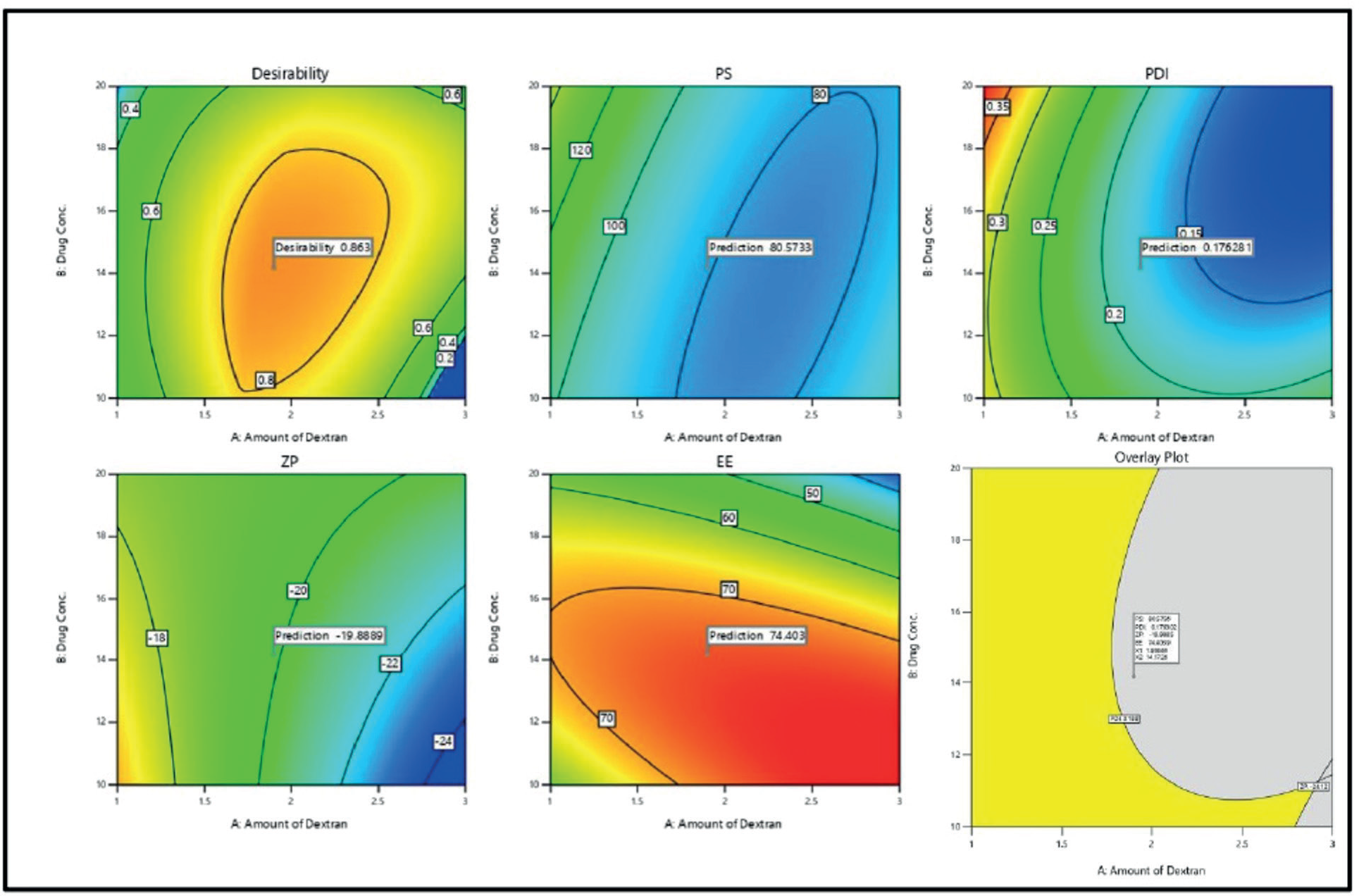
The desirability function of Derringer was applied to optimize factors influencing response parameters. Accordingly, the responses were translated into a desirability scale and combined through mean geometry functions using intensive search, leading to a global desirability value. The optimal configuration (F opt solution) involving the drug is 14.17 % w/v, with dextran sulphate at 1.90 %, and a homogenization time of 4.78 minutes, resulting in a D value of 0.863. By adjusting the specified values for Critical Quality Attributes (CQAs) like PS, PDI, ZP, and EE, we were able to improve the potential for customized graphics. The design space and overlay plot were shown in Figure 5. Three checkpoints were indispensable for the validation endeavour of this project to ascertain its strength and formulate it. The expected mean values for size, as given in Suppl. Table III showed 80.56 nm with a PDI of 0.176; ZP was -19.88 and EE 74.40, respectively, while the measured mean values corresponded to 89.8 ±9.5 nm, 0.284 ± 0.108, ZP -18.6 ± 1.2, and EE 72.86 ± 1.54.

Graphical depiction of overlay and Desirability plot (Grey color zone denoted feasible region) plot.
Evaluation of PS, PDI, ZP, and EE
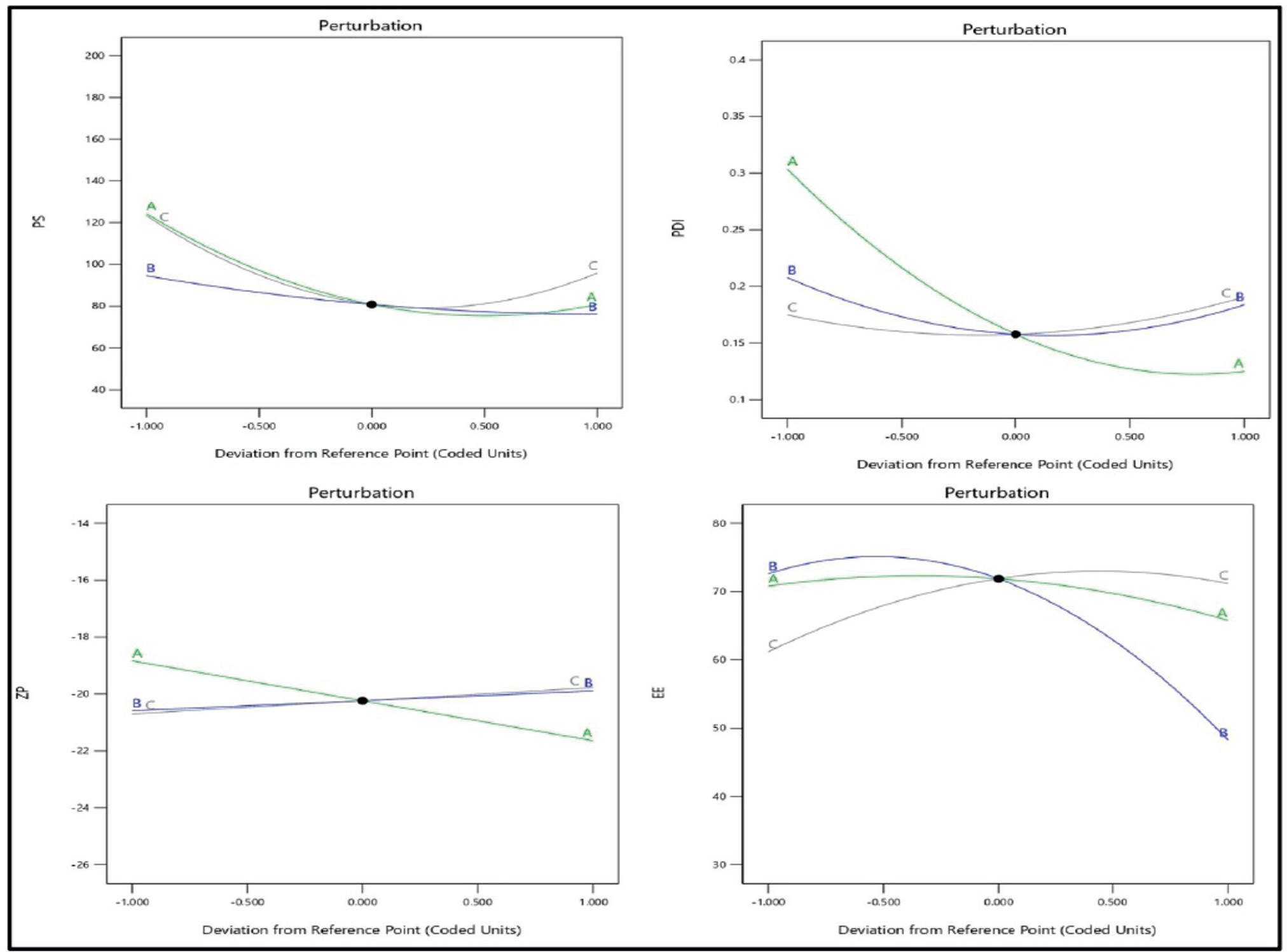
The nanobubbles displayed consistent size and uniformity, as evidenced by the particle size (PS) of 89.8 ± 9.5 nm and polydispersity index of 0.284 ± 0.108, indicating a homogeneous system (PDI < 0.3). The ZP of the optimized NBs was found to be -18.6 ± 1.2 mV, highlighted the stabilizing effect of steric stabilizers surrounding the particles, which is essential for stability based on the double electric layer (DEL) theory. Encapsulation efficiency (EE) was calculated to be 72.86 ± 1.54% with a drug loading of 33.57± 2.10 %. Figure 6 provided a visual representation of the particle size, PDI, and ZP. Also, the perturbation plots PS, PDI, ZP, and EE were depicted in Supplementary Figure 1.

Scanning Electron Microscopy (SEM)
Figure 7 depicted the exterior properties of the formulation and the drug. The drug in its initial state displayed a broad range of particle sizes and irregular cubic shapes with micrometre-sized particles. Conversely, when the drug was transformed into nanobubbles, it produced spherical nanoparticles consistently measuring below 200 nm, as verifi ed by the zeta sizer. This research showcased the successful achievement of nanosizing the drug through nanobubble formation, resulting in particles within the nanometre scale and exhibiting a low polydispersity index (PDI).

SEM image of pure drug; (A and B); Nanobubbles (NBs)(C and D) (magnification of pure drug 30.0kv 8.1mm x 800 60 pa and for Nanobubbles the magnifications was 15kv 5.3 mm x 10.0k).
Fourier Transform Infra-Red Spectroscopy (FTIR)
Analyzing the nano-formulation, excipients, and simple IRO IR spectra was used to determine component compatibility, as shown in Figure 8. 400 - 4000 cm-1 was the scanning range used. The spectra of the IRO exhibits peaks near 3511 cm-1 (OH vibrations), 2940 cm-1 (symmetric CH2 stretching), 2876 cm-1 (CH stretching), 1747 cm-1 1686 cm-1, 1661 cm-1 1611 cm-1, and 1417 cm-1 (characteristic of IRO C=O and C-C vibrations). The peaks at 1452 and 1435 cm-1 are assigned to methyl groups and were observed with peaks at 1216 and 1160 cm-1 due to CH in-plane bending vibrations. Notably, 1106, 1065, and 1007 cm-1 were attributed to stretching ring vibrations bunch C-C and C-N of the IRO drug. In the case of dextran sulphate, it showed distinct peaks at (2926.11, 1610.61, 1157.33, 1078.24, 916.22 and 1321. 28 cm-1) corresponding to OH stretching vibrations, CH stretching, bound water, C-O-C glycosidic bond stretching, C-O stretch of pyranose ring, glycosidic linkage and also CH bending. As no new peaks were seen in the NBs preparation and PM it clearly indicated no chemical interaction between the stabilizer and drug (Iliescu et al., 2014).

A) Overlay of FTIR analysis A: IRO-PD (black color strokepure drug); B: Dextran sulphate (red color stroke -); C: Physical mixture-PM (Blue color stroke); D) Formulation NBs (green color stroke).
DSC
DSC assessment was accomplished to evaluate the thermal characteristics of the drug, dextran sulphate, and the nanobubbles (Figure 9). The pure drug exhibited a clear endothermic peak at 270.98°C, signifying its melting point, indicating its crystalline characteristics. The thermograph of dextran sulphate exhibited peaks at 295.6°C. In the thermographs physical mixture peak was seen at 145.71 °C. In the optimized nanobubbles, a peak was emerged at 183.66°C indicated a minor shift in the melting point of the drug and its confinement within the polymeric structure due to weak intermolecular interactions between the drug and the polymer (Bhaskaran et al., 2022).

A) Overlay of DSC thermograms of i) PD (black color stroke); ii) Dextran sulphate (red color stroke); iii) PM-physical mixture (blue color stroke); iv) Formulation (optimized NBs green color stroke).
XRD
The XRD patterns are depicted in Figure 10. The IRO is showing stable diffraction peaks (2 θ scattered angles of 10.68, 12.18, 19.94, 17.26, 24.53, and 25.81°) indicating the IRO crystalline nature. Earlier authors reported the similar diffraction peaks for the drug under study. However, in the nanobubbles, the characteristic diffraction peaks of drug vanished, suggested the pure drug may have formed a solid-state complex at a molecular level in the nanobubbles (Sachdeva et al., 2023).

A) Overlay of DSC thermograms of i) PD (black color stroke); ii) Dextran sulphate (red color stroke); iii) PM-physical mixture (blue color stroke); iv) Formulation (optimized NBs green color stroke).
In vitro drug release
Figure 11 depicts the dissolution profiles of PD and NBs without and with sonic assistance. Nanobubbles released considerably more medication than a normal drug solution using ultrasound. The cumulative drug release (CDR) at 8 h was 16.39 ± 3.02%, 40.64 ± 6.3%, and 74.58 ± 6.28 % for PD, NBs subjected to without acoustic, and with acoustic, respectively. By 48 h, over 97.84 ± 8.18% was released from nanobubbles with acoustic support and 66.78 ± 7.29% without acoustic, but in the case of the pure drug, only 35.02 ± 4.25% of the drug was released. Release of drug from NBs is attributed to the collapse cavitation caused by acoustic waves, leading to disruption of structures with rapid drug release. With meticulous control of acoustic waves, its non-invasive nature, offers accurate drug delivery and with targeting abilities. Under the influence of ultrasound, the oscillation of bubbles can trigger the shell to open, thereby aiding in the release of drugs. The study aligned with previous findings on nanobubble stability under varying temperature conditions. The acoustic streaming flow generated by bubble oscillation regulated the movement of detached materials, which was influenced by both the radial excursion and the duration of the ultrasound pulses (Din, Jin, Choi, 2021).

In-vitro drug release of plain drug, drug loaded nanobubbles with and without ultrasound aid.
Stability studies
IRO-loaded nanobubbles (NBs) underwent stability assessments at various storage conditions (4 °C, 25 °C, and 40 °C) for 0, 1 month, two months, and three months (Table IV). A minute variation in drug content and EE confirmed robustness when NBs stored at low temperatures, indicating protection against degradation. Nevertheless, at elevated temperatures, a drop in EE % from 72.86± 1.54 to 67.63± 4.37% indicated structural disruption. Throughout the experiment, the PS of the formulation is less than 100 nm, and the zeta potential is around -17.6 ± 1.4 mV, highlighting the stability and uniformity of IRO nanobubbles. Storage in a polyethylene (PE) pouch led to a faster drop in number concentration compared to a glass bottle. Hydrogen bonding interactions were emphasized as critical factors in forming bulk nanobubbles and their exceptional longlasting stability (Ponnaganti, Babu, Palanati, 2022).
Pharmacokinetic studies
The time concentration profile after drug administration of PD and NBs was plotted (Figure 12). The PK statistics in Table V revealed that the NBs had meaningfully higher Tmax, Cmax (**p < 0.001), AUC024 (**p < 0.001), and AUC0-∞ (**p < 0.001) as compared to the pure drug data. The bioanalytical chromatogram showed a retention time of 10.2 min and 8.8 min for drug and for internal standard (3-methylxanthine) with peak plasma around 3.5 min respectively (Suppl. Figure 2).

The optimized formulation reached a maximum level (Cmax) of 7.35 times higher, while the area under the curve (AUC0-t) was 7.955 times higher than the free drug. In-vivo studies revealed a progressive drug release from the nanobubble preparation with extended half-life. Comparing the data to the free drug, oral bioavailability has significantly improved. This finding suggested a notable improvement in oral bioavailability compared to the free drug. The enhanced bioavailability can be related to the more drug circulation facilitated by the NBs system.
DISCUSSION
NBs are gaining prominence as a formulation stratagem owing to their targeting capability. In this study, NBs of IRO loaded in dextran sulphate were produced using the emulsion method. The optimization of NBs was carried out through the BBD. The process in which nanodroplets in perfluoro pentane transition from liquid to vapor upon exposure to ultrasound waves is called acoustic droplet vaporization. Perfluoro pentane undergoes a phase transition that turns nanodroplets into nanobubbles with high ultrasonic (US) wave reflectivity. The echogenic qualities of perfluoro pentane make them visible in ultrasonography images (Ponnaganti, Babu, Palanati, 2022). The NBs were developed using dextran sulphate polymer containing unbound carboxylic end chains using perfluoro pentane for the inner core and PLGA as the outer shell (NavarroBecerra, Borden, 2023). The term "ultrasound" describes mechanical vibrations or pressure waves that exhibit compressional and rarefactional pressure fluctuations and have frequencies equivalent to or higher than the human hearing threshold (20 kHz) (Messerschmidt et al., 2023).
Ultrasound effects primarily involved two mechanisms: cavitation and sonoporation. The cavitation effect played a crucial role in reducing the size of bubbles, whereas the sonoporation effect facilitates the uptake of these reduced bubbles. NBs therapy with ultrasound-assisted method aids in the localization of drugs while alleviating side effects. Dextran-sulphatestabilized nanobubbles, synthesized in the laboratory, are now under investigation for drug delivery due to their physical and surface properties. Dextran sulphate is highly recommended in numerous pharmaceutical applications like implantation of bone and in controlled release of drug, due to its biocompatibility and biodegradability properties.
The proposed design used a quadratic model for PS, PDI, ZP, and EE. The positive coefficient of the model indicates that the variables are positively correlated, which means increased values of the associated variable lead to increased drug encapsulation. Stabilizer concentration has affected particle size, PDI, and ZP. However, when it came to EE, a high stabilizer concentration led to a decrease in drug entrapment. Dextran sulphate, as a polymer along with lipid stabilizers, has a potential to impede drug entrapment in nanobubbles due to its competition for surface adsorption and the subsequent increase in solution viscosity. FTIR studies confirmed compatibility between drug and excipients. In DSC and XRD studies, no distinct drug peak was found in the formulation, which means crystalline drug material is absent (Chinna Babu et al., 2012). SEM analysis showed there are smooth, spherical nanobubbles that are homogeneous. Release of the drug happened by collapse cavitation induced by an acoustic wave, leading to the disruption of nanobubble structures that allow quick release of the drug. Stability studies under ultrasound indicated that the gas core was transformed from nanodroplets into bubbles, known as acoustic droplet generation. (Navarro-Becerra, Borden, 2023). The study aligned with previous findings on nanobubble stability under varying temperature conditions. The temperature-dependent behaviour observed in particle size, ZP, and entrapment emphasized understanding nanobubble characteristics in diverse environmental conditions for practical drug delivery applications. Different polymer materials submerged in nanobubble dispersions exhibited varied effects on NB number concentrations due to hydrophobic interactions. In in-vivo experiments, a gradual drug release was seen to occur from the formulation, leading to the increased half-life of the drug with increased AUC. The results thus confirm that nanobubbles significantly improve oral bioavailability compared to the free drug. The improved bioavailability of NBs with dextran sulphate is achieved through various mechanisms. These include better drug encapsulation within dextran sulphate nanodroplet, longer circulation time due to the protective coating of dextran sulphate, and increased uptake by target cells or tissue.
CONCLUSION
This research introduced an innovative approach to improve the solubility of irinotecan (IRO) nanobubbles. By conducting a thorough investigation, the study demonstrated that nanobubbles significantly enhanced the release of drug, indicating their potential as a new and smart delivery system. Response surface methodology ensured precise control over the size distribution, resulting in improved uniformity. Additionally, drugloaded nanobubbles exhibited an exceptional stability and dissolution in the gastrointestinal tract compared to traditional drug suspensions, suggested a longer drug halflife and increased effectiveness. These findings highlighted a promising role of dextran sulphate nanobubbles as a formulation loaded drug for cancer therapy, offering notable benefits such as faster dissolution rates, sustained, targeted drug release, and improved oral bioavailability.
ACKNOWLEDGEMENT
We acknowledge no one.
DATA AVAILABILITY STATEMENT
The data supporting the findings of this study are available from the corresponding author upon reasonable request.
REFERENCES
- Abdelnabi D, Lastakchi S, Watts C, Atkins H, Hingtgen S, Valdivia A, et al Local administration of irinotecan using an implantable drug delivery device stops high-grade glioma tumor recurrence in a glioblastoma tumor model. Drug Deliv Transl Res. 2024;14(11):3070-3088.
- Baroni S, Argenziano M, La Cava F, Soster M, Garello F, Lembo D, et al Hard-Shelled Glycol Chitosan Nanoparticles for Dual MRI/US Detection of Drug Delivery/Release: A Proof-of-Concept Study. Nanomaterials (Basel). 2023 Aug 1;13(15):2227.
- Basak D, Arrighi S, Darwiche Y, Deb S. Comparison of Anticancer Drug Toxicities: Paradigm Shift in Adverse Effect Profile. Life (Basel). 2021 Dec 29;12(1):48.
-
Bhaskaran NA, Jitta SR, Salwa, Cheruku S, Kumar N, Kumar L. Orally delivered solid lipid nanoparticles of irinotecan coupled with chitosan surface modification to treat colon cancer: Preparation, in-vitro and in-vivo evaluations. Int J Biol Macromol. 2022 Jun 30;211:301315. doi: 10.1016/j.ijbiomac.2022.05.060.
» https://doi.org/10.1016/j.ijbiomac.2022.05.060. - Campos FL, de Alcântara Lemos J, Oda CMR, de Oliveira Silva J, Fernandes RS, Miranda SEM, et al IrinotecanLoaded Polymeric Micelles as a Promising Alternative to Enhance Antitumor Efficacy in Colorectal Cancer Therapy. Polymers (Basel). 2022 Nov 14;14(22):4905.
-
Cavalli R, Bisazza A, Giustetto P, Civra A, Lembo D, Trotta G, et al Preparation and characterization of dextran nanobubbles for oxygen delivery. Int J Pharm. 2009 Nov 3;381(2):160-5. doi: 10.1016/j.ijpharm.2009.07.010. Epub 2009 Jul 17. PMID: 19616610.
» https://doi.org/10.1016/j.ijpharm.2009.07.010. - Chabot GG. Clinical pharmacokinetics of irinotecan. Clin Pharmacokinet. 1997 Oct;33(4):245-59.
-
Chen F, Huang G, Huang H. Preparation and application of dextran and its derivatives as carriers. Int J Biol Macromol. 2020 Feb 15;145:827-834. doi: 10.1016/j. ijbiomac.2019.11.151.
» https://doi.org/10.1016/j.ijbiomac.2019.11.151 - Chinna Babu P, Sundaraganesan N, Sudha S, Aroulmoji V, Murano E. Molecular structure and vibrational spectra of Irinotecan: a density functional theoretical study. Spectrochim Acta A Mol Biomol Spectrosc. 2012;98:1-6.
- Din FU, Jin SG, Choi HG. Particle and Gel Characterization of Irinotecan-Loaded Double-Reverse Thermosensitive Hydrogel. Polymers (Basel). 2021 Feb 13;13(4):551.
- Duan B, Zhao Y, Bai J, Wang J, Duan X, Luo X, et al Colorectal Cancer: An Overview. In: Morgado-Diaz JA, editor. Gastrointestinal Cancers. Brisbane (AU): Exon Publications. 2022; Sep 30. Chapter 1.
- Duan L, Yang L, Jin J, Yang F, Liu D, Hu K et al Micro/ nano-bubble-assisted ultrasound to enhance the EPR effect and potential theranostic applications. Theranostics. 2020 Jan 1;10(2):462-483.
-
Ette EI. Stability and performance of a population pharmacokinetic model. J Clin Pharmacol. 1997 Jun;37(6):486-95. doi: 10.1002/j.1552-4604.1997. tb04326.x. PMID: 9208355.
» https://doi.org/10.1002/j.1552-4604.1997.tb04326.x - Fujita K, Kubota Y, Ishida H, Sasaki Y. Irinotecan, a key chemotherapeutic drug for metastatic colorectal cancer. World J Gastroenterol. 2015 Nov 21;21(43):12234-48.
- Gagliardi A, Giuliano E, Venkateswararao E, Fresta M, Bulotta S, Awasthi V, et al Biodegradable Polymeric Nanoparticles for Drug Delivery to Solid Tumors. Front Pharmacol. 2021 Feb 3;12:601626.
- Iliescu RI, Andronescu E, Ghitulica CD, Voicu G, Ficai A, Hoteteu M. Montmorillonite-alginate nanocomposite as a drug delivery system--incorporation and in vitro release of irinotecan. Int J Pharm. 2014 Mar 25;463(2):184-92.
- Kancheva M, Aronson L, Pattilachan T, Sautto F, Daines B, Thommes D, et al Bubble-Based Drug Delivery Systems: Next-Generation Diagnosis to Therapy. J Funct Biomater. 2023 Jul 17;14(7):373.
- Karimi M, Solati N, Amiri M, Mirshekari H, Mohamed E, Taheri M, et al Carbon nanotubes part I: preparation of a novel and versatile drug-delivery vehicle. Expert Opin Drug Deliv. 2015 Jul;12(7):1071-87.
- Kciuk M, Marciniak B, Kontek R. Irinotecan-Still an Important Player in Cancer Chemotherapy: A Comprehensive Overview. Int J Mol Sci. 2020 Jul 12;21(14):4919.
- Kishore MK, Jaya PD, Basava Rao VV. Chitosan nanobubbles development and evaluation for the delivery of Sunitinib-an anticancer agent. Int J App Pharm. 2022;14(6):58-67.
- Kümler I, Sørensen PG, Palshof J, Høgdall E, SkovriderRuminski W, Theile S, et al Oral administration of irinotecan in patients with solid tumors: an open-label, phase I, dose escalating study evaluating safety, tolerability and pharmacokinetics. Cancer Chemother Pharmacol. 2019 Jan;83(1):169-178.
- Laizure SC, Herring V, Hu Z, Witbrodt K, Parker RB. The role of human carboxylesterases in drug metabolism: have we overlooked their importance? Pharmacotherapy. 2013 Feb;33(2):210-22.
- Lintoiu-Ursut B, Tulin A, Constantinoiu S. Recurrence after hepatic resection in colorectal cancer liver metastasis -Review article-. J Med Life. 2015;8 Spec Issue (Spec Issue):12-4.
- Mahdy MS, Azmy AF, Dishisha T, Mohamed WR, Ahmed KA, Hassan A, et al Irinotecan-gut microbiota interactions and the capability of probiotics to mitigate Irinotecanassociated toxicity. BMC Microbiol. 2023 Mar 2;23(1):53.
- Malatesta L, Cosco D, Paolino D, Cilurzo F, Costa N, Di Tullio A, et al Simultaneous quantification of Gemcitabine and Irinotecan hydrochloride in rat plasma by using high performance liquid chromatography-diode array detector. J Pharm Biomed Anal. 2018 Sep 10;159:192-199.
- Messerschmidt V, Ren W, Tsipursky M, Irudayaraj J. Characterization of Oxygen Nanobubbles and In Vitro Evaluation of Retinal Cells in Hypoxia. Transl Vis Sci Technol. 2023 Feb 1;12(2):16.
- Milano G, Innocenti F, Minami H. Liposomal irinotecan (Onivyde): Exemplifying the benefits of nanotherapeutic drugs. Cancer Sci. 2022 Jul;113(7):2224-2231.
- Mirón-Barroso S, Correia JS, Frampton AE, Lythgoe MP, Clark J, Tookman L, et al Polymeric Carriers for Delivery of RNA Cancer Therapeutics. Noncoding RNA. 2022;8(4):58.
- Navarro-Becerra JA, Borden MA. Targeted Microbubbles for Drug, Gene, and Cell Delivery in Therapy and Immunotherapy. Pharmaceutics. 2023 May 30;15(6):1625.
- Nicolas J, Mura S, Brambilla D, Mackiewicz N, Couvreur P. Design, functionalization strategies and biomedical applications of targeted biodegradable/biocompatible polymer-based nanocarriers for drug delivery. Chem Soc Rev. 2013 Feb 7;42(3):1147-235.
- Patel K, Shah S, Patel J. Solid dispersion technology as a formulation strategy for the fabrication of modified release dosage forms: A comprehensive review. Daru. 2022 Jun;30(1):165-189.
- Ponnaganti M, Babu AK, Palanati M. Formulation and optimization of Ceritinib loaded nanobubbles by BoxBehnken Design. Int J App Pharm. 2022;14(4):219-226.
- Rahmani S, Park TH, Dishman AF, Lahann J. Multimodal delivery of irinotecan from microparticles with two distinct compartments. J Control Release. 2013 Nov 28;172(1):239245.
- Ramasundaram S, Saravanakumar G, Sobha S, Oh TH. Dextran Sulfate Nanocarriers: Design, Strategies and Biomedical Applications. Int J Mol Sci. 2022 Dec 26;24(1):355.
- Sachdeva B, Sachdeva P, Negi A, Ghosh S, Han S, Dewanjee S, et al Chitosan Nanoparticles-Based Cancer Drug Delivery: Application and Challenges. Mar Drugs. 2023 Mar 28;21(4):211.
- Sohaib M, Shah SU, Shah KU, Khan NR, Irfan MM et al Physicochemical Characterization of Chitosan-Decorated Finasteride Solid Lipid Nanoparticles for Skin Drug Delivery. Biomed Res Int. 2022 Aug 6;2022:7792180.
-
Stewart CF, Zamboni WC, Crom WR, Houghton PJ. Disposition of irinotecan and SN-38 following oral and intravenous irinotecan dosing in mice. Cancer Chemother Pharmacol. 1997;40(3):25965. doi: 10.1007/s002800050656. PMID: 9219511.
» https://doi.org/10.1007/s002800050656.
Edited by
-
Associate Editor:
Marlus Chorilli

 Thumbnail
 Thumbnail