
 Biological potential and chemical profile of Carapanauba (Aspidosperma excelsum benth) extracts
Abstract
This study aimed to determine the chemical composition and biological activities of extracts from commercial samples of Aspidosperma excelsum stem bark. The extracts underwent preliminary phytochemical screening, chromatographic fractionation, and spectrometric and spectroscopic analyses, including mass spectrometry and nuclear magnetic resonance. Antioxidant and antimicrobial activities were also evaluated. Phytochemical screening revealed the presence of alkaloids, anthrones, triterpenes, and steroids. Fractionation led to the isolation and structural identification of the indole alkaloids carapanaubine and O-acetylyohimbine. APCI-MS/MS analysis of the crude extracts identified three additional indole alkaloids: spruceanumine B, 10-methoxydihydrocorinanteol, and 11-methoxy-yohimbine. In antimicrobial assays, the hexane extract exhibited moderate antifungal activity against Candida parapsilosis (MIC = 400 µg/mL), while the other extracts displayed weak activity (MIC = 1600 µg/mL). Against Candida albicans, all extracts demonstrated weak antifungal activity, and none showed antibacterial activity at the tested concentrations. Total phenolic content was low, ranging from 31.25 to 138.06 ± 0.9 mg GAE/g. The hexane extract lacked antioxidant activity, while the chloroform, ethyl acetate, and methanol extracts exhibited relatively high activity in the ABTS•+ assay (801.88 ± 42.22 to 936.33 ± 61.28 μM TE) compared to Trolox (500–1000 μM TE). However, all extracts displayed low activity in the DPPH• assay. These findings suggest that A. excelsum warrants further investigation to explore the antioxidant and antimicrobial properties of its identified constituents.
Keywords:
Apocynaceae; Indole alkaloids; Antioxidant; Antimicrobial
INTRODUCTION
Amid the rich biodiversity of the Amazon, species of the Apocynaceae Juss. family stand out for their economic significance, particularly in wood and latex production, as well as their abundance of bioactive compounds (Oliveira et al., 2009b; Di Stasi & Hiruma-Lima, 2002). This family, comprising 424 genera and 4,600 species, is predominantly found in tropical and subtropical regions (Bhadane et al., 2018). Notable genera Alstonia, Aspidosperma, Rauwolfia, Vinca, Tabernaemontana, Mandevilla, Hancornia, Nerium, Strophantus, Catharanthus, Allamanda, Thevetia, Himatanthus (Plumeria), and Wrightia (Di Stasi, Hiruma-Lima, 2002).
Apocynaceae species produce a diverse range of chemical compounds—primarily alkaloids, terpenes, steroids, flavonoids, glycosides, lactones, and hydrocarbons—many of which have medicinal properties (Bhadane et al., 2018). For instance, alkaloids such as reserpine, ajmalicine, ajmaline, and serpentine, extracted from Rauwolfia serpentina, are widely used in treating hypertension and cardiac arrhythmias (Arora & Madan, 1956).
Among these, the genus Aspidosperma Mart. & Zucc. is particularly noteworthy, comprising approximately 300 species (Almeida et al., 2019). In Brazil, 68 species are recognized, commonly known as peroba, guatambu, pau-pereiro, carapanauba, pequiá, quina, taroba, and amargoso (Lorenzi, 1998). This genus is phytochemically distinguished by the dominance of indole-type alkaloids, known for their structural diversity and pharmacological potential (Lopes, 2019).
In traditional medicine, infusions from the leaves or bark of Aspidosperma species are used to treat various ailments, including leishmaniasis, malaria, uterine and ovarian inflammation, diabetes, liver disease, stomach disorders, cancer, fever, and rheumatism (Pereira et al., 2007; Almeida et al., 2019). Biological studies have demonstrated its therapeutic properties, including antitumor, antiplasmodial, and antihypertensive activities (Henrique et al., 2010).
Studies have shown that Aspidosperma species, including A. marcgravianum, A. desmanthum, and A. nitidum, exhibit antimicrobial activity against Staphylococcus aureus, Staphylococcus epidermidis, Escherichia coli, and Pseudomonas aeruginosa. The metabolic extract of A. marcgravianum demonstrated a minimum inhibitory concentration (MIC) of 3.12 µg/ mL against S. aureus, 12.5 µg/mL against S. epidermidis and E. coli, and 50 µg/mL against P. aeruginosa. Additionally, aqueous extracts of A. desmanthum and A. nitidum exhibited an MIC of 50 µg/mL against S. epidermidis and E. coli, while methanolic extracts displayed MICs of 25 µg/mL and 50 µg/mL against S. aureus and 50 µg/mL against S. epidermidis and E. coli (Araújo, 2022).
One of the most common Aspidosperma species in the Amazon basin is Aspidosperma excelsum Benth., with A. marcgravianum Woodson and A. nitidum Benth. ex Müll.Arg recognized as synonyms (IPNI, 2024). Known locally as carapanauba, carapanúba-preta, sapopema, and sapopemba, this species has been traditionally used to treat gastrointestinal disorders, bronchitis, inflammation, fever, diabetes, cancer, and particularly malaria (Trindade et al., 2016). These medicinal properties are largely attributed to its rich content of indole alkaloids, with 22 such compounds isolated to date (Pereira et al., 2007). Key alkaloids identified in A. excelsum include yohimbine, O-acetylyohimbine, excelsinin, O-acetylexcelsinin, and 16-epi-excelsinin (Benoin et al., 1967; Burnell, Nguyên, 1971).
Given its medicinal significance, A. excelsum is widely sold throughout the Amazon region. Considering the high alkaloid content of Aspidosperma species and their extensive use in traditional medicine, this study aims to characterize the chemical profile and evaluate the antimicrobial and antioxidant activities of extracts from A. excelsum stem bark.
MATERIAL AND METHODS
Plant Material and Extract Preparation
Dried stem bark of Aspidosperma excelsum (carapanauba) was commercially sourced from the municipality of Itacoatiara, Amazonas (3° 8′ 31″ S, 58° 26′ 33″ W). Botanical identification was performed by analyzing stem characteristics—such as shape, color, surface texture, and additional structures—compared to descriptions in virtual herbaria and identification keys.
A total of 261.98 g of pulverized plant material underwent sequential cold extraction using 500 mL of organic solvents over a 24-hour period per solvent at room temperature (27 ± 1 °C). The solvents used were hexane, chloroform, ethyl acetate, and methanol, each with a total volume of 1500 mL. After extraction, the filtrates were concentrated using a rotary evaporator (FISATOM, Model 801) under reduced pressure at 40 °C and 40 RPM, then transferred to glass bottles. This process yielded extracts of varying polarity, labeled as hexane (EH-Aex), chloroform (EC-Aex), ethyl acetate (EA-Aex), and methanol (EM-Aex).
Chemical Analysis
Phytochemical Prospection
The crude extracts were analyzed for major metabolite classes following the methodology described by Matos (1997). Qualitative tests, including chemical reactions and thin-layer chromatography (TLC), were conducted to detect anthocyanins, anthocyanidins, flavonoids, leucoanthocyanidins, catechins, flavones, alkaloids, anthraquinones, anthrones, coumarins, phenols, tannins, triterpenes, steroids, and saponins. The specific conditions for each test are outlined in Table I.
Chromatographic Fractionation
The chloroform extract (EC-Aex, 2 g) was fractionated using column chromatography (30 × 300 mm, VIDROLABOR) with Merck silica gel 60 (55.55 g) as the stationary phase. Elution was performed with binary solvent mixtures (90:10) of hexane, dichloromethane, ethyl acetate, and methanol, yielding 158 fractions. These fractions were analyzed via thin-layer chromatography (TLC) on Merck silica gel sheets (2 mm), using dichloromethane-methanol (9:1) as the mobile phase. Detection was performed under UV light at 254 and 366 nm, followed by Dragendorff’s reagent and 10% sulfuric acid staining.
Fractions 1–4 were further fractionated using Merck silica gel 60 and eluted with hexane, dichloromethane, ethyl acetate, and methanol, yielding 38 additional fractions. Fraction 1-4.6 (12.6 mg) was processed using a Pasteur pipette column with dichloromethane-methanol (9:1), resulting in the isolation of compound Fr2ClAex1-4.6.1, which was analyzed via atmospheric pressure chemical ionization mass spectrometry (APCI-MS) and hydrogen nuclear magnetic resonance (¹H-NMR).
Fraction 52 (25.4 mg) was further fractionated using Merck silica gel 60 (500 mg) with ethyl acetate, yielding two fractions. The fraction identified as CFr1ClAex52 was crystalline and subsequently analyzed by APCI-MS and ¹H-NMR.
Mass Spectrometry (APCI-MS)
Mass spectra of the crude extracts and fractions (1 mg/mL) were obtained using direct infusion in an LCQ Fleet™ ion trap mass spectrometer (THERMO SCIENTIFIC) in both positive and negative ion modes. The APCI ionization source scanned m/z values from 100 to 1000 Da, with a probe voltage of 4.5 kV (positive mode) and 3.8 kV (negative mode), using nitrogen at 30 psi. The source temperature was maintained at 250 °C, with lens and capillary voltages of 35 V and 75 V, respectively. Spectra were analyzed using X-Calibur® 2.0.7 software.
Nuclear Magnetic Resonance (¹H-NMR)
¹H-NMR spectra were acquired on a Bruker Avance III HD spectrometer (500.13 MHz) equipped with a 5-mm BBFO Plus SmartProbe™ with a magnetic field gradient in the Z direction. Samples were dissolved in deuterated chloroform (CDCl3) with tetramethylsilane (TMS) as the internal reference. Chemical shifts (δ) were reported in parts per million (ppm), and data processing was conducted using MestReNova software.
In Vitro Antimicrobial Activity
The minimum inhibitory concentration (MIC) was determined using the broth microdilution method according to Clinical and Laboratory Standards Institute (CLSI) guidelines. Extract concentrations ranged from 1600 to 1.56 µg/mL, tested in duplicate against standard American Type Culture Collection (ATCC) strains: Staphylococcus aureus (ATCC 25923), Streptococcus pneumoniae (ATCC 49619), Enterococcus faecalis (ATCC 29212), Escherichia coli (ATCC 25922), Klebsiella pneumoniae (ATCC 700603), Burkholderia cepacia (ATCC 17759), Candida albicans (ATCC 80193), and Candida parapsilosis (ATCC 22019).
Extracts were diluted (1:2) in RPMI-1640 broth, and 100 µL of the inoculum (2.5 × 10³ cells/mL) was added to sterile 96-well microplates. Positive control wells contained both culture medium and bacterial inoculum, while negative controls contained only the culture medium. Microplates were incubated at 35 °C for 24 hours, and MIC values were recorded as the lowest concentration (µg/mL) that inhibited 50% of microbial growth compared to the control.
Antioxidant Activity and Total Phenolic Content
Total Phenolic Content (TPC)
TPC was quantified spectrophotometrically in triplicate (BEL PHOTONICS, UV-M51) using the Folin-Ciocalteu reagent. Extracts (200 µL, 1 mg/mL) were mixed with 1500 µL of Folin-Ciocalteu reagent and 6% sodium carbonate, incubated for 90 minutes in the dark, and measured at 725 nm. Results were expressed as mg gallic acid equivalents per gram of sample (GAE mg/g), based on a gallic acid standard curve (31.25–250 µg/mL).
Antioxidant Capacity
The antioxidant potential of A. excelsum extracts was evaluated using DPPH• (2,2-diphenyl-1-picrylhydrazyl) and ABTS+ (2,2-azinobis (3-ethylbenzothiazoline-6-sulfonic acid)) radical scavenging assays on a UV/ VIS spectrophotometer (BEL PHOTONICS, UV-M51). Trolox (100–1500 μM) was used as a reference antioxidant, with results expressed in µM Trolox equivalents (µM TE).
The DPPH• assay was conducted following Molyneux (2004), with modifications for spectrophotometric analysis at 515 nm. Extracts (100 µL, 1 mg/mL) were added to 3900 µL of a methanolic DPPH• solution (100 µM; Abs = 0.996) in triplicate and incubated in the dark for 30 minutes. The Trolox standard curve was y = -0.0006x + 0.923 (R² = 0.9999).
The ABTS+ assay followed Re et al. (1999) with extracts (30 µL, 1 mg/mL) added to 3000 µL of the ABTS+ radical solution (Abs = 0.700) in triplicate, incubated in the dark for 6 minutes, and measured at 734 nm. The Trolox standard curve was y = -0.0003x + 0.9179 (R² = 0.9992).
RESULTS AND DISCUSSION
Yield of Aspidosperma excelsum Bark Extracts
The yield of Aspidosperma excelsum stem bark extracts ranged from 0.20% to 6.24%, as shown in Table II. The lowest yield (0.20%) was obtained with hexane, the least polar solvent, while the highest yield (6.24%) was achieved with methanol, the most polar solvent used.
Chemical Analysis
Phytochemical Screening of Extracts
Phytochemical screening of Aspidosperma excelsum stem bark extracts revealed the presence of alkaloids, anthrones, triterpenes, and steroids. Alkaloids were detected in all extracts, aligning with previous reports that highlight indole-type alkaloids as chemotaxonomic markers of the Aspidosperma genus (Pereira et al., 2007).
Steroids and triterpenes were present in the hexane, chloroform, ethyl acetate, and methanol extracts, consistent with findings in the ethanolic extract of Aspidosperma subincanum (Alves, 2007; Santos et al., 2009). Similarly, a mixture of steroids (stigmasterol, β-sitosterol, and campesterol) has been reported in Aspidosperma macrocarpon (Aquino, 2012). Anthrones were exclusively detected in the chloroform extract, corroborating previous reports of these compounds in Aspidosperma pyrifolium Mart. leaf extracts (Barbosa, 2014).
In a phytochemical screening conducted by Alves (2007), powdered stem bark of A. subincanum tested positive for steroids, triterpenoids, saponins, tannins, alkaloids, coumarins, and resins, but negative for flavonoids. Similarly, A. excelsum hydroethanolic extract has been reported to contain alkaloids, saponins, reducing sugars, phenols, and tannins, while lacking quinones, terpenes, and flavonoids (Gomes, 2011).
Notably, differences in phytochemical composition within the same species were observed, likely due to environmental factors such as soil composition and climate, which can influence metabolite concentrations (Mendonça et al., 2015). Additionally, solvent selection plays a crucial role in determining which compounds are preferentially extracted (Simões et al., 2010).
The bioactive compounds identified in plant extracts exhibit diverse biological properties. Alkaloids possess antibacterial, antifungal, antiplasmodial, and antitumor activities, while tannins exhibit antioxidant effects. Saponins have demonstrated antimicrobial, antiparasitic, and antitumor properties, and phenolic compounds contribute to antioxidant activity (Simões et al., 2010).
Chemical Constituents Identified by APCI-MS
Atmospheric pressure chemical ionization mass spectrometry (APCI-MS) identified several metabolites in the extracts, particularly alkaloids, without requiring prior isolation. Sequential mass spectrometry (MS2) further confirmed the presence of these compounds by comparing spectral data with previously reported findings on A. excelsum and other Aspidosperma species. Table III summarizes the identified molecules.
In the hexane extract (EH-Aex), fragmentation of the quasi-molecular ion m/z 439 [M+H]+ exhibited characteristic patterns of spruceanumine B (Figure 1). The peak at m/z 379 resulted from the loss of acetic acid (CH3CO2H, 60 Da). Further fragmentation at m/z 351 involved cleavage of the E-ring via a Retro-Diels-Alder reaction, leading to a 28 Da loss corresponding to ethene (CH2=CH2). These fragmentation patterns align with those reported for plumerane alkaloids, such as 21-oxo-aspidoalbidine, previously isolated from Aspidosperma exalatum (Brown et al., 1966). This compound has also been identified in the hexane extract fractions of A. spruceanum seeds and in the methanolic extract of A. spruceanum stem bark (Oliveira, 2008).

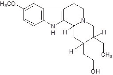
The mass spectrum of the methanolic extract revealed ions at m/z 329 and 385 [M+H]+, previously documented in the literature. Fragmentation of the m/z 329 ion generated peaks at m/z 286, 251, 214, and 168. The m/z 286 peak resulted from the loss of an isopropyl group (43 Da), while the m/z 251 peak corresponded to a 78 Da loss due to aromatic ether cleavage with hydrogen migration. These data are consistent with the structure of 10-methoxydihydrocorinanteol (Figure 2), an alkaloid previously isolated from the stem bark of Aspidosperma dicolor A. DC (Dastoor et al., 1967). Upon fragmentation, the protonated ion m/z 385 [M+H]+, assigned to 11-methoxy-yohimbine (Figure 3), generated peaks at m/z 174 and 159. The fragmentation process involved a substantial 211 Da loss, attributed to the elimination of a terpene moiety via Retro-Diels-Alder cleavage, followed by C-ring fragmentation (Araújo, 2022; Miet et al., 1977). The m/z 159 peak represented a 15 Da loss due to the elimination of a methyl group (-CH3), a characteristic fragmentation pattern used to identify adjacent methoxyl groups in the structure (Tavares et al., 2005). This compound was confirmed in alkaloidal fractions of A. marcgravianum and A. nitidum stem barks through mass spectrometry (Araújo, 2022) and was previously isolated from the methanolic extract of A. spruceanum stem bark and seeds (Oliveira et al., 2009a).


Structural Characterization of Sample Fr2ClAex 1-4.6.1
The sample Fr2ClAex1-4.6.1 (4 mg) was identified as a yellow/orange amorphous solid. APCI-MS analysis in positive mode displayed a dominant peak at m/z 429 (m.e. 428). Sequential mass analysis produced fragments at m/z 397, 369, 353, and 220, consistent with the fragmentation pattern of the alkaloid carapanaubine (C23H28N2O6). The m/z 397 peak resulted from a 32 Da loss, corresponding to methanol elimination (CH3OH). Subsequent losses of 28 Da (CO) and 16 Da (CH4) generated the m/z 369 and 353 peaks, respectively. The m/z 220 fragment was formed via an alternative fragmentation pathway, involving a 209 Da loss due to C-ring cleavage (Figure 4).

¹H-NMR spectra of Fr2ClAex 1-4.6.1 revealed signals indicative of methoxyl, carbomethoxy, aromatic, methyl, methylene, and methine hydrogens. Integration of the signals confirmed the presence of 28 hydrogen atoms within the 1.13-7.49 ppm range, which were compared with literature data (Table IV).
Key spectral features included:
-
Two singlets at δH 3.87 and 3.85, each integrating for three hydrogens, corresponding to two methoxyl groups;
-
Two singlets at δH 6.82 and 7.07, integrating for one hydrogen each, with para coupling (J = 0 Hz), indicating a disubstituted benzene ring at the ortho and meta positions;
-
The most deshielded signal at δH 7.49 (H17), assigned to a hydrogen bonded to an sp² carbon;
-
A signal at δH 3.68, integrating for three hydrogens, attributed to an ether methyl group;
-
Methine hydrogen signals at δH 1.71, 2.20, and 2.35 (multiplets); δH 4.23 (dd, J = 6.8, 1.3 Hz); and δH 1.13 (d, J = 6.8 Hz);
-
Eight methylene hydrogen signals at δH 1.69 (dt, J = 3.7, 4.90, 13.25 Hz), 2.26 (dd, J = 2.23, 6.60, 8.30 Hz), 2.84 (dd, J = 11.5, 6.1 Hz), 2.31 (d, J = 11.20 Hz), 2.40 and 2.34 (dd, J = 7.6, 2.1 Hz), and δH 2.10 (m) and 1.25 (m).
The combined spectral data, summarized in Table IV, confirm that Fr2ClAex1-4.6.1 (solvent: deuterated chloroform) corresponds to carapanaubine (solvent: deuterated methanol) (Figure 5). This indole alkaloid has been previously isolated from Aspidosperma nitidum Benth and other species of the Rauwolfia genus within the Apocynaceae family (Sales, 2019; Kumar et al., 2016).
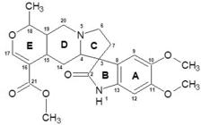
Proposed structure for the alkaloid carapanaubine based on 1H RMN data for sample Fr2ClAex 1-4.6.1.
Structural Characterization of Sample CFr1ClAex52
The sample CFr1ClAex52 (6 mg) was isolated as a white amorphous solid. APCI-MS analysis in positive mode revealed a protonated molecular ion peak [M + H]+ at m/z 397 (m.e. 396), consistent with the molecular formula C23H28N2O4. Sequential mass analysis produced a fragment at m/z 365, corresponding to the loss of a methanol molecule (Figure 6), and another at m/z 313. These fragmentation patterns align with the reported mass spectrum of the alkaloid O-acetylyohimbine (Nascimento et al., 2018).

The ¹H-NMR spectrum of CFr1ClAex52 displayed singlets at δH 2.07 and 3.74, each integrating for three hydrogens, corresponding to the methyl groups in the -OCOCH3 and -COOCH3 functional groups, respectively. A multiplet at δH 3.96, integrating for one hydrogen, was assigned to H-17 within the CHOCOCH3 moiety. Additionally, a singlet at δH 7.57, integrating for one hydrogen, was attributed to the hydrogen attached to the nitrogen atom in the indole nucleus.
Comparison with literature data (Table V) confirmed that the compound is O-acetylyohimbine (Figure 7), an alkaloid previously isolated from the bark and roots of Aspidosperma excelsum (Benoin et al., 1967; Verpoorte et al., 1983).
¹H-NMR data (500.13 MHz; d-chloroform) for the CFr1ClAex52 fraction compared with literature data for O-acetylyohimbine (solvent: deuterated chloroform)

Antimicrobial Activity
The classification of antimicrobial activity followed the criteria established by Holetz et al. (2002), with modifications. Activity was categorized as good for MIC < 100 μg/mL, moderate for MIC between 100 and 500 μg/mL, weak for MIC between 500 and 1600 μg/mL, and inactive if no inhibition was observed at the highest concentration tested (MIC > 1600 μg/mL).
Analysis of the results revealed that the extracts exhibited no inhibitory activity against the tested bacteria (MIC > 1600 μg/mL). Although no specific data exist regarding the antibacterial activity of Aspidosperma excelsum extracts, other species within the Aspidosperma genus, including A. marcgravianum—a synonym for the species under investigation—have been evaluated for such potential.
For instance, Oliveira and colleagues (2009b) reported that ethanolic extracts of A. polyneurum, A. dispermum, and A. pyrifolium bark were inactive against S. aureus, B. subtilis, and P. aeruginosa (MIC > 1000 μg/mL). In contrast, A. tomentosum exhibited weak antibacterial activity against S. aureus and B. subtilis, with MIC values of 1000 and 500 μg/mL, respectively (Pessini, 2015). Additionally, the methanolic extract of A. marcgravianum bark demonstrated good antibacterial activity against S. aureus (MIC = 3.125 μg/mL), S. epidermidis (MIC = 12.5 μg/mL), P. aeruginosa (MIC = 50 μg/mL), and E. coli (MIC = 12.5 μg/mL) (Araújo, 2022).
Regarding antifungal activity (Table VI), the hexane extract was inactive (MIC > 1600 μg/mL), while the other extracts displayed weak activity against Candida albicans (MIC = 1600 μg/mL). The EH-Aex sample exhibited moderate activity against C. parapsilosis, showing antifungal effects in the first three dilutions (MIC = 400 μg/mL). The EC-Aex (MIC = 1600 μg/mL), EA-Aex (MIC = 800 μg/mL), and EM-Aex (MIC = 800 μg/mL) extracts exhibited weak activity against C. parapsilosis.
Similar findings were reported by Pessini (2015), who investigated the antifungal activity of bark fractions from Aspidosperma species, including A. macrocarpon and A. pyrifolium, against the same fungal strains. Other studies have also documented the inactivity of crude ethanolic extracts from the branches and leaves of A. ramiflorum and A. olivaceum against S. aureus and C. albicans (Agripino et al., 2004), as well as the roots, stems, and bark of A. tomentosum against both Candida species (Pessini, 2015).
Among the metabolites identified in the chemical characterization of the extracts, alkaloids stand out for their antifungal potential due to their ability to destabilize biological membranes (Henrique et al., 2010). However, the antifungal activity observed in this study was not promising, which may be attributed to the specific chemical composition of the extracts and the microorganisms tested.
Endo et al. (2010) suggested that Candida albicans biofilm formation could contribute to resistance or reduced susceptibility to antifungal agents, including plant extracts. Therefore, further evaluation of the antifungal potential of extracts and isolated compounds from Aspidosperma excelsum against other fungal species is necessary.
The literature reports that the alkaloids ramiflorine A and B, isolated from the crude methanolic extract of A. ramiflorum, exhibit significant antimicrobial activity against Cryptococcus neoformans, with MIC values ranging from 3.12 to 12.5 µg/mL and 12.5 to 25 µg/mL, respectively (Souza et al., 2006). Additionally, ethanolic extracts from A. polyneuron bark have demonstrated antimicrobial activity against Proteus mirabilis (Granato et al., 2005). These findings underscore the importance of exploring variations in chemical composition and testing against a broader range of microorganisms.
Assessment of Total Phenolic Content and Antioxidant Activity
Table VI presents the total phenolic content (TPC) and antioxidant activity of the tested extracts, evaluated using the DPPH• and ABTS•+ assays.
Analysis of the results indicates that the samples have a low total phenolic content. The EH-Aex extract exhibited a value below 31.25 mg GAE/g, while the other extracts ranged from 120.89 ± 2.26 to 138.06 ± 0.9 mg GAE/g.
Although phenolic compounds are widely recognized for their biological properties, they are primarily associated with antioxidant activity (Rossa, 2013). However, no correlation was observed between phenolic content and antioxidant activity in either of the assays conducted.
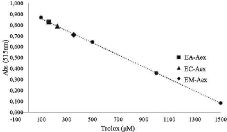
The antioxidant potential of A. excelsum extracts was evaluated using the DPPH• and ABTS•+ assays, both of which measured radical reduction capacity based on a Trolox calibration curve.
In the DPPH• assay, all extracts exhibited low radical scavenging activity. At a concentration of 1 mg/ mL, EA-Aex, EC-Aex, and EM-Aex displayed activity below 500 μM Trolox, while EH-Aex showed even lower activity, below the 100 μM Trolox equivalent (Figure 8).

In the ABTS•+ assay, all extracts except EH-Aex exhibited antioxidant activity. The EC-Aex extract had the highest radical-reducing capacity (936.33 ± 61.28 μM TE), followed by EM-Aex (876.33 ± 20.09 μM TE) and EA-Aex (801.88 ± 42.22 μM TE). These values indicate antioxidant activity comparable to Trolox, ranging between 500 and 1000 μM (Figure 9).

The literature on the antioxidant potential of A. excelsum is limited, but studies on other Aspidosperma species have been conducted. Santos (2016) reported significant DPPH• radical inhibition (83.68%) in the dichloromethane fraction of A. pyrifolium seed extracts, with an IC50 of 133.49 ± 2.22 μg/mL. Araújo (2022) observed low inhibition percentages in bark extracts from A. desmanthum, A. marcgravianum, and A. nitidum, suggesting that species within this genus are rich in monoterpenoid indole alkaloids, which are synthesized in response to oxidative stress. Since oxidative stress influences the concentration of bioactive alkaloids, and the DPPH• assay measures antioxidant reactivity based on the presence of hydroxyl-bearing molecules, the low activity observed in this study may reflect a lower concentration of these molecules attached to aromatic rings in the extracts.
These findings underscore the importance of using multiple assays to assess antioxidant activity, as different methodologies may yield varying results. Alam and colleagues (2023) emphasized that antioxidant potential should not be determined using a single method, as each assay evaluates different mechanisms of antioxidant action.
In this study, the ABTS•+ assay measured the ability of an antioxidant to reduce the ABTS•+ cation, while the DPPH• assay assessed the capacity of molecules to donate protons (H⎕) (Araújo, 2022). Additionally, the ABTS•+ assay detects both hydrophilic and lipophilic compounds, whereas DPPH• is limited to compounds soluble in organic media (Daroncho, 2012). Since the analyzed extracts are rich in alkaloids, the higher activity observed in the ABTS•+ assay compared to DPPH• is expected, as alkaloids contain nitrogen atoms and free electron pairs that contribute to ABTS•+ reduction (Vizzotto, Krolow, Weber, 2010).
No correlation between antioxidant activity and phenolic content was observed, reinforcing the idea that antioxidant potential cannot be attributed solely to phenolic compounds. Structural characterization of active compounds is essential to fully understand their antioxidant properties (Tavares, Ramos, 2008).
CONCLUSION
The preliminary phytochemical analysis of Aspidosperma excelsum confirmed the presence of alkaloids, anthrones, triterpenes, and steroids. Mass spectrometry of the hexane and methanol extracts identified indole alkaloids such as spruceanumine B, 10-methoxydihydrocorinanteol, and 11-methoxyyohimbine, previously reported in other Aspidosperma species.
NMR analysis of a chloroform extract fraction enabled the structural elucidation of two indole alkaloids—carapanaubine and O-acetylyohimbine— marking their first report in this species.
Biological assays revealed that all extracts were inactive against the tested bacteria, while the hexane extract exhibited moderate antifungal activity against Candida parapsilosis. The other extracts displayed weak antimicrobial activity. In antioxidant assessments, the hexane extract was inactive against ABTS•+, while all extracts exhibited low DPPH• scavenging activity.
Future studies will further explore the antioxidant potential of A. excelsum extracts using the FRAP assay and β-carotene/linoleic acid autoxidation test. Additionally, the chemical composition of A. excelsum will be further investigated through the isolation and identification of bioactive compounds with significant antioxidant and antimicrobial properties. These efforts will contribute to expanding scientific knowledge of the bioactive potential of A. excelsum.
ACKNOWLEDGEMENTS
The authors acknowledge Programa de Pós-Graduação em Ciência e Tecnologia para Recursos Amazônicos (PPGCTRA – UFAM) for its support (EDITAL N. 003/2020 – PAINTER (01.02.016301.03897/2022-80) and EDITAL N. 008/2022 – KUNHÃ (01.02.016301.03977/2022-36)).
DATA AVAILABILITY STATEMENT
All data is available within the article or its supplementary materials
REFERENCES
- Agripino DG, Lima MCE, Silva MR, Meda CI, Bolzani VS, Cordeiro I, et al. Screening of Brazilian plants for antimicrobial and dna-damaging activities: I. Atlantic rain forest. Ecological station juréia-itatins. Biota Neotrop. 2004;4:1-15.
- Alam MN, Bristi NJ, Rafiquzzaman, M. Review on in vivo and in vitro methods evaluation of antioxidant activity. Saudi Pharm J. 2013;21(2):143-152.
- Almeida VL, Silva CG, Silva AF, Campana PRV, Foubert K, Lopes JCD, et al. Aspidosperma species: A review of their chemistry and biological activities. J Ethnopharmacol. 2019;231:125–140.
- Alves NM. Estudo farmacognóstico e da toxicidade experimental (aguda e subaguda) do guatambu (Aspidosperma subincanum Mart.). [Master’s Dissertation]. Brasília: Universidade de Brasília; 2007.
- Aquino PGV. Estudo químico e atividades anti-hipertensiva e antioxidante de Aspidosperma macrocaroum Mart. (Apocynaceae). [Master’s Dissertation]. Maceió: Universidade Federal de Alagoas; 2012.
- Araújo BRO. Investigação do perfil de alcaloides de espécies de Aspidosperma ssp por LC-MS. [Master’s Dissertation]. Manaus: Universidade Federal do Amazonas; 2022.
- Arora RB, Madan BR. Antiarrhythmics. VI. Ajmaline and serpentine in experimental cardiac arrhythmias. J Pharmacol Exp Ther. 1956;117(1):62-67.
- Barbosa DP. Estudo fitoquímico e atividades leishmanicida e antioxidante de Aspidosperma pyrifolium Mart. (Apocynaceae). [Master’s Dissertation]. Escola de Enfermagem e Farmácia, Programa de Pós Graduação em Ciências Farmacêuticas, Universidade Federal de Alagoas, Maceió; 2014.
- Benoin PR, Burnell RH, Riedina JD. Alkaloids of Aspidosperma excelsum Benth. Can J Chem. 1967;45:725-730.
- Bhadane BS, Patil MP, Maheshwari VL, Patil RH. Ethnopharmacology, phytochemistry, and biotechnological advances of family Apocynaceae: A review. Phytother Res. 2018;32(7):1181–1210.
- Brown KS, Sanchez LWE, Figueiredo AA, Ferreira-Filho JM. Unusual Mass Spectral Fragmentation of 21 -Oxoaspidoalbidine-T ype Alkaloids. J Am Chem Soc. 1966;88(21):4984-4989.
- Burnell RH, Nguyên TS. Alpha-yohimbine from Aspidosperma excelsum Phytochemistry. 1971;10:895.
-
Daroncho M. Quantificação da atividade antioxidante através de análises pelos métodos DPPH e ABTS. 2012. Available at <http://www.unifra.br/eventos/seminarionutricao2012/Trabalhos/4392.pdf>. Accessed on Jan. 10, 2024.
» http://www.unifra.br/eventos/ seminarionutricao2012/Trabalhos/4392.pdf - Dastoor NJ, Gorman AA, Schmid H. Uber die Alkaloide von Aspidosperma discolor A. DC. Helv Chim Acta. 1967;50(1):213–231.
- Di Stasi LC, Hiruma-Lima CA. Plantas Medicinais na Amazônia e na Mata Atlântica. 2nd ed. São Paulo: Editora UNESP, p.372-393; 2002.
- Endo EH, Cortez DA, Ueda-Nakamura T, Nakamura CV, Dias Filho BP. Potent antifungal activity of extracts and pure compound isolated from pomegranate peels and synergism with fluconazole against Candida albicans. Res Microbiol. 2010;61(7):161:534–540.
- Gomes LFS. Abordagem ftoquímica, determinação da atividade antiplasmódica in vitro e avaliação preliminar da toxicidade do extrato hidroetanólico das cascas de Aspidosperma excelsum Benth (Apocynaceae). [Master’s Dissertation]. Belém: Instituto de Ciências da Saúde, Faculdade Farmácia, Universidade Federal do Pará; 2011.
- Granato D, Nunes DS, Mattos PP, Rios EM, Glinski A, Rodrigues LC, et al. Chemical and biological evaluation of rejects from the wood industry. Braz Arch Biol Technol. 2005;48:237-241.
- Henrique MC, Nunomura SM, Pohlit AM. Alcaloides indólicos de cascas de Aspidosperma vargasii e A. desmanthum Quim Nova. 2010;33(2):284-287.
- Holetz FB, Pessini GL, Sanches NR, Cortez DAG, Nakamura, CV, Dias Filho BP. Screening of some plants used in the Brazilian folk medicine for the treatment of infectious diseases. Mem Inst Oswaldo Cruz. 2002;97(7):1027-1031.
-
International Plant Names Index (IPNI). Published on the Internet http://www.ipni.org, The Royal Botanic Gardens, Kew, Harvard University Herbaria & Libraries and Australian National Herbarium. [Retrieved 02 July 2024].
» http://www.ipni.org - Kumar S, Singh A, Bajpai V, Srivastava M, Singh BP, Kumar B. Structural characterization of monoterpene indole alkaloids in ethanolic extracts of Rauwolfia species by liquid chromatography with quadrupole time-of-flight mass spectrometry. J Pharm Anal. 2016;6(6):363-373.
- Lopes EV. Alcaloides indólicos monoterpenoídicos com atividade antimalárica de carapanaúba (Aspidosperma excelsum Benth). Tese (Doutorado em Química) - Universidade Federal do Amazonas, Manaus, 2019, 190f.
- Lorenzi H. Árvores brasileiras: manual de identificação e cultivo de plantas arbóreas nativas do Brasil. 2nd ed. São Paulo: Instituto Plantanarum; 1998.
- Matos FJA. Introdução à Fitoquímica Experimental 2. ed. Fortaleza: Edições UFC, 1997, 141p.
- Mendonça ACAM, Silva MAP, Andrade AO, Alencar SR, Machado ME. Prospecção fitoquímica de Psychotria colorata (Willd. Ex. R. & S.) Müll. Arg. E P. Hoffmannseggiana (R. & S.) Müll. Arg. Cad Cult Ciênc. 2015;13(2):7-16.
- Miet C, Croquelois G, Poisson J. Structure de deux methoxy-yohimbines, isolees de Rauwolfia capuroni Phytochemistry. 1977;16(6):803–805.
- Molyneux P. The Use of the Stable Free Radical Diphenylpicryl-Hydrazyl (DPPH) for Estimating Anti-Oxidant Activity. Songk-lanakarin J Sci Technol. 2004;26:211–219.
- Nascimento MS, Pina NDPV, Silva ASB, Gomes LFDS, Vasconcellos F, Brandão GC, et al. In vitro antiplasmodial activity and identification, using tandem LC-MS, of alkaloids from Aspidosperma excelsum, a plant used to treat malaria in Amazonia. J Ethnopharmacol. 2018;228:9-109.
- Oliveira VB, Vieira IJC, Braz-Filho R, Mathias L, Lopes NP, Crotti AEM, et al. Spruceanumines A and B, novel plumeran indole alkaloids from Aspidosperma spruceanum (Apocynaceae). J Braz Chem Soc. 2009a;20(4):753–759.
- Oliveira VB, Freitas MSM, Mathias L, Braz-Filho R, Vieira IJC. Atividade biológica e alcaloides indólicos do gênero Aspidosperma (Apocynaceae): Uma revisão. Rev Bras Pl Med. 2009b;11(1):92-99.
- Oliveira VB. Alcalóides indólicos de Aspidosperma spruceanum (APOCYNACEAE). [Doctoral Dissertation]. Rio de Janeiro: Universidade Estadual do Norte Fluminense Darcy Ribeiro, 2008.
- Pereira MM, Jácome RLR, Alcântara AFDC, Alves RB, Raslan DS. Alcalóides indólicos isolados de espécies do gênero Aspidosperma (Apocynaceae). Quim Nova. 2007;30(4):970-983.
- Pessini GL. Atividade antimicrobiana e estudo químico bioguiado de espécies de Aspidosperma [Doctoral Dissertation]. Maceió: Instituto de Química e Biotecnologia-Universidade Federal de Alagoas; 2015.
- Rossa UB. Produtividade e compostos foliares de erva-mate sob efeitos de luminosidade e fertilização. Tese (Doutorado em Engenharia Florestal) – Universidade Federal do Paraná, Curitiba, 2013, 208f.
- Sales MLF. Estudo fitoquímico de Aspidosperma nitidum (Benth). [Master’s Dissertation]. Universidade Federal do Amazonas; 2019.
- Santos SPD. Alcaloides indólicos de Aspidosperma pyrifolium: estudo fitoquímico e dados espectroscópicos. [Master’s Dissertation]. Natal: Centro de Ciências Exatas e da Terra, Universidade Federal do Rio Grande do Norte; 2016.
- Santos SR, Rangel ET, Lima JC, Silva RM, Lopes L, Noldin VF, et al. Toxicological and phytochemical studies of Aspidosperma subincanum Mart. stem bark (Guatambu). Pharmazie. 2009;64(12):836-839.
- Simões CMO, Schenkel EP, Gosmann G, Mello JCP, Mentz LA, Petrovick PR. Farmacognosia da planta ao medicamento. 6ª ed. Porto Alegre/Florianópolis: Editora UFRS/ UFS; 2010.
- Souza ACM, Souza LKH, Silva MRR, Oliveira CMA, Kato L, Silva CC, et al. Propriedades antifúngicas dos alcalóides de Aspidosperma ramiflorum: In: 29º Reunião Anual da Sociedade Brasileira de Química. Águas de Lindóia, São Paulo, Adaltech; 2006.
- Tavares JF, Barbosa-Filho JM, Da Silva MS, Maia JGS, Da-Cunha EVL. Alkaloids and volatile constituents from the stem of Fusaea longifolia (Aubl.) Saff. (Annonaceae). Rev Bras Farmacogn. 2005;15(2):115-118.
- Tavares MSS, Ramos MIL. Atividade Antioxidante de Frutos do Cerrado e do Pantanal, do Estado de Mato Grosso do Sul: Padronização de Metodologias. 2008 Accessed on Dec. 18, 2023.
- Trindade RCS, Kikuchi TYS, Silva RJF, Vale VV, Oliveira AB, Dolabela MF, et al. Estudo farmacobotânico das folhas de Aspidosperma excelsum Benth. (Apocynaceae). Rev Fitos. 2016;10(3):238-253.
- Verpoorte R, Kos-Kuyck E, Tsoi ATA, Ruigrok CLM, De Jong G, Svendsen AB. Medicinal Plants of Surinam Ill: Antimicrobially Active Alkaloids from Aspidosperma excelsum Planta Med. 1983; 48:283-289.
- Vizzotto M, Krolow ACR, Weber GEB. Metabólitos secundários encontrados em plantas e sua importância; Pelotas: Embrapa Clima Temperado, 2010. 16 p.
Edited by
-
Associated Editor:
Camila Manoel Crnkovic

 Source: Authors (2024).
 Source: Authors (2024).
 Source: Authors (2024)
 Source: Authors (2024).
 Source: Authors (2024).
 Source: Authors (2024).
 Source: Authors (2024).
 Source: Authors (2024).
 Source: Authors (2024).