 Clinoptilolite inhibits αvβ1 integrin-mediated matrix metalloproteinase up-regulation in PC3 cells
Abstract
Prostate cancer changes from an androgen-dependent to an independent type during its progression, and metastasis greatly limits treatment options and survival rates. Matrix metalloproteinases (MMPs) play an essential role in inflammation, malignant cell proliferation, invasion, and metastasis by inducing matrix degradation. Endogenous tissue inhibitors of MMPs (TIMPs) limit the activities of MMPs. The integrin signalling regulates cancer cell adhesion, proliferation, and migration by promoting MMP activation. Clinoptilolite is a micronized type of zeolite, which is a hydrated aluminosilicate. It is used as a chelation and detoxification agent to remove heavy metals from the body. This study investigated the effects of clinoptilolite on the expression and/or activity of gelatinases (MMP-2/MMP-9), MMP-14, TIMP-2, and αvβ1 integrin in PC3 cells. PC3 cells were incubated with/without clinoptilolite (25 mg/ml) for 24 h. The expression and/or activity of MMP-2 and MMP-9 were determined by gelatin zymography. The expression levels of MMP-14, TIMP-2, and αvβ1 were examined by immunostaining. Clinoptilolite inhibited MMP-2 activity and MMP-14 expression while up-regulating TIMP-2 expression, and it decreased MMP-2/TIMP-2 and MMP-14/ TIMP-2 ratios, which are critical for MMP/TIMP balance. Clinoptilolite also down-regulated αvβ1 integrin expression. Clinoptilolite may be useful in prostate cancer treatment due to its αvβ1 integrin-mediated MMP inhibitory effects.
Keywords:
Prostate cancer; PC3; Clinoptilolite; Integrin; Matrix metalloproteinase.
INTRODUCTION
Prostate cancer is the fourth most common type of cancer in men worldwide (Sandhu et al., 2021). Despite recent developments in early diagnosis and traditional treatments, the progression of prostate cancer has not clearly changed (Quinn et al., 2017). In the initial period of the disease, tumour growth emerges as androgen-dependent. Transformation of the cancer cell into an androgen-independent type makes it resistant to chemotherapy and dramatically reduces the likelihood of survival. Due to the limited treatment options available against androgen-independent forms of prostate cancer, new approaches are required (Deng et al., 2014; Jiménez et al., 2006; Quinn et al., 2017).
Matrix metalloproteinase enzymes (MMPs) are a large family of Zn2+-dependent enzymes that degrade extracellular matrix (ECM) proteins (Nagase, Visse, Murphy, 2006). MMPs are considered important regulators and biological markers of inflammation (Fingleton, 2017). The enzymatic activities of MMPs are controlled by endogenous tissue inhibitors of MMPs (TIMPs) binding to Zn2+ in the active site of enzymes (Nagase, Visse, Murphy, 2006). MMP up-regulation triggers acute and chronic inflammatory processes and leukocyte migration/activation, and it contributes significantly to many stages of cancer pathogenesis (Hadler-Olsen, Winberg, Uhlin-Hansen, 2013; Nissinen, Kähäri, 2014). MMPs, in particular gelatinases (MMP-2 and MMP-9), have been demonstrated to have an active role in prostate cancer (Alaseem et al., 2019; Ilhan et al., 2022; Ogut et al., 2016). TIMPs regulate ECM turnover by inhibiting the activity of MMPs in the oncologic microenvironment of the prostate. MMP/TIMP imbalance is crucial in tumour growth, angiogenesis, and metastasis (Gong, Chippada-Venkata, Oh, 2014). MMP-2 expression/activity is preferentially promoted by MMP-14 and inhibited by TIMP-2 (Onursal et al., 2023).
Integrins are a family of transmembrane adhesion receptors that mediate cell growth, proliferation, and migration by interacting with ECM proteins and stimulating MMPs (Morozevich et al., 2009; Reed et al., 2015; Yang et al. 2012). In many studies, expression levels of integrins were reported to exhibit a positive correlation with cancer progression (Niland, Eble, 2020; Sil, Chatterjee, 2015; Yang et al., 2012). Consistently, αvβ1 integrin signalling has been demonstrated to induce the expression of MMP-14 and MMP-2 and trigger cancer cell metastasis (Hu et al., 2006; Yang et al., 2013). However, a limited number of studies have reported that αvβ1 integrin up-regulation suppresses tumour activity due to its inhibitory effect on cancer cell colonization and metastasis (Schaffner, Ray, Dontenwill, 2013; Schirner et al., 1998).
Zeolite is a natural mineral that is classified as a hydrated aluminosilicate. The primary building units of zeolite are AlO4 and SiO4 tetrahedra (Kraljević Pavelić et al., 2018; Mastinu et al., 2019). Clinoptilolite is the micronized powder form of zeolite (particle size of <10 μm). It is the most popular type of zeolite because of its prevalence, wide range of applications, and homogeneity (Kraljević Pavelić et al., 2018). In recent years, clinoptilolite has been used in different oral formulations (in powder, capsule, or liquid form) as a detoxification agent to remove heavy metals, radioactive particles, or pesticides from the body. The safety of clinoptilolite products for human consumption was approved by the US Food and Drug Administration (FDA), and it was placed on the FDA’s GRAS (Generally Recognized as Safe) List (Inglezakis, Zorpas, 2012). Furthermore, the safety of clinoptilolite in medical applications in vivo and its positive medical effects have been reported (Kraljević Pavelić et al., 2018; Mastinu et al., 2019).
Even though many studies have been published related to applications of clinoptilolite, there is minimal research on its medical effects. In studies on various human cancer types, such as colon, cervix, breast, liver, pancreas cancers, and mouse fibrosarcoma, clinoptilolite was used as an adjuvant, and it was reported to potentiate anti-cancer doxorubicin (Pavelić et al., 2001; Zarkovic et al., 2003). In addition, clinoptilolite was demonstrated to reduce hepatotoxicity induced by doxorubicin treatment by eliminating NF-κB, TNF-α, and IL-1β-mediated inflammation and apoptosis in rat liver cancer cells (Yapislar et al., 2016). In addition, it was reported that clinoptilolite treatment for 28 days of mice injected with melanoma cells showed immunostimulatory and anti-metastatic effects by increasing the number of macrophages and their superoxide anion production and by enabling the translocation of p65 (NFκB subunit) into the nucleus of spleen cells (Pavelić et al., 2002). In all these studies, the effects of clinoptilolite as an adjuvant and after chemotherapy were tested with anti-cancer agents either in cancer cell lines or in cancer cell-injected animals. However, no study has investigated the potential anti-inflammatory and anti-cancer effects of clinoptilolite only on human prostate cancer cells. Furthermore, the effects of clinoptilolite on MMPs, TIMPs, and integrins have never been investigated in prostate cancer.
In the light of this, this study investigated the effects of clinoptilolite, a chelating agent, on the expression and/or activity of gelatinases, MMP-14, TIMP-2, and αvβ1 integrin in PC3 human prostate cancer cells.
MATERIAL AND METHODS
Preparation of clinoptilolite solution
For the preparation of clinoptilolite solutions with different concentrations (15, 25 and 50 mg/ml), 90 mg, 150 mg and 300 mg clinoptilolite (Sigma, Saint Louise, USA) were weighed (A&D Company HR-120, Japan) respectively and sterilized in an autoclave (HMC Hirayama Hiclave HV-50 L, Japan). Each clinoptilolite sample was then added to three separate Falcon tubes containing 6 ml of cell culture medium (Dulbecco’s Modified Eagle Medium, DMEM) supplemented with 1% L-glutamine (Lonza, Belgium) and 1% penicillin/ streptomycin (Gibco, UK). Each Falcon tube was incubated on a horizontal shaker by shaking for 18 h. Then, the mixture in each Falcon tube was centrifuged at 5200 g for 10 min (Minifuge RF Heraeus Sepatech, Germany). After centrifugation, the DMEM on top of the sediment in each tube was separated by aspirating with a pipette. The obtained mediums (DMEM treated with different concentrations of clinoptilolite) were then used with PC3 cells from the clinoptilolite group.
Cell culture
A PC3 androgen-independent human prostate cancer cell line was used for this study. The PC3 cell line was kindly gifted by Prof P. Kirmizibayrak, Ege University, Izmir, Turkey (the cell line was originally obtained from the American Type Culture Collection, ATCC, USA). Cells that were removed from a freezer at -80°C were quickly placed in a warm water bath (Memmert Oilbath Model One 10, Germany) at 37°C until they were thawed (1-2 min). Then, the cells were plated onto 10 cm culture plates (Sarstedt Tc Dishes, Germany) using automatic pipettes (Biohit Midi Pro, UK). The cells were routinely cultured in DMEM supplemented with 10% foetal bovine serum (Biowest, South America), 1% penicillin/streptomycin (5 mg/ ml), and 1% L-glutamine (200 mM) in a humidified atmosphere containing 5% CO2 at 37°C (Thermo Scientific, USA).
After the cells had become monolayered and confluent in the plate, they were counted in a Thoma cell counting chamber (Marienfeld, Germany) with a trypan blue solution (Thermo Fisher Scientific, USA) to take a certain number of cells into a new culture while passaging and to determine the density of the cell suspension. Counted cells were suspended in clinoptilolite-treated and untreated DMEM and plated (500,000 cells in 2 ml per well) in 6-well plates (Sarstedt 6-Well Standard Flat Bottom, Germany). The plates were then incubated for 24 hours in an incubator containing 95% humidity and 5% CO2 at 37°C. In the clinoptilolite group, DMEM (2 ml/well) pre-incubated with clinoptilolite (25 mg/ml) was used. In the control group, standard DMEM in the same volume was used. After the cells were taken out of the incubator, their densities and adherence were checked using an inverse microscope (Leica, Germany). Afterwards, the culture mediums in the plates were collected with pipettes, placed in 1.5 ml Eppendorf tubes on ice, and centrifuged for 3 minutes at 270 g to remove cell debris. Then, they were stored in a -80°C freezer (Nuaire Glacier Ultralow Freezers, Japan) to be used in gelatin zymography experiments.
First low (0.05%) and then high (0.25%) concentrations of trypsin (Lonza, Belgium) were added to the cells in the plates, and then the cells were placed in the incubator to be raised. Phosphate-buffered saline (PBS) (ATCC, Manassas, USA) was added, and the cells were transferred to 1.5 ml Eppendorfs (SSI Bio, USA). They were centrifuged (Minifuge RF Heraeus Sepatech, Germany) for 5 minutes at 5000 g. The supernatant was removed. Then, they were centrifuged at 10,000 g twice for 1 and 3 minutes, and the supernatant was removed again. Pellets remaining in the Eppendorfs were stored in a freezer at -80°C for later use.
WST-1 assay
Water-soluble tetratetrazolium-1 (WST-1) is a cell proliferation reagent, and the WST-1 assay detects cell proliferation and cytotoxicity. To determine the cytotoxic effects of DMEM treated with three concentrations of clinoptilolite (15, 25, and 50 mg/ml) on PC3 cells, a commercial WST-1 assay kit (Roche, Mannheim, Germany) was used following the protocol recommended by the manufacturer.
Briefly, standard DMEM or DMEM treated with one of three concentrations (15, 25, 50 mg/ml) of clinoptilolite were added into 96 well plates, and then 50,000 cells were plated per well. For the total volume of 100 μl that was applied, 10 μl of reagent from the kit was added, incubated for 1 hour, and then allowed to mix by gentle shaking for 1 minute on a horizontal shaker (Heidolph, Germany). Afterwards, the absorbance was measured using a spectrophotometer (Varioscan Thermo Scientific, Finland) at 420 nm. Thus, the changes induced by clinoptilolite on cell proliferation compared to the control group were observed, and effective clinoptilolite concentration was determined.
Protein isolation
To each Eppendorf tube containing the pre-collected cell pellets that were stored at -80°C, 200 μl of SDS lysis buffer [2% SDS (w/v) (Applichem, Darmstadt, Germany), 16% glycerol (v/v) (Applichem, Darmstadt, Germany) and 50 mM Tris (pH 6.8) (Applichem, Darmstadt, Germany)] was added, and cell lysates were suspended by pipetting each Eppendorf three times every 10 minutes and then centrifuged at 15,616 g for 30 min (Beckman Coulter Microfuge 22R Centrifuge, USA) at 4°C. Supernatants were transferred to new Eppendorfs, and the obtained protein extracts were stored at -80°C.
Measurement of total protein concentration
To normalize the protein amount in the cell medium samples to be loaded on zymogram gels, total protein amounts in the pre-obtained cell extracts were determined. For this purpose, a commercial kit (Thermo Fisher Scientific, USA) compatible with the bicinchoninic acid (BCA) protein assay, namely a copper-based colorimetric protein assay, was used following the manufacturer’s instructions. As the protein standard, bovine serum albumin was used. After an equal amount of BCA reagent was added onto the blind, standards, and samples in the 96-well plate, the plate was kept at 37°C for 30 minutes, and their absorbance was measured at 562 nm against to blind using a spectrophotometer.
Using the measured absorbance values against standard concentrations, a standard graph was drawn. The protein amount (μg/ml) in the samples was determined by calculating the concentration corresponding to the measured absorbance value using the standard graph equation. Each sample and standard were measured twice, and the mean was used for the statistical analyses.
Gelatin zymography
Ready-to-use commercial gelatin zymography gels (Norvex, Thermo Fisher Scientific, USA) containing 10% gelatin were used for gelatin zymography. Conditioned cell mediums collected after incubation with standard DMEM or 25 mg/ml clinoptilolite-treated DMEM for 24 hours were used as zymography samples and loaded into the gels. For sample preparation, conditioned medium samples containing 25 μg of protein were mixed with 5× sample loading buffer (Norvex Hi-Density TBE, Thermo Fisher Scientific, USA) and 20× inductive agent (Fermentas, Thermo Fisher Scientific, USA) and loaded into the wells of the gels. Molecular weight marker (Opti-Protein Marker-G252, ABM, Richmond, Canada) was also loaded into one of the wells. Afterwards, the electrophoresis system was connected to an electrophoresis power supply (Labnet Power Station 200, USA) and run at a constant 90V in a 4°C cold room for about 3 hours. After the electrophoresis, as in our previous study (Onursal et al., 2023), the manufacturer’s protocol was followed. Stained gels were scanned using a scanner (Epson Scanner, USA). In captured images, optic densities, band areas of MMP-2, and expression levels of MMP-2 after normalization relative to control were determined quantitatively using Image J software (Image J 1.46r, National Institutes of Health, USA) (Hu, Beeton, 2010).
Immunohistochemical staining
Coverslips were fixed with 4% paraformaldehyde (PFA) for 20 minutes and washed with 1× PBS three times. After fixation, permeabilization was done with 0.1%-1% Triton X-100 for 15 minutes at room temperature (RT). Then, blocking was performed with a blocking solution (Reagent A, Invitrogen-Plus Broad Spectrum). After primer antibodies MMP-2 (bs0412R, 1:100), MMP-14 (PA5-13183, 1:25), TIMP-2 (bs 10395R, 1:100), and αvβ1 integrin (bs2016R, 1:100) were diluted in blocking buffer, samples were incubated overnight at 4°C. The secondary antibody (Reagent B and C, Invitrogen-Plus Broad Spectrum) was incubated for 45 minutes at RT, the signal was visualized with 3,3’-diaminobenzidine (DAB) (Roche 11718096001), and sections were counterstained with haematoxylin. Finally, imaging was performed using light microscopy (Euromex Trino-Ox3035, Oxion HDMI High Definition Colour Camera-VC3036, ImageFocus4 Software).
Statistical analysis
All data are expressed as mean ± SEM; p≤0.05 was considered statistically significant. The “n” symbol was used for samples obtained from culture plates from different passages. GraphPad Prism 5.03 (San Diego California, USA) software was used for the statistical analyses. For the analysis of the data from the WST-1 assay, one-way ANOVA and Tukey’s multiple comparison test and, for the comparison of zymography data, paired Student’s t-test were applied.
RESULTS
Effect of clinoptilolite on cell proliferation
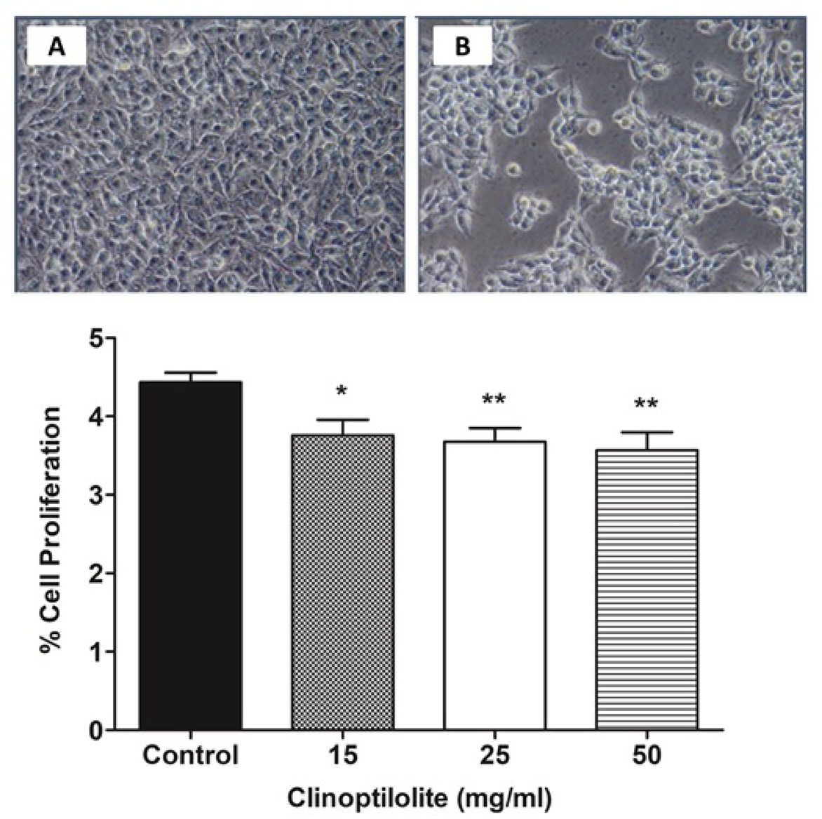
The effects of clinoptilolite treatment in different concentrations (15, 25 or 50 mg/ml) for 24 hours on cell proliferation in PC3 cells were studied using a WST-1 assay kit. Analysis showed that clinoptilolite treatment inhibited cell proliferation significantly in all applied concentrations and that 25 mg/ml is an effective (median) clinoptilolite concentration (Figure 1). This concentration was used in subsequent experiments.
Appearance of PC3 cells from control and clinoptilolite (25 mg/ml) groups and antiproliferative effects of clinoptilolite treatment in different concentrations (15, 25 or 50 mg/ml) for 24 h in PC3 cells. A: Control, B: Clinoptilolite. *p≤0.05, **p≤0.01 control vs. clinoptilolite, one-way ANOVA and Tukey’s multiple comparison test (n=7).
Effects of clinoptilolite on expression and activities of gelatinase enzymes
The expression and activity of gelatinases (MMP-2, MMP-9) in PC3 cells incubated in the presence or absence of clinoptilolite (25 mg/ml) for 24 hours were determined using the gelatin zymography method.
Gelatin zymography analysis revealed that 25 mg/ ml clinoptilolite treatment for 24 hours reduced MMP-2 expression significantly compared to the control. However, the pro-MMP-2 band was not observed in either control or clinoptilolite-treated cells (Figure 2). Furthermore, pro or active MMP-9 bands were also not observed in either group.
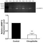
Representative zymogram image showing the effects of clinoptilolite (25 mg/ml) treatment for 24 h on aMMP-2 expression in PC3 cells. MW: Molecule weight marker, kDa: kilo Dalton, Cont.: Control, Clin.: Clinoptilolite, aMMP-2: Active MMP-2. **p≤0.01, control vs. clinoptilolite, paired Student’s t-test (n=4).
Effects of clinoptilolite on expression of MMP-14, TIMP-2 and ratios
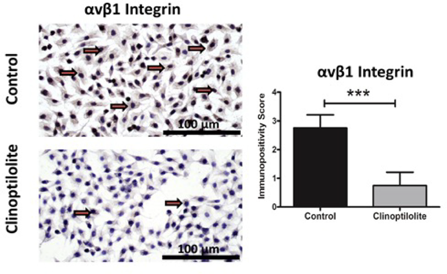
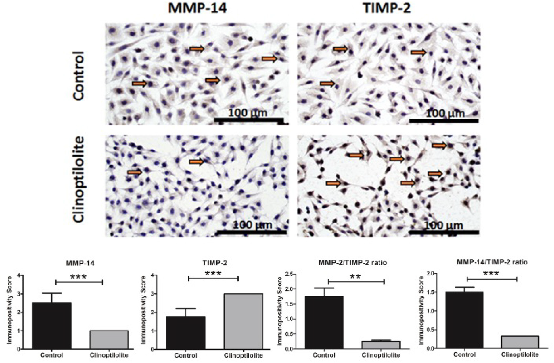
In immunohistochemical analysis, an increase was found in the expression of TIMP-2, the tissue inhibitor of MMP-2, in the clinoptilolite group compared to the control group. In contrast, a significant decrease was observed in the expression of αvβ1 integrin and MMP-14, the MMP-2 activator (Figure 3, Figure 4) As expected, MMP-2/TIMP-2 and MMP-14/TIMP-2 ratios were significantly decreased in the clinoptilolite group compared to the control group (Figure 4).
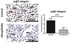
Effects of clinoptilolite (25 mg/ml) treatment for 24 h on the expression of αvβ1 integrin in PC3 cells. ***p≤0.001 control vs. clinoptilolite, paired Student’s t-test (n=4).
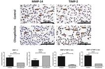
Effects of clinoptilolite (25 mg/ml) treatment for 24 h on the expression of MMP-14, TIMP-2, and MMP-2/TIMP-2, MMP-14/ TIMP-2 ratios in PC3 cells. **p≤0.01, ***p≤0.001 control vs. clinoptilolite, paired Student’s t-test (n=4).
DISCUSSION
This study investigated the potential anti-inflammatory and anti-cancer effects of clinoptilolite on gelatinases (MMP-2, MMP-9), MMP-14, TIMP-2, and αvβ1 integrin in PC3 cells. We demonstrated for the first time that clinoptilolite inhibits MMP-2 activity and MMP-14 expression while up-regulating TIMP-2 expression, significantly decreasing MMP-2/TIMP-2 and MMP-14/TIMP-2 ratios and down-regulating αvβ1 integrin expression in PC3 cells.
In the last 15 years, in experimental studies have indicated that synthetic MMP inhibitors can inhibit MMP activity by chelating with Zn2+ in the active site of MMPs and thus prevent both inflammation and cancer cell proliferation (Lokeshwar, 2011; Overall, Kleifeld, 2006). Second-generation synthetic MMP inhibitors are still being tested on various cancer types (Alaseem et al., 2019). Some of these inhibitors have been demonstrated to inhibit the formation, growth, and metastasis of tumour cells in prostate cancer (Lokeshwar, 2011; Ogut et al., 2016). Among these inhibitors, low-dose doxycycline is known to have an MMP inhibitor effect via chelation (Bench, Jeremias, Brown, 2011). Indeed, our group showed that doxycycline inhibits MMP activation, inflammation, and proliferation in PC3 cells (Ogut et al., 2016).
Considering previous evidence, we hypothesized that clinoptilolite may have MMP inhibitor activity in PC3 cells due to its chelator feature. Our results showed that clinoptilolite inhibited the activity of MMP-2 (which plays an important role in inflammation and proliferation) in PC3 cells. In our experiment conditions, MMP-2 activity in the control PC3 cells was considerably high since pro-MMP-2 transformed completely into active MMP-2. However, clinoptilolite significantly down-regulated MMP-2 activity due to its strong MMP inhibitory effect. On the other hand, pro and active MMP-9 bands were not observed in the zymogram in the present study. This finding, which is in parallel with our previous study, suggests that MMP-2 expression/activity is more prominent compared to MMP-9 expression/activity at the 24th hour under our experimental conditions in PC3 cells (Onursal et al., 2023).
We also studied the potential effects of clinoptilolite on MMP-14, which is the activator of MMP-2 and endogenous inhibitor TIMP-2 in PC3 cells. The individual expression levels or balance between these three markers may be a determinant of the prognosis of prostate cancer. MMP-14 up-regulation results in the activation of MMP-2 and indicates tumour cell metastasis and worse prognosis (Têtu et al., 2006). Once MMP-2 is activated, a trimer of pro-MMP-2, MMP-14, and TIMP-2 is formed. In this triad, TIMP-2 expression is critical for the control of MMP-2 activity. Insufficient TIMP-2 expression causes pro-MMP-2 to not be localized to the cell surface, while excess TIMP-2 reduces the level of free MMP-14 (Alcazar, Cousins, Marin-Castaño, 2007). MMP-14 is associated with increased ECM proteolysis, angiogenesis, cell migration, and invasion in many cancer types (Chang et al., 2016; Niland, Eble, 2020). Similarly, various cancer studies in humans have reported a relationship between the high proteolytic activity of MMP-14 and active MMP-2 levels (Figueira et al., 2009; Yosef, Arkadash, Papo, 2018). The correlation between the two MMPs and TIMP-2 makes them therapeutically important targets for the prevention or treatment of cancer. Therefore, in our study, we focused on gelatinases, MMP-14, and TIMP-2 as therapeutical targets and examined whether clinoptilolite affects these targets in androgen-independent PC3 prostate cancer cells.
We observed that clinoptilolite treatment downregulated MMP-2 and MMP-14 expression in correlation but up-regulated TIMP-2 expression - probably to compensate for high MMP-2 activity. In parallel with our results, MMP-2 and TIMP-2 expression levels were reported to change in opposite ways as a response to the treatment of inflammatory conditions such as atherosclerosis and cancer (Ogut et al., 2016; Onursal et al., 2023).
Additionally, we examined the ratios of MMP-14/ TIMP-2 and MMP-2/TIMP-2, which are critical in the final MMP/TIMP balance (Onursal et al., 2023). Both MMP-14/TIMP-2 and MMP-2/TIMP-2 ratios reduced with clinoptilolite treatment compared to the control in PC3 cells, which points at the prominent inhibitory effect of clinoptilolite on MMP-2 and MMP-14. The favourable effects of clinoptilolite on these critical ratios associated with cancer prognosis suggest that clinoptilolite may be useful for the treatment of prostate cancer.
Integrins are known to interact with ECM proteins and signalling pathways and to modulate tumour progression by mediating the expressions of specific MMPs (Niland, Eble, 2020). However, the expression pattern of αvβ1 integrin in various types of cancers is controversial. Some studies have reported that αvβ1 integrin in the upstream signalling pathway of MMPs up-regulates the expression and/or activity of MMP-2 and MMP-14, thus triggering chronic inflammation, invasion, and metastasis of cancer cells (Niland, Eble, 2020; Yang et al., 2012). In parallel with this view, αvβ1 integrin signalling was shown to trigger inflammation and cancer progression by stimulating the focal adhesion kinase signalling pathway and activating MMP-2 in glioma cells (Hu et al., 2006). Similarly, αvβ1 integrin signalling was reported to promote inflammation and cancer progression by mediating MMP-14 up-regulation in colorectal cancer (Yang et al., 2012).
Conversely, several studies on various cancer cell lines have reported that αvβ1 integrin may behave as a tumour suppressor gene in cancer progression (Schaffner, Ray, Dontenwill, 2013). Another study reporting that overexpression of αvβ1 inhibits the colonization and metastasis of colon cancer cells supports this evidence (Schirner et al., 1998). The discrepancy in these studies related to the roles of αvβ1 integrin may be attributed to the different stages and/or types of cancer cells (Hou et al., 2020).
With this contradictory information, in this study we investigated whether αvβ1 integrin signalling mediates the MMP inhibitor effects of clinoptilolite in PC3 cells. Our data revealed that clinoptilolite significantly down-regulated αvβ1 integrin expression consistently with MMP-2 and MMP-14 expression or activity while up-regulating TIMP-2 expression in PC3 cells. Clinoptilolite may therefore exert anti-inflammatory and anti-cancer effects in prostate cancer by inhibiting the αvβ1 integrin-mediated expression and/or activity of MMP-2 and MMP-14 due to its chelating properties.
Although new strategies are developed daily in cancer treatment and clinoptilolite has great potential in biomedical treatment, its mechanisms of action in human cancers have not yet been fully elucidated. This study may contribute to understanding the underlying mechanism of the anti-inflammatory and anti-cancer action of clinoptilolite in prostate cancer. However, further studies investigating the effects of clinoptilolite on other MMPs, integrins, and other upstream or downstream pathways and other types of cancer may help to illuminate the comprehensive molecular mechanisms in clinoptilolite action and enable discovery of innovative, targeted cancer therapies.
In conclusion, clinoptilolite may be useful in prostate cancer treatment as an anti-inflammatory and anti-cancer agent due to its αvβ1 integrin-mediated MMP inhibitory effects.
ACKNOWLEDGEMENTS
We kindly thank Ege University Faculty of Pharmacy FABAL Laboratories for supporting this research.
DATA AVAILABILITY STATEMENT
All data is available within the article or its supplementary materials.
REFERENCES
- Alaseem A, Alhazzani K, Dondapati P, Alobid S, Bishayee A, Rathinavelu A. Matrix Metalloproteinases: A challenging paradigm of cancer management. Semin Cancer Biol. 2019;56:100-115.
- Alcazar O, Cousins SW, Marin-Castaño ME. MMP-14 and TIMP-2 overexpression protects against hydroquinone-induced oxidant injury in RPE: implications for extracellular matrix turnover. Invest Ophthalmol Vis Sci. 2007;48(12):5662-5670.
- Bench TJ, Jeremias A, Brown DL. Matrix metalloproteinase inhibition with tetracyclines for the treatment of coronary artery disease. Pharmacol Res. 2011;64(6):561-566.
- Chang JH, Huang YH, Cunningham CM, Han KY, Chang M, Seiki M, et al. Matrix metalloproteinase 14 modulates signal transduction and angiogenesis in the cornea. Surv Ophthalmol. 2016;61(4):478-497.
- Deng X, He G, Liu J, Luo F, Peng X, Tang S, et al. Recent advances in bone-targeted therapies of metastatic prostate cancer. Cancer Treat Ver. 2014;40(6):730-738.
- Figueira RC, Gomes LR, Neto JS, Silva FC, Silva ID, Sogayar MC. Correlation between MMPs and their inhibitors in breast cancer tumor tissue specimens and in cell lines with different metastatic potential. BMC Cancer. 2009;9(20):1-11.
- Fingleton B. Matrix metalloproteinases as regulators of inflammatory processes. Biochim Biophys Acta Mol Cell Res. 2017;1864(11 Pt A):2036-2042.
- Gong Y, Chippada-Venkata UD, Oh WK. Roles of matrix metalloproteinases and their natural inhibitors in prostate cancer progression. Cancers. 2014;6(3):1298-1327.
- Hadler-Olsen E, Winberg JO, Uhlin-Hansen L. Matrix metalloproteinases in cancer: their value as diagnostic and prognostic markers and therapeutic targets. Tumour Biol. 2013;34(4):2041-2051.
- Hou J, Yan D, Liu Y, Huang P, Cui H. The Roles of Integrin α5β1 in Human Cancer. Onco Targets Ther. 2020;13:13329-13344.
- Hu B, Jarzynka MJ, Guo P, Imanishi Y, Schlaepfer DD, Cheng SY. Angiopoietin 2 induces glioma cell invasion by stimulating matrix metalloprotease 2 expression through the alphavbeta1 integrin and focal adhesion kinase signaling pathway. Cancer Res. 2006;66(2):775-783.
- Hu X, Beeton C. Detection of functional matrix metalloproteinases by zymography. J Vis Exp. 2010;45(e2445):1-5.
- Ilhan S, Dilekci G, GunerA, Bektas H. Newly Synthesized Benzimidazoles Inhibit Vascular Endothelial Growth Factor and Matrix Metalloproteinase-2 and -9 Levels in Prostate Cancer Cells. Anticancer Agents Med Chem. 2022;22(11):2109-2115.
- Inglezakis VJ, Zorpas AA. Handbook of natural zeolites: Bentham Science Publishers; 2012.
- Jiménez JA, Kao C, Raikwar S, Gardner TA. Current status of anti-angiogenesis therapy for prostate cancer. Urol Oncol. 2006;24(3):260-268.
- Kraljević Pavelić S, Simović Medica J, Gumbarević D, Filošević A, Pržulj N, Pavelić K. Critical Review on Zeolite Clinoptilolite Safety and Medical Applications in vivo. Front Pharmacol. 2018;9(1350):1-15.
- Lokeshwar BL. Chemically modified non-antimicrobial tetracyclines are multifunctional drugs against advanced cancers. Pharmacol Res. 2011;63(2):146-150.
- Mastinu A, Kumar A, Maccarinelli G, Bonini SA, Premoli M, Aria F, et al. Zeolite Clinoptilolite: Therapeutic Virtues of an Ancient Mineral. Molecules. 2019;24(8):1-15.
- Morozevich G, Kozlova N, Cheglakov I, Ushakova N, Berman A. Integrin alpha5beta1 controls invasion of human breast carcinoma cells by direct and indirect modulation of MMP-2 collagenase activity. Cell Cycle. 2009;8(14):2219-2225.
- Nagase H, Visse R, Murphy G. Structure and function of matrix metalloproteinases and TIMPs. Cardiovasc Res. 2006;69(3):562-573.
- Niland S, Eble JA. Hold on or Cut? Integrinand MMP-Mediated Cell-Matrix Interactions in the Tumor Microenvironment. Int J Mol Sci. 2020;22(238):1-41.
- Nissinen L, Kähäri VM. Matrix metalloproteinases in inflammation. Biochim Biophys Acta. 2014;1840(8):2571-2580.
- Ogut D, Reel B, Gonen Korkmaz C, Arun MZ, Cilaker Micili S, Ergur BU. Doxycycline down-regulates matrix metalloproteinase expression and inhibits NF-κB signaling in LPS-induced PC3 cells. Folia Histochem Cytobiol. 2016;54(4):171-180.
- Onursal C, Reel B, Bintepe C, Guzeloglu M, Ersoy N, Bagriyanik A. Pioglitazone inhibits oxidative stress, MMP-mediated inflammation and vascular dysfunction in high glucose-induced human saphenous vein grafts. J Diabet Complications. 2023;37(4):1-8.
- Overall CM, Kleifeld O. Tumour microenvironment - opinion: validating matrix metalloproteinases as drug targets and anti-targets for cancer therapy. Nat Rev Cancer. 2006;6(3):227-239.
- Pavelić K, Hadzija M, Bedrica L, Pavelić J, Dikić I, Katić M, et al. Natural zeolite clinoptilolite: new adjuvant in anticancer therapy. J Mol Med. 2001;78(12):708-720.
- Pavelić K, Katic M, Sverko V, Marotti T, Bosnjak B, Balog T, et al. Immunostimulatory effect of natural clinoptilolite as a possible mechanism of its antimetastatic ability. J Cancer Res Clin Oncol. 2002;128(1):37-44.
- Quinn DI, Sandler HM, Horvath LG, Goldkorn A, Eastham JA. The evolution of chemotherapy for the treatment of prostate cancer. Ann Oncol. 2017;28(11):2658-2669.
- Reed NI, Jo H, Chen C, Tsujino K, Arnold TD, DeGrado WF, et al. The αvβ1 integrin plays a critical in vivo role in tissue fibrosis. Sci Transl Med. 2015;7(288):1-17.
- Sandhu S, Moore CM, Chiong E, Beltran H, Bristow RG, Williams SG. Prostate cancer. Lancet. 2021;398(10305):1075-1090.
- Schaffner F, Ray AM, Dontenwill M. Integrin α5β1, the Fibronectin Receptor, as a Pertinent Therapeutic Target in Solid Tumors. Cancers. 2013;5(1):27-47.
- Schirner M, Herzberg F, Schmidt R, Streit M, Schöning M, Hummel M, et al. Integrin alpha5beta1: a potent inhibitor of experimental lung metastasis. Clin Exp Metastasis 1998;16(5):427-435.
- Sil H, Chatterjee A. Integrin alpha-V beta-3-matrix metalloproteinase-2 (MMP-2), cross-talk. J Cancer Ther. 2015;6(09):793-802.
- Têtu B, Brisson J, Wang CS, Lapointe H, Beaudry G, Blanchette C, et al. The influence of MMP-14, TIMP-2 and MMP-2 expression on breast cancer prognosis. Breast Cancer Res. 2006;8(3):1-9.
- Yang B, Gao J, Rao Z, Shen Q. Clinicopathological and prognostic significance of α5β1-integrin and MMP-14 expressions in colorectal cancer. Neoplasma. 2013;60(3):254-261.
- Yang B, Gao J, Rao Z, Shen Q. Clinicopathological significance and prognostic value of MMP-13 expression in colorectal cancer. Scand J Clin Lab Invest. 2012;72(6):501-505.
- Yapislar H, Taskin E, Ozdas S, Akin D, Sonmez E. Counteraction of Apoptotic and Inflammatory Effects of Adriamycin in the Liver Cell Culture by Clinopitolite. Biol Trace Elem Res. 2016;170(2):373-381.
- Yosef G, Arkadash V, Papo N. Targeting the MMP-14/MMP-2/integrin α(v)β(3) axis with multispecific N-TIMP2-based antagonists for cancer therapy. J Biol Chem. 2018;293(34):13310-13326.
- Zarkovic N, Zarkovic K, Kralj M, Borovic S, Sabolovic S, Blazi MP, et al. Anticancer and antioxidative effects of micronized zeolite clinoptilolite. Anticancer Res. 2003;23(2b):1589-1595.
Edited by
-
Associate Editor:
Michelle Carvalho