 Protective effects of bromelain in LPS-induced acute lung injury
Abstract
Acute lung injury (ALI) is a severe disease that can cause extensive lung damage and lead to death. Elevated levels of inflammatory cytokines and associated inflammation and oxidative damage irreversibly damage the lung tissue, leading to death. This study aimed to utilize the powerful antioxidant properties of bromelain (BRO) in ALI to determine its beneficial effects. We used an LPS-induced ALI model in rats, and 40 male specimens were divided into five groups: Healthy, Healthy+100BRO, ALI, ALI + 50BRO, and ALI + 100BRO. At the end of the study, histopathological and biochemical analyses of the lung tissue samples were performed. The results showed that tumor necrosis factor, interleukin-1, interleukin-6, and malondialdehyde levels increased in the ALI group and that these levels decreased dose-dependently with BRO treatment. We also demonstrated that superoxide dismutase levels, which decreased in the ALI group, increased dose-dependently in the BRO treatment groups. Histopathologically, significant improvement was detected in the BRO treatment groups. As a result of this research, it is suggested that BRO should be considered as supportive therapy in ALI. However, more detailed experimental and clinical studies on this subject are needed.
Keywords:
Acute lung injury; Bromelain; Inflammation; Rat
INTRODUCTION
Acute lung injury (ALI) is an acute diffuse inflammatory lung injury caused by various pathogenic factors (Tang et al., 2021). A large number of neutrophils infiltrating the lungs after various pathogenic factors increase proinflammatory cytokines and free oxygen radicals (reactive oxygen species), which leads to pulmonary microvascular endothelial and epithelial damage and plays a role in the development of ALI (Wan et al., 2018). ALI and its more severe form, acute respiratory distress syndrome, are important causes of morbidity and mortality. Despite decades of research, the mortality rate ranges from 22% to 40% (Mowery, Terzian, Nelson, 2020). Numerous drugs are used in the treatment of ALI, but a truly radical cure has yet to be found (Zhang, Zhao, Yang, 2022).
Many pulmonary and non-pulmonary conditions can cause ALI, but the most common cause is primary pneumonia due to bacterial, viral, or fungal causes (Matthay, Zemans, 2011). The second most common cause is severe sepsis, which may be associated with a source of infection such as pneumonia or peritonitis (Matthay, Zemans, 2011). Lung infection induced by lipopolysaccharide (LPS), a key component of the cell wall of gram-negative bacteria, is among the most prevalent causes of ALI (Tang et al., 2021). LPS is often used to model experimental ALI (Wan et al., 2018). LPS is preferred in ALI induction over a CLP-induced model because the standard dose of LPS application induces a more standard ALI model than a CLP model with its many variables such as intestine content, ligation errors, and/or puncture differences (Zhou et al., 2023). Proinflammatory cytokines such as the tumor necrosis factor (TNF-α), interleukin-1 (IL-Ιβ), and interleukin-6 (IL-6) mediate the initiation and regulation of inflammatory responses in ALI (Song et al., 2021). Studies have shown that TNF-α release occurs rapidly when the lung is exposed to LPS (Jiang et al., 2015). TNF-α induces the inflammatory cascade and is an important parameter in the severity of lung injury (Cinar et al., 2019). TNF-α also leads to an increase in free oxygen radicals (Cadirci et al., 2019). IL-6 exacerbates the body’s inflammatory response by enhancing the synthesis and release of acute-phase reactants (Chen et al., 2015a). IL-1β, a crucial mediator in the acute phase of inflammation, plays a role in the repair of alveolar epithelial tissue (Chen et al., 2015b). Due to the interaction of LPS with the TLR4 receptor, free oxygen radicals and proinflammatory cytokines are released. This leads to lipid peroxidation and causes cell and organ damage (Meng et al., 2018). Parameters used as markers of lipid peroxidation in ALI include superoxide dismutase (SOD) and malondialdehyde (MDA) levels. Previous ALI studies indicated that free oxygen radical accumulation, which occurs due to the systemic inflammatory response, plays a vital role in tissue damage (Pendyala et al., 2009). ALI is thought to be prevented by pharmacologic agents that inhibit or scavenge the formation of free oxygen radicals (Cinar et al., 2019). Therefore, using an agent that modulates inflammation and has antioxidant properties in treating ALI may be useful. In prior studies, bromelain (BRO) attracted attention due to its antioxidant and anti-inflammatory role.
BRO is a proteolytic enzyme containing a sulfhydryl group derived from the Ananas comosus plant (Hikisz, Bernasinska-Slomczewska, 2021). It possesses antiinflammatory, cardioprotective, immunomodulatory, anticoagulant, and antioxidant activities (Zhou et al., 2017; El-Demerdash et al., 2022; Hu et al., 2022; Agostinis et al., 2015; Juhasz et al., 2008). Many clinical studies have shown that BRO is safe and effective against various medical conditions such as sinusitis, osteoarthritis, surgical wounds, cardiovascular health, and digestive system diseases (Della Volpe et al., 2022; Gupta et al., 2022; Shoham et al., 2021; Jayachandran, Khobre, 2017; Ley et al., 2016). In vivo studies examining the effects of BRO on lung tissue can also be found. A previous study showed that BRO has protective effects in cadmium-induced lung toxicity (Rafiei-Asl et al., 2021). BRO has also been shown to be therapeutic in allergic airway disease (Secor et al., 2012). In addition, a recent study has demonstrated that BRO may synergistically reduce benzopyrene-induced lung carcinogenesis associated with inflammation and oxidative stress (Majumder et al., 2021).
No studies on the effects of BRO in ALI were found in the literature. Therefore, this study aimed to evaluate the possible impact of BRO on oxidative and inflammatory markers via biochemical and histopathological methods in an LPS-induced ALI model in rats.
MATERIAL AND METHODS
Forty male Wistar albino rats weighing between 240 and 280 g were used in this study. The animals were obtained from the Atatürk University Experimental Research and Application Centre experimental animal laboratory. During the experiment, the rats were given water and pellet feed ad libitum. They were housed in groups in the laboratory at normal room temperature (22 °C) and fed before the experiment.
Experimental protocols on the rats
BRO was administered orally to the experimental group specimens. The manufacturer’s instructions were followed to render the powdered substances into a saline solution. The same solvent was similarly administered to the Healthy group.
The rats were randomly divided into the following five groups:
-
Healthy group: the healthy rats received saline solution (0.9% NaCl) as a vehicle (n = 8)
-
Healthy + 100BRO group: the healthy rats were given 100 mg/kg of BRO (n = 8)
-
ALI group: rats with ALI (n = 8)
-
ALI + 50BRO group: the rats with ALI received 50 mg/kg of BRO (n = 8)
-
ALI + 100BRO group: the rats with ALI were administered 100 mg/kg of BRO (n = 8)
BRO was administered to the Healthy + 100BRO, ALI + 50BRO, and ALI + 100BRO groups via oral gavage at the doses (Şehirli et al., 2021) mentioned above. The rats were given these doses 1 h after induction of ALI with LPS (Shaban et al., 2023).
After placing the rats in a supine position, a midline cervical incision was made in the skin. To reduce tissue damage, the fascia, sternocleidomastoid muscle, and parathyroid glands were meticulously excised using cotton swabs, facilitating surgical access to the trachea. A 26G sterile insulin injector was introduced into the intratracheal space via the tracheal cartilage. After retracting the injector’s piston to allow air to enter, 5 mg/kg of LPS (Sigma-Aldrich, L2880, LPS from Escherichia coli O55:B5), diluted in isotonic NaCl, was gradually administered into the trachea. After the injection, the rats were rotated along three axes to ensure the even distribution of LPS throughout the lungs. The skin was then closed using an Appose ULC 35 W AutoSuture Slim Body Skin Stapler (Covidien, Dublin, Ireland) (Aydin et al., 2022).
All groups were anesthetized via intraperitoneal administration of a ketamine and xylazine combination 12 h after induction of ALI. Cardiac blood and lung tissue samples were collected from the specimens, and they were then euthanized. One half of each lung was perfused with saline and stored at -80 °C for biochemical and molecular analyses; the other half was fixed in a 10% formalin solution for histopathological examination. The blood samples were collected in serum tubes and centrifuged at 4 °C and 4000 rpm for 10 min. The resulting supernatants were collected, frozen at -20 °C, and placed in an ultra-cooler at -80 °C for future biochemical parameter measurement. On the day of the study, the supernatants were removed from the cooler, brought to room temperature, and the experiments were performed.
Biochemical analyses
The collected lung tissues were first homogenized. A TissueLyser II grinder jar set (QIAGEN, Hilden, Germany) was used for this. Centrifugation was then performed (Aydin et al., 2022). The TNF-α, IL-1, and IL-6 contents were analyzed in the lung tissues via enzyme-linked immunosorbent assay (ELISA) following the manufacturer’s instructions. The MDA and SOD contents were analyzed via the manual method.
SOD manual method analysis
SOD activity was measured according to the method described by Sun et al. (1988). The estimation was based on the production of superoxide anion (O2-) generated from the reaction between xanthine and xanthine oxidase, which then reacts with nitro blue tetrazolium to yield formazan dye. SOD activity was subsequently quantified by assessing the extent of inhibition of this reaction at 560 nm.
MDA manual method analysis
MDA levels were measured following the protocol established by Ohkawa, Ohishi, and Yagi (1979). The measurement principle is based on assessing the absorbance at 532 nm of the pink compound formed from the reaction of MDA with thiobarbituric acid, which results from lipid peroxidation.
Histopathological procedure
Histopathological specimens were immediately fixed in a 10% formalin solution for 48 h. After fixation, the tissues were routinely processed and embedded in paraffin. Sections of 3 μm were obtained from the tissue blocks, stained with hematoxylin and eosin (H&E), and photographed for histopathological evaluation using a light microscope equipped with a digital camera. For the histopathological assessment of the edema area, alveolar wall thickness and inflammatory cell infiltration were evaluated based on the previous literature (Ugan et al., 2018).
Statistical analysis
IBM SPSS Statistics 20 (IBM Corp., Armonk, NY, USA) was used to compare the data. The results were evaluated via a one-way analysis of variance and compared using Tukey’s test; p < 0.05 was considered significant.
RESULTS
Biochemical analysis results
IL-6 results
As shown in Figure 1, the IL-6 level in the ALI group was significantly increased compared to the Healthy group in the measurements made via ELISA (p < 0.001). The IL-6 levels were significantly lower in the ALI + 50BRO and ALI + 100BRO groups than in the ALI group (p < 0.001). The BRO-induced decrease in the IL-6 levels was dose-dependently increased in the ALI groups (Figure 1A).

IL-6 (a), IL-1β (b), and TNF-α (c) levels in rat lung tissue. *comparison according to the Healthy group; #comparison according to the ALI group; ***significant difference at p < 0.001 compared to the Healthy group; ###significant difference at p < 0.001 compared to the ALI group.
IL-1β results
As seen in Figure 2, the IL-Ιβ levels in the ALI group were significantly increased compared to the Healthy group in the measurements made via ELISA (p < 0.001). In the ALI + 50BRO and ALI + 100BRO groups, the IL-Ιβ results were significantly decreased compared to the ALI group (p < 0.001). Among the ALI groups, the decrease in the IL-1β levels was higher in the ALI + 100BRO group (p < 0.001) (Figure 1B).

SOD activity and MDA level in rat lung tissue. *comparison according to the Healthy group; #comparison according to the ALI group; ***significant difference at p < 0.001 compared to the Healthy group; ###significant difference at p < 0.001 compared to the ALI group; **significant difference at p < 0.01 compared to the Healthy group; ##significant difference at p < 0.01 compared to the ALI group.
TNF-α results
As shown in Figure 3, ELISA measurements indicated that TNF-α levels in the ALI group were significantly elevated compared to those in the Healthy group (p < 0.001). BRO treatment reduced TNF-α levels compared to the ALI group. The decrease in the TNF-α levels in the ALI groups was higher in the ALI + 100BRO group (Figure 1C).

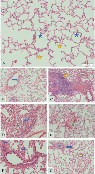
Histopathological examination of the H&E sections of the Healthy group (A), in which no pathological findings were observed histomorphologically (blue star: alveolar space, yellow star: alveolar sac), Healthy + BRO100 group (B), which did not reveal any pathologies in the respiratory and circulatory units of the lung (blue arrow: ordinary bronchial epithelium), ALI group (C, D, E), in which areas of interalveolar hemorrhage were also observed (yellow arrow: intense inflammation around the vascular structure; blue arrow: parenchymal edema and inflammation; green arrow: intra-alveolar hemorrhage), ALI + 50 group (F) (blue arrows: mildly edematous, inflamed lung parenchyma), and the ALI + 100 group (g) (blue arrows: intact bronchioles, yellow arrow: mildly edematous parenchymal area).
SOD results
The SOD enzyme level in the ALI group was significantly decreased compared to the Healthy group. BRO treatment significantly increased SOD levels in both the ALI + 50BRO (p < 0.01) and ALI + 100BRO (p < 0.001) groups compared to the ALI group. The best SOD activity was realized in the ALI + 100BRO group compared to the ALI group (p < 0.001) (Figure 2A).
MDA results
As shown in Figure 4, the measurements revealed that MDA levels in the ALI group were significantly higher than those in the Healthy group (p < 0.001). BRO treatment significantly decreased the MDA levels at both doses compared to the ALI group (p < 0.001). The ALI + 100BRO group had the highest decrease in MDA levels (Figure 2B).
Histopathological results
In the histopathological examination of H&E sections in the Healthy and Healthy + 100BRO groups, the bronchi, terminal bronchioles, respiratory bronchioles, alveolar sacs, interalveolar septa, arteries, veins, and capillary vasculature in the lung tissue were examined in detail (Figures 3A, 3B). In the ALI group, advanced edema and inflammation were observed around the vascular structures. The interalveolar septum of the lung parenchyma was enlarged with diffuse parenchymal edema. Areas of interalveolar hemorrhage were also observed (Figures 3C, 3D, 3E). In the ALI + 50BRO group, edema and inflammation were observed in the H&E sections. However, the findings were more attenuated than those of the ALI group (Figure 3F). Interalveolar septa were observed to be relatively close to normal due to decreased edema in the parenchyma. In the ALI + 100BRO group, edematous areas around the vessels, submucosa, and terminal bronchioles were significantly reduced. Alveolar septum thickening was found to be normal enough to be considered healthy. Inflammation cells were rarely seen in the tissue. In general, a histologic appearance similar to the Healthy group was observed in this group (Figure 3G).
DISCUSSION
In this study, the effects of BRO—increasingly popular because of its antioxidant and anti-inflammatory properties, on LPS-induced ALI were examined through TNF-α, IL-1β, IL-6, SOD, and MDA levels. In ALI induced by LPS, increased TNF- α, IL-1β, and IL-6 cytokine levels and increased MDA levels, along with oxidative damage, decreased with BRO administration. The levels of SOD, an important part of the antioxidant system, increased in the Healthy + 100BRO, ALI + 50BRO, and ALI + 100BRO groups compared to the ALI group. Histopathological examination showed that BRO administration decreased inflammation and pulmonary edema.
ALI is an acute diffuse inflammatory lung injury in which the endothelial and epithelial barriers of the lung are disrupted. An intense influx of neutrophils into the lungs occurs with increased free oxygen radicals and proinflammatory mediators (Zhang, Zhao, Yang, 2022). The severe inflammatory process occurring in ALI and the resulting high levels of TNF- α, IL-1β, and IL-6 cytokines in the body are positively correlated with the morbidity and mortality of the disease (Mowery, Terzian, Nelson, 2020). Control of excessive inflammatory response and uncontrolled production of free oxygen radicals are critical therapeutic targets in treating ALI (Ge et al., 2020). Numerous mouse models have demonstrated that antioxidant substances can alleviate the severity of ALI (Kellner et al., 2017). Therefore, recent studies have focused on substances with antioxidant or anti-inflammatory properties for treating the disease. When clinical studies were examined, substances with antioxidant properties such as melatonin (Kang et al., 2022), mitoquinone (Zhan et al., 2022), obacunone (Li et al., 2022), and quercetin (Sang et al., 2022) were tested in ALI, and significant results were obtained. BRO, an antioxidant substance, has also come to the forefront in recent studies.
As a proteolytic enzyme obtained from Ananas comosus, BRO is classified within the Bromeliaceae family (Didamoony et al., 2022) and is a protease enzyme containing different thiol groups; these groups protect cells against oxidative stress caused by free radicals due to their sulfhydryl content (Saptarini, Rahayu, Herawati, 2019). In this way, BRO acts as an antioxidant by inhibiting lipid peroxidation and scavenging free oxygen radicals (Khazaeel et al., 2022). It has also been shown to have hepatoprotective (Hu et al., 2020), neuroprotective (Kumar et al. , 2022), and pulmonoprotective (Rafiei-Asl et al., 2021) properties. A prior study observed that antioxidant enzymes increased and free oxygen radicals decreased with BRO application in rats with liver damage (El-Demerdash et al., 2022). In another study, the authors found that SOD levels increased and MDA levels decreased after BRO administration to mice given pesticides (Agarwal et al., 2016). In the current study, the SOD level increased, and the MDA level decreased with BRO administration in LPS-induced ALI.
In the pathogenesis of ALI, the accumulation of free oxygen radicals and the increase in proinflammatory cytokines play an essential role. Free oxygen radicals can activate various transcription factors involved in inflammatory pathways. One is NF-ΚB, an important transcription factor (Sul, Ra, 2021). Activation of NF-ΚB results in elevated levels of proinflammatory cytokines, contributing to the progression of ALI (Li, Verma, 2002). Research has shown that BRO reduces inflammatory cytokine levels by inhibiting NF-ΚB translocation, resulting in an anti-inflammatory effect (Hong et al., 2021). BRO has been found to suppress the activity of important cytokines and molecules associated with the inflammatory process in LPS-induced inflammation models in studies involving human (Bhui et al., 2012) and rat (Habashi et al., 2016) cell cultures. Another study found that administering BRO led to reduced levels of TNF-α, IL-1β, IL-6, and MDA alongside elevated SOD levels in rats with ocular toxicity (Okkay et al., 2023).
In the present study, BRO significantly decreased elevated TNF-α, IL-1β, and IL-6 levels in LPS-induced ALI. The histopathological examination revealed perivascular edema, inter alveolar septum thickening, submucosal edema, and severe inflammatory areas in the ALI group. These were improved in the Healthy + 100BRO, ALI + 50BRO, and ALI + 100BRO groups. Considering this information, the biochemical and histopathological analyses demonstrated that BRO elevates antioxidant enzymes and lowers inflammatory cytokine levels, including TNF-α, IL-1β, and IL-6 in ALI. Accordingly, we recommend that BRO should, at a minimum, be considered as supportive therapy in ALI. However, more detailed experimental and clinical studies on this subject are needed.
The study’s limitations include the lack of western blot analyses to determine protein levels directly in lung tissue and the fact that we only evaluated BRO’s effects on one ALI model.
ACKNOWLEDGMENTS
We would like to thank Prof. Dr. Zekai Halici for his scientific contributions. We also thank Atatürk University’s Scientific Research Projects Coordination Unit for supporting this study (grand number TAB-2022-10681).
DATA AVAILABILITY STATEMENT
Data available from the corresponding author upon reasonable request.
REFERENCES
- Agarwal S, Chaudhary B, Bist R. Bacoside A and bromelain relieve dichlorvos induced changes in oxidative responses in mice serum. Chem Biol Interact. 2016;254:173-178.
- Agostinis C, Zorzet S, De Leo R, Zauli G, De Seta F, Bulla R. The combination of N-acetyl cysteine, alphalipoic acid, and bromelain shows high anti-inflammatory properties in novel in vivo and in vitro models of endometriosis. Mediat Inflamm. 2015;1:918089.
- Aydin P, Magden ZBA, Uzuncakmak SK, Halici H, Akgun N, Mendil AS, et al. Avanafil as a novel therapeutic agent against LPS-induced acute lung injury via increasing CGMP to downregulate the TLR4-NF-κB-NLRP3 inflammasome signaling pathway. Lung. 2022;200:561-572.
- Bhui K, Tyagi S, Srivastava AK, Singh M, Roy P, Singh R, et al. Bromelain inhibits nuclear factor kappa-B translocation, driving human epidermoid carcinoma A431 and melanoma A375 cells through G 2/M arrest to apoptosis. Mol Carcinog. 2012;51(3):231-243.
- Cadirci E, Ugan RA, Dincer B, Gundogdu B, Cinar I, Akpinar E, et al. Urotensin receptors as a new target for CLP induced septic lung injury in mice. Naunyn Schmiedebergs Arch Pharmacol. 2019;392:135-145.
- Cinar I, Sirin B, Aydin P, Toktay E, Cadirci E, Halici I, et al. Ameliorative effect of gossypin against acute lung injury in experimental sepsis model of rats. Life Sci. 2019;221:327-334.
- Chen T, Guo Q, Wang H, Zhang H, Wang C, Zhang P, et al. Effects of esculetin on lipopolysaccharide (LPS)-induced acute lung injury via regulation of RhoA/Rho Kinase/NF-κB pathways in vivo and in vitro. Free Radic Res. 2015;49(12):1459-1468.
- Chen T, Mou Y, Tan J, Wei L, Qiao Y, Wei T, et al. The protective effect of CDDO-Me on lipopolysaccharide-induced acute lung injury in mice. Int Immunopharmacol. 2015;25(1):55-64.
- Della Volpe A, De Luca P, De Lucia A, Martines F, Piroli P, D’Ascanio L, et al. A. Single-center-single-blinded clinical trial to evaluate the efficacy of a nutraceutical containing boswellia serrata, bromelain, zinc, magnesium, honey, tyndallized lactobacillus acidophilus and casei to fight upper respiratory tract infection and otitis media. Healthcare. 2022;10(8):1526.
- Didamoony MA, Atwa AM, Abd El-Haleim EA, Ahmed LA. Bromelain ameliorates D-galactosamine-induced acute liver injury: role of SIRT1/LKB1/AMPK, GSK3p/Nrf2 and NF-κB p65/TNF-α/caspase-8, -9 signalling pathways. J Pharm Pharmacol. 2022;74(12):1765-1775.
- El-Demerdash FM, Hussien DM, Ghanem NF, Al-Farga AM. bromelain modulates liver injury, hematological, molecular, and biochemical perturbations induced by aluminum via oxidative stress inhibition. Biomed Res Int. 2022;1:5342559.
- Okkay IF, Okkay U, Bayram C, Cicek B, Sezen S, Aydin IC, et al. Bromelain protects against cisplatin-induced ocular toxicity through mitigating oxidative stress and inflammation. Drug Chem Toxicol. 2023;46(1):69-76.
- Ge X, Meng X, Fei D, Kang K, Wang Q, Zhao M. Lycorine attenuates lipopolysaccharide-induced acute lung injury through the HMGB1/TLRs/NF-κB pathway. 3 Biotech. 2020;10:1-10.
- Gupta AA, Kambala R Bhola N, Jadhav A. Comparative efficacy of bromelain and aceclofenac in limiting post-operative inflammatory sequelae in surgical removal of lower impacted third molar: A randomized controlled, triple blind clinical trial. J Dent Anesth Pain Med. 2022;22(1):29.
- Habashi SA, Sabouni F, Moghimi A, Majd SA. Modulation of lipopolysaccharide stimulated nuclear factor kappa B mediated iNOS/NO production by bromelain in rat primary microglial cells. Iran Biomed J. 2016;33-40.
- Hikisz P, Bernasinska-Slomczewska J. Beneficial properties of bromelain. Nutrients. 2021;13(12):4313.
- Hong JH, Kim MR, Lee BN, Oh WM, Min KS, Im YG, et al. Anti-inflammatory and mineralization effects of bromelain on lipopolysaccharide-induced inflammation of human dental pulp cells. Medicina. 2021;57(6):591.
- Hu PA, Chen CH, Guo BC, Kou YR, Lee TS. Bromelain confers protection against the non-alcoholic fatty liver disease in male C57bl/6 mice. Nutrients. 2020;12(5):1458.
- Hu PA, Wang SH, Chen CH, Guo BC, Huang JW, Lee TS. New mechanisms of bromelain in alleviating non-alcoholic fatty liver disease-induced deregulation of blood coagulation. Nutrients. 2022;14(11): 2329.
- Jayachandran S, Khobre P. Efficacy of bromelain along with trypsin, rutoside trihydrate enzymes and diclofenac sodium combination therapy for the treatment of TMJ osteoarthritis—A randomised clinical trial. J Clin Diagn Res. 2017;11(6):ZC09
- Jiang Q, Yi M, Guo Q, Wang C, Wang H, Meng S, et al. Protective effects of polydatin on lipopolysaccharide-induced acute lung injury through TLR4-MyD88-NF-κB pathway. Int Immunopharmacol. 2015;29(2):370-376.
- Juhasz B, Thirunavukkarasu M, Pant R, Zhan L, Penumathsa SV, Secor ER, et al. Bromelain induces cardioprotection against ischemia-reperfusion injury through Akt/FOXO pathway in rat myocardium. Am J Physiol Heart Circ Physiol. 2008;294(3):1365-1370.
- Kang JY, Xu MM, Sun Y, Ding ZX, Wei YY, Zhang DW, et al. Melatonin attenuates LPS-induced pyroptosis in acute lung injury by inhibiting NLRP3-GSDMD pathway via activating Nrf2/HO-1 signaling axis. Int Immunopharmacol. 2022;109:108782.
- Kellner M, Noonepalle S, Lu Q, Srivastava A, Zemskov E, Black SM. ROS signaling in the pathogenesis of Acute Lung Injury (ALI) and Acute Respiratory Distress Syndrome (ARDS). Adv Exp Med Biol. 2017;105-137.
- Khazaeel K, Rad OR, Jamshidian J, Tabandeh MR, Mohammadi G, Atashfaraz A. Effect of bromelain on sperm quality, testicular oxidative stress and expression of oestrogen receptors in bisphenol-A treated male mice. Andrologia. 2022;54(11):e14584.
- Kumar R, Kumar R, Sharma N, Khurana N, Singh SK, Satija S, et al. Pharmacological evaluation of bromelain in mouse model of Alzheimer’s disease. Neurotoxicology. 2022;90:19-34.
- Ley CM, Ni Q, Liao X, Gao HL, Robinson N. Bromelain and cardiovascular risk factors in diabetes: An exploratory randomized, placebo controlled, double blind clinical trial. Chin J Integr Med. 2016;22:728-737.
- Li J, Deng S hua, Li J, Li L, Zhang F, Zou Y, et al. Obacunone alleviates ferroptosis during lipopolysaccharide-induced acute lung injury by upregulating Nrf2-dependent antioxidant responses. Cell Mol Biol Lett. 2022;27(1):29.
- Li Q, Verma IM. NF-κB regulation in the immune system. Nat Rev Immunol. 2002;2(10):725-734.
- Majumder D, Debnath R, Nath P, Libin Kumar KV, Debnath M, Tribedi P, et al. Bromelain and Olea europaea (L.) leaf extract mediated alleviation of benzo(a)pyrene induced lung cancer through Nrf2 and NFκB pathway. Environ Sci Pollut Res Int. 2021;28(34):47306-47326.
- Matthay MA, Zemans RL. The acute respiratory distress syndrome: Pathogenesis and treatment. Annu Rev Pathol Mech Dis. 2011;6(1):147-163.
- Meng L, Li L, Lu S, Li K, Su Z, Wang Y, et al. The protective effect of dexmedetomidine on LPS-induced acute lung injury through the HMGB1-mediated TLR4/NF-ΚB and PI3K/Akt/mTOR pathways. Mol Immunol. 2018;94:7-17.
- Mowery NT, Terzian WTH, Nelson AC. Acute lung injury. Curr Probl Surg. 2020;57(5):100777.
- Ohkawa H, Ohishi N, Yagi K. Assay for lipid peroxides in animal tissues by thiobarbituric acid reaction. Anal Biochem. 1979;95(2):351-358.
- Pendyala S, Usatyuk P V., Gorshkova IA, Garcia JGN, Natarajan V. Regulation of NADPH oxidase in vascular endothelium: The role of phospholipases, protein kinases, and cytoskeletal proteins. Antioxid. Redox Signal. 2009;11(4):841-860
- Rafiei-Asl S, Khadjeh G, Jalali SM, Jamshidian J, Rezaie A. Protective effects of bromelain against cadmium-induced pulmonary intoxication in rats: a histopathologic and cytologic study. Arch Razi Inst. 2021;76(5):1427-1436.
- Sang A, Wang Y, Wang S, Wang Q, Wang X, Li X, et al. Quercetin attenuates sepsis-induced acute lung injury via suppressing oxidative stress-mediated ER stress through activation of SIRT1/AMPK pathways. Cell Signal. 2022;96:10363.
- Saptarini N, Rahayu D, Herawati I. Antioxidant activity of crude bromelain of pineapple (Ananas comosus (L.) Merr) crown from Subang district, Indonesia. J Pharm Bioallied Sci. 2019;11(4):551-555.
- Secor ER Jr, Shah SJ, Guernsey LA, Schramm CM, Thrall RS. Bromelain limits airway inflammation in an ovalbumin-induced murine model of established asthma. Altern Ther Health Med. 2012; 18(5):9-17.
- Shaban P, Honari N, Erfanian N, Hosseini_M, Safarpour H, Nasseri S. Anti-inflammatory effects of ziziphus jujube Mill on LPS-induced acute lung injury in mice. Iran J Allergy Asthma Immunol. 2023;22(3):281-289
- Shoham Y, Shapira E, Haik J, Harats M, Egozi D, Robinson D, et al. Bromelain-based enzymatic debridement of chronic wounds: Results of a multicentre randomized controlled trial. Wound Repair Regen. 2021;29(6):899-907.
- Song D, Zhao M, Feng L, Wang P, Li Y, Li W. Salidroside attenuates acute lung injury via inhibition of inflammatory cytokine production. Biomed Pharmacother. 2021;142:111949.
- Sul OJ, Ra SW. Quercetin prevents lps-induced oxidative stress and inflammation by modulating nox2/ros/nf-kb in lung epithelial cells. Molecules. 2021;26(22):6949.
- Sun Y, Oberley LW, Li Y. A simple method for clinical assay of superoxide dismutase. Clin Chem. 1988;34(3):497-500.
- Şehirli AÖ, Sayiner S, Saytekin G, Velioğlu-Öğünç. Protective effect of bromelain on corrosive burn in rats. Burns. 2021;47(6):1352-1358.
- Tang J, Xu L, Zeng Y, Gong F. Effect of gut microbiota on LPS-induced acute lung injury by regulating the TLR4/NF-κB signaling pathway. Int Immunopharmacol. 2021;91:107272.
- Ugan RA, Cadirci E, Halici Z, Toktay E, Cinar I. The role of urotensin-II and its receptors in sepsis-induced lung injury under diabetic conditions. Eur J Pharmacol. 2018;818:457-469.
- Wan L, Meng D, Wang H, Wan S, Jiang S, Huang S, et al. Preventive and therapeutic effects of thymol in a lipopolysaccharide-induced acute lung injury mice model. Inflammation. 2018;41:183-192.
- Zhan P, Lu X, Li Z, Wang WJ, Peng K, Liang NN, et al. Mitoquinone alleviates bleomycin-induced acute lung injury via inhibiting mitochondrial ROS-dependent pulmonary epithelial ferroptosis. Int Immunopharmacol. 2022;113:109359.
- Zhang C, Zhao Y, Yang X. Azilsartan attenuates lipopolysaccharide-induced acute lung injury via the Nrf2/HO-1 signaling pathway. Immunol Res. 2022;70(1):97-105.
- Zhou G, Xie D, Fan R, Yang Z, Du J, Mai S, et al. Comparison of Pulmonary and Extrapulmonary Models of Sepsis-Associated Acute Lung Injury. Physiol Res. 2023;72(6):741-752.
- Zhou Z, Wang L, Feng P, Yin L, Wang C, Zhi S, et al. Inhibition of Epithelial TNF-alpha Receptors by Purified Fruit Bromelain Ameliorates Intestinal Inflammation and Barrier Dysfunction in Colitis. Front Immunol. 2017;8:1468.