 Recent advances in paclitaxel polymer nanomedicine delivery for cancer therapy
Abstract
Paclitaxel (PTX) is a widely used anti-tumor drug in cancer therapy. However, its poor water solubility restricts its application in therapeutic procedures. Furthermore, the Cremophor® in the formulation can cause various side effects, leading to non-specific drug distribution in tumors and normal tissues. To overcome these limitations, researchers have explored numerous drug delivery systems to enhance the solubility of PTX and achieve targeted delivery. One such strategy involves using polymer nanoparticles, tiny particles composed of polymers that encapsulate and stabilize PTX, thereby improving its solubility and bioavailability. In addition, these polymer nanoparticles can enhance the therapeutic effect of PTX through passive targeting and active targeting mechanisms. This review first highlights the challenges of paclitaxel in current applications, focusing on current synthetic advances in PTX and drug resistance; subsequently, various approaches to deliver PTX using polymer Nanoparticles are discussed. Finally, current challenges and prospects are reviewed. Our goal was to demonstrate how these different nanocarriers can improve the therapeutic efficiency of PTX as a chemotherapeutic agent.
Keywords:
Paclitaxel; Cancer treatment; Drug delivery; Polymer.
INTRODUCTION
Paclitaxel (PTX) is an essential member of the paclitaxel family, a diterpene alkaloid isolated from the Pacific Yew tree by Wall and Wani from the Research Triangle Institute in 1967 (Figure 1) (Abu Samaan et al., 2019). The fundamental structure of PTX consists of four oxetane rings and an ester side chain. In the C13 chain, the C3' amide acyl group acts as the active moiety of the compound, while the hydroxyl group at the C2' position enhances its function. These key critical groups were retained in developing active derivatives of PTX, such as doxorubicin and cabazitaxel (Alavi, Nokhodchi, 2022). Since its approval by the FDA in 1992, PTX has become one of the clinic's more commonly used anticancer drugs (Figure 2 for the research process of PTX). PTX has demonstrated significant therapeutic efficacy in treating various tumors, especially cancer early-stage (Ji et al., 2021; Zhu, Chen, 2019). In addition, it can also be used to alleviate the symptoms and improve the quality of life of patients with advanced cancer (Hashem et al., 2022). Clinically, PTX is widely used in the treatment of breast cancer, ovarian cancer, non-small cell lung cancer, Kaposi's sarcoma, and other cancers etc (Alavi, Nokhodchi, 2022). By interfering with the usual standard assembly and stabilization of intracellular microtubule structure, PTX blocks the mitotic process of cancer cells, thus inhibiting their proliferation and ultimately leading to their apoptosis (Das et al., 2021). Due to its wide clinical application and remarkable efficacy, the World Health Organisation (WHO) has also included PTX in the list of essential medicines to ensure its accessibility and rational use worldwide (Sofias et al., 2017). However, it should be noted that PTX also has some side effects, such as bone marrow suppression, neuropathological reactions, and allergic reactions (Cai et al., 2020).


However, due to its poor water solubility and binding to plasma proteins, PTX has a wide distribution in the body, leading to the use of Cremophor® (polyoxyethylene castor oil, CrEL) as a solubilizing agent for drug delivery (Xiao et al., 2021). Unfortunately, CrEL causes severe side effects, such as allergic reactions, nephrotoxicity, neurotoxicity, and cardiotoxicity, which are distressing to patients. The structure of PTX lacks functional groups, making its solubility enhancement one of the main challenges (Yi et al., 2021). Researchers have recently improved PTX delivery by utilizing drug delivery systems, such as polymeric nanoparticles, hydrogels, and liposomes. Among them, polymer nanoparticles have attracted much attention (Zare et al., 2021). These polymers encapsulate and stabilize PTX, and nanoparticles comprise polymers that encapsulate and stabilize PTX and enhance its solubility and stability. Delivery nanoparticles enable more precise drug delivery by modifying particle size (Arruda et al., 2023), surface properties, and drug release rate (Chen et al., 2022). Additionally, polymeric Nanoparticles can actively target tumor cells by modifying targeting ligands on their surface (Colby et al., 2017). Researchers have also explored other approaches to improve PTX's solubility and efficiency, such as using hydrogels and liposomes (Ding, Zhang, 2017). The application of these drug delivery systems is expected to enhance the effectiveness of PTX in tumor treatment and reduce its side effects on patients (Ferrari et al., 2021). In conclusion, employing drug delivery systems, especially polymeric Nanoparticles, can overcome PTX's poor solubility and side effect issues, improving its effectiveness and efficiency in tumor therapy (Jain et al., 2020). These studies provide new ideas and methods to optimize the therapeutic regimen of PTX further to optimize the therapeutic regimen of PTX (Jiang et al., 2021).
Among the nanodrug delivery systems delivering PTX (Table I), lipid nanoparticles are considered the most biocompatible (Khoury et al., 2023). This is because the raw materials used for lipid nanoparticles are typically natural or synthetic lipid compounds (Miguel et al., 2022a). Most of the FDA-approved Nanoparticles for cancer therapy belong to lipid nanoparticles. Lipid Nanoparticles are highly biocompatible, and their transmembrane transport efficiency is high (Mustafa et al., 2023). Owing to their resemblance to cell membranes, lipid Nanoparticles can cross cell membranes more effectively for drug internalization and release (Niznansky et al., 2022). In comparison, inorganic Nanoparticles possess lower biocompatibility and may present potential side effects (Shazly et al., 2023). Nonetheless, because of their minute diameters, inorganic Nanoparticles can enter capillaries following intravenous injection and bypass cellular obstacles for successful drug delivery. Polymeric Nanoparticles can be modified by modification of their surface functional groups to gain a wide range of functionalities and improve their pharmacokinetic properties (Vale et al., 2020). Polymers from different sources can be blended to form copolymers with two structural units, resulting in well-characterized Nanoparticles. In conclusion, lipid Nanoparticles have the advantage of high biocompatibility and transmembrane transport efficiency in delivering PTX (Velhal et al., 2022). In contrast, inorganic Nanoparticles can use cellular bypass to enter the capillaries due to their small size (Voci et al., 2021). Polymeric Nanoparticles can acquire various functions and improve their pharmacokinetic properties by modifying the surface functional groups(Wang et al., 2022). These different types of Nanoparticles provide various options for PTX delivery (Table II).
MECHANISMS OF PTX TREATMENT OF TUMORS
Unlike other anti-tumor drugs, PTX is a microtubule stabilizer that does not directly act on cellular DNA and RNA. It binds to intracellular β-microtubule protein subunits and promotes their polymerization to form stable microtubule structures(Xu et al., 2016). Microtubules play an essential role in cell division, maintaining cellular morphology and chromosome segregation, thus affecting the cell cycle. PTX targets microtubule proteins, and the primary microtubule assembly process and microtubule inhibitors' main binding sites are shown in Figure. 3. PTX binds to amino acid 31 and amino acids 217-231 at the N-terminus of β-microtubule proteins, aggregating microtubules into clusters and bundles(Cui et al., 2020). This prevents the normal reorganization of microtubule networks by inhibiting multimerization, leading to microtubule stabilization. The dynamic equilibrium of microtubulin and microtubulin dimers is disrupted, causing the spindle to lose its normal function(Windmeier, Gressner, 1997). This, in turn, results in the inability of chromosomes to move toward the poles, thereby halting malignant tumor cell growth at the G2 and M phases. This cell cycle arrest triggers cellular dysfunction and activates spindle checkpoints, ultimately leading to apoptosis at low concentrations or necrosis at high concentrations of cancer cells (Lohan-Codeco et al., 2022).
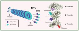
The assembly process of microtubules and the primary binding sites of microtubule inhibitors.
Moreover, PTX exerts additional effects beyond its stabilizing influence on microtubules (Mahabady et al., 2022). On the one hand, it can alter cell motility and angiogenesis without directly affecting cell proliferation. On the other hand, it can regulate mitochondrial respiration and intracellular apoptotic signaling by changing the conformation of mitochondrial proteins. Furthermore, PTX stimulates immunogenic cell death and enhances the production of CD8+ T cells, thereby influencing the immunosuppressive microenvironment and cytotoxic activity within tumors.
In 2021, Christina et al. discovered a new mechanism of PTX's anticancer effect; PTX does not exert its anti-tumor effect by blocking the mitosis of cancer cells but by causing chromosome disaggregation on the multipolar spindle of cancer cells, which increases the chromosome instability and thus exerts its anticancer effect. Meanwhile, the baseline rate of chromosomal instability could be a predictive biomarker of PTX response (Nawara et al., 2021).
In summary, PTX acts as an anti-tumor agent against cancer cells by interfering with microtubule dynamics and cancer's other mechanisms. This makes PTX one of the most important drugs for the treatment of many types of cancer, including breast, ovarian, and non-small cell lung cancer.
MAJOR CHALLENGES FOR PTX IN THE TREATMENT OF TUMORS
PTX is a white crystalline powder with low bioavailability (<8%) and suboptimal pharmacokinetics upon administration (Chen et al., 2021). According to the Biopharmaceutics Classification System (BCS), PTX is classified as a Class IV chemical drug due to its high lipophilicity (Log P = 3.96), low water solubility (0.3-0.5 µg/mL), as well as the need for recrystallization post-dilution (Gangannapalle et al., 2023). Furthermore, PTX is subject to exocytosis mediated by P-glycoprotein (P-gp), a transporter protein that facilitates the drug's excretion from cells, thereby reducing its concentration in the body. To overcome these problems, the earliest commercial form of PTX (Taxol®) was an intravenous dosage form, achieved by dissolving PTX in equal parts of anhydrous ethanol and CrEL. Anhydrous ethanol and CrEL effectively increase the solubility of PTX, facilitating its distribution in the body (Lu et al., 2022). The introduction of the intravenous infusion form provides an effective way to cope with the limitations of PTX's drug properties, allowing it to perform its anti-tumor effects in the clinic to perform its anti-tumor effects in clinic better (Mendoza, Ibarra, 2023).
In addition to its low bioavailability, PTX faces three significant challenges in clinical use: low extraction efficiency, severe side effects, and drug resistance during treatment (Moghbeli et al., 2023). Despite being a naturally occurring compound, the demand for PTX currently requires more work to satisfy due to the limited availability of its sources. As a result, alternative approaches have been explored to develop more cost-effective and efficient methods for addressing the issue of PTX supply. These mainly encompass chemical total synthesis, semi-synthesis, fungal production, and plant cell culture (Mostafa-Hedeab et al., 2022).
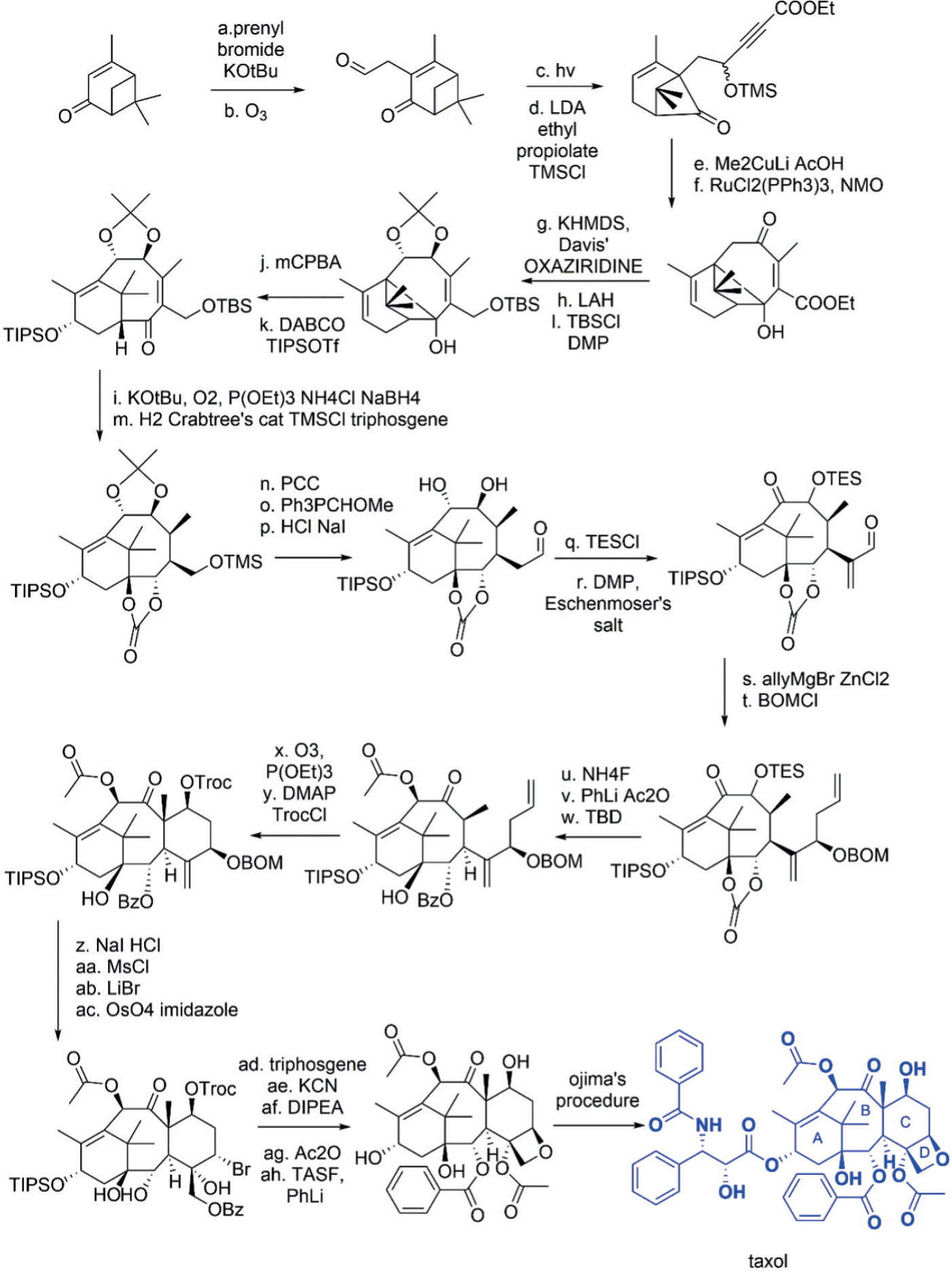
PTX, with its 6-8-6 carbocyclic structure, has intrigued chemists worldwide since the early 20th century (Ramezanpour et al., 2023). The total synthesis of PTX is divided into three stages: the construction of the mother nucleus skeleton, the functional group reactions and modifications of the skeleton, and finally, the addition of the side-chain phenyl isoserine to complete the synthesis (Rehman et al., 2022). This process is intricate, labor-intensive, time-consuming, and inefficient (Wang et al., 2023; Zhou et al., 2022; Zhou et al., 2023). To date, Wender’s total synthesis method represents the shortest route for the total synthesis of PTX (McKinlay, Waymouth, Wender, 2016). Beginning with compound 1, a series of reactions led to the formation of compound 7, marking the completion of the AB ring synthesis (Min et al., 2023). Subsequently, the C-3 position reaction and oxidation reaction resulted in the formation of compound 10. Adding alcohol and formaldehyde through condensation reactions produced compound 12 ring rings, signifying the C ring's establishment. Further, the bromination of C-5, C-4, and C-20, followed by ozonation, led to compound 13 and ultimately to compound 13. The completion of the oxygen-containing D ring was achieved through these reactions. Finally, the acetylation of C-10 and the chain chain chain side chain addition were performed to obtain PTX. The synthetic route is illustrated in Figure 4 (Min et al., 2023).

Wender's Full Synthetic Route (The total synthesis route is divided into three main processes: the synthesis of the parent nucleosome of paclitaxel, the modification of the skeleton by functional group reaction, and the addition of phenylisoserine to the side chain to complete the total synthesis).
The total synthesis route is lengthy, and the yield is relatively low, making it essential to discover a more economical and straightforward synthesis method (Utsugi, Kamada, Nakada, 2008). After conducting a multitude of experiments, researchers found that 10-diacetyl berry gibberellin III, which structurally resembles PTX, can be synthesized through a mere few conversions followed by the introduction of a side chain (refer to Figure 5) (Shimada et al., 2003). Furthermore, the content of 10-deacetyl berry gibberellin III was found to be higher than that of both P. horsetail pine (Pinus massoniana, Chinese red pine, horsetail pine) and Cephalotaxus sinensis (Chinese rough tephrosia), both of which belong to the Pinaceae family (Petrignet et al., 2011). The content of 10-diacetyl berry gibberellin III in horsetail pine and Chinese rough tephrosia is 10-30 times higher than that of PTX; thus, commercially available PTX from Bristol-Meyers Squibb was produced by this semisynthetic approach (Min et al., 2023).

However, this method has a significant drawback. The purification process is rather challenging. Additionally, harnessing endophytic bacteria for PTX production is an increasingly promising strategy (Min et al., 2023). As of now, Aspergillus flavipes (185 µg/mL), Aspergillus oryzae (95 µg/mL), Aspergillus aculeatinus (1.3 mg/ mL), Metarizium anisopliae (0.16 mg/L), and Aspergillus fumigatus (1.6 g/mL) are considered to be the most proficient PTX microbial synthesis strains (Wang et al., 2021a). The biosynthetic route of PTX involves a 19-step enzymatic reaction (refer to Figure. 6) that is divided into three stages (Ramirez-Estrada et al., 2016): (i) the generation of bacitracin III from the PTX scaffold; (ii) The formation of the side-chain phenylalanine coenzyme A; and (iii) The linkage of the C13 acyl groups of the bacitracin III backbone to the side-chain, followed by modification. The detailed process is as follows: Firstly, the Methylerythritol phosphate (MEP) pathway synthesizes the diterpene co-precursor Geranylgeranyl pyrophosphate (GGPP), which is then converted into the PTX skeleton, PTX diene [taxa-4(5),11(12)-diene], by Taxadiene synthase (TS). This diene is further hydroxylated or isomerized by several P450 enzymes (at the C1, C2, C5, C7, C9, C10, and C13 positions) to produce bacitracin III (Sanchez-Munoz et al., 2020). Secondly, phenylalanine aminotransferase (PAM) converts β-hydroxypropyl pyrophosphate into β-phenylpropyl pyrophosphate, the precursor of GGPP. PAM is then converted into β-Phenylalanine-Coenzyme A ligase (PCL), which combines PCL with acetyl coenzyme A to generate β-phenylalanyl CoA. Thirdly, baccatin III 3-amino-3-phenylpropanoyl-transferase (BAPT) catalyzes the acylation of baccatin III at the C13 position with β-phenylalanyl CoA as the acyl donor, producing β-phenylalanyl baccatin III. This is followed by the acylation of baccatin III at the C13 position by Taxane 2'-α-hydroxylase (T2'αH) and 3'-N-deoxy taxol3'-N-deoxy taxol 3'-N-phenyl taxol transferase (DBTNPT), which catalyze the formation of β-phenylalanyl baccatin III, respectively. The baccatin III side chain is hydroxylated at the C2' position and benzoylated at the C3' position, ultimately leading to the formation of PTX. Currently, 14 enzymatic steps in the PTX biosynthetic pathway have been successfully resolved, including six hydroxylases (T2αH, T5αH, T7βH, T10βH, T13αH, and T2'OH), five transferase enzymes (BAPT, TAT, TBT, DBA, and DBTNBT), two types of cyclohydrolases (TS1 and TS2), one mutase (PAM), and one β-phenylalanyl-CoA PCL. Of these, 12 enzymes were identified before 2004, whereas PCL, T2'OH, and the class II cyclase TS2 have only recently been discovered. However, the sequence of oxidative modifications at the C1 and C9 positions, the formation of the 4,5-epoxypropane ring, and the different hydroxylation reactions remain unclear (Howat et al., 2014).

Paclitaxel biosynthetic route (solid line is known synthetic pathway; dashed line is putative synthetic pathway).
In addition, plant cell culture is an attractive alternative for producing PTX as it provides a more sustainable and environmentally friendly source than the traditional extraction method from the Pacific yew tree (Buyel, 2018). In 2019, Xu et al. optimized a suspension culture system of southern red bean tree cells. They found that the optimal medium for suspension culture of southern red bean tree was B5+0.4 mg/L 2,4-D+ 0.3 mg/L NAA+1.2-1.6 mg/L KT+30.0g/L sucrose (pH 5.8), and the optimal inoculum amount was 0.09 g/mL(Fridlender, Kapulnik, Koltai, 2015). In commercial production, Phyton Biotech, as a pioneer of this technology, has operated the world's most extensive plant cell fermentation (PCF) plant in Germany (Ji et al., 2023). Phyton Biotech, a pioneer in this technology, has been operating Germany's world's largest PCF facility for medicinal plants for more than 10 years (Kornel, Nadile, Tsiani, 2022). With a production capacity of 75,000 L and an annual production capacity of more than 100 kg of paclitaxel, the company is a leader in its field (Perez-Matas et al., 2023; Yang, Mao, Tan, 2020)
The complexity of cancer cell resistance mechanisms is reflected in the challenges associated with PTX therapy (Behroozaghdam et al., 2022). PTX can disrupt the mitotic process of cancer cells and trigger cell death. Still, cancer cells can enhance their malignant characteristics through cell viability responses, allowing them to survive and develop resistance to chemotherapy- one of the primary reasons for PTX treatment failure (Bhardwaj, Bikal, Sachdeva, 2023). Studies have identified PTX resistance as a multifactorial mechanism involving modifications or alterations to various molecular components within the target molecule. These changes include increased drug efflux or decreased drug entry into cancer cells, mutations in the genes encoding αand β-microtubule proteins, abnormal cellular metabolism, overactive DNA repair activity (altered p53 status), modifications in apoptosis-regulating molecules (e.g., Bcl-2, Bcl-xL, Bax), excessive production of cytokines (e.g., IL-6), elevated levels of cellular senescence, autophagy, and tumor microenvironmental conditions, among others. These mechanisms may interact, collectively contributing to the developing resistance to PTX in cancer cells (Brala et al., 2023).
PTX resistance is associated with changes in the expression of various oncogenes and proteins(Hashemi et al., 2022). Among them, the most widely studied is the overexpression of members of the ATP-binding cassette (ABC) family, particularly ABCB1, also known as multidrug resistance protein 1 (MDR1) or multidrug transporter protein P-gp 1(Hashemi et al., 2023). MDR1 is a transmembrane ATP-dependent efflux pump that lowers the intracellular levels of hydrophobic, harmful chemicals (e.g., PTX), reducing the response to death-inducing stimuli. In addition to MDR1, other genes and proteins associated with PTX resistance include FOXM1, KANK1, GALNT14, LRG1, ALDH3A1, PIK3R3, WEE2, NF-кB, TKTL1, HER2, β-catenin, and HIF-1. Abnormal expression of these genes and proteins may influence cellular sensitivity to PTX, contributing to the development of drug resistance (Mahabady et al., 2022).
Another well-studied association with PTX resistance is FOXM1 in the FOX transcription factor superfamily (Maharati, Moghbeli, 2023). FOXM1 is a transcription factor that promotes cell proliferation by regulating genes at the G1/S and G2/M checkpoints in the cell cycle. However, FOXM1 expression is upregulated during cancer development, leading to uncontrolled cell division and genomic instability, promoting aberrant cell proliferation. In addition, FOXM1 tends to bind to the promoter structural domains of related genes, stimulating the overexpression of the transporter molecule ABCC5. This drug efflux pump expels chemotherapeutic drugs, such as PTX, from the cell, thereby reducing the effectiveness of these drugs and generating drug resistance (Manthalkar, Ajazuddin, Bhattacharya, 2022). In conclusion, upregulation of FOXM1 can increase PTX resistance through multiple pathways, including promoting cell proliferation, leading to genomic instability, and stimulating the overexpression of ABCC5 (Miguel et al., 2022b). Further study of FOXM1 and its regulated genetic and molecular mechanisms can help us better understand the occurrence and development of PTX resistance and provide new targeting strategies for treatment (Sousa-Pimenta et al., 2023).
PTX resistance is often associated with dysregulated expression of various oncogenes. One such oncogene, KANK1, plays a role in cell mobility by regulating cytoskeleton formation while inhibiting cell growth and promoting apoptosis, which has anti-cancer effects (Villegas et al., 2023b). During chemoresistance, specific oncogenes are upregulated, contributing to drug resistance. For instance, the GALNT14 protein enhances the glycosylation of P-gp, reduces its degradation sensitivity, and increases its expression, resulting in increased cellular efflux activity and a decrease in the concentration of chemotherapeutic molecules in cancer cells (Vitiello et al., 2022). Additionally, the ALDH3A1 protein partially inhibits the growth of reactive aldehydes produced during membrane lipid peroxidation, aiding cellular detoxification. Furthermore, the PIK33 protein promotes resistance by activating the P13K/AKT/NF-B signaling pathway, stimulating the phosphorylation of AKT protein kinase B, inhibiting apoptosis, and enhancing cell proliferation, ultimately leading to chemoresistance in cells (Wang, Aguilar, Ojima, 2022).
Prolonged exposure to PTX can activate nuclear factor-κB (NF-κB) excessively, a crucial oncogene associated with cell survival, proliferation, invasion, angiogenesis, and metastasis, as well as chemotherapy resistance( Wang, Li, Feng, 2023). Moreover, TKTL1, another oncogene, is overexpressed in cancer cells and plays a vital role in tumor-specific glucose metabolism. This, in turn, influences the therapeutic impact of PTX and promotes cell proliferation and tumor growth. Interactions between different oncogenes also occur, such as the relationship between HER2 (a membrane tyrosine kinase) and β-linker protein (a protein involved in maintaining physical homeostasis)(Wang et al., 2022). Elevated PTX expression has been linked to the acquisition of stem-cell-like properties in cancer cells, acceleration of tumor progression and metastasis, and resistance to breast cancer. Furthermore, interactions between various protein/protein complexes and oncogenes contribute significantly to the development of PTX resistance(Wu et al., 2023). For instance, in proteasome and hypoxia-inducible factor 1 (HIF-1) signaling, increased proteasome activity leads to a notable reduction in HIF-1 expression. This, in turn, triggers the expression of specific growth factors, growth factor receptors, and hypoxic conditions, all of which are associated with angiogenesis and the production of proteins related to cancer cell survival and PTX resistance(Yang et al., 2023). In summary, these studies reveal the intricate interplay of multiple factors and mechanisms during cancer development, which is crucial for obtaining a deeper understanding of cancer progression and the efficacy of drug therapy.
Non-coding transcripts have a significant role in cancer and are linked to crucial biological processes, including chemotherapy resistance, metastasis, and prognosis. For instance, the lncRNA HOTAIR is excessively expressed in various cancers, disrupting cellular and biochemical processes and enhancing drug resistance, metastatic capacity, cell proliferation, and invasiveness(Zhao et al., 2022). These effects, in turn, negatively impact the survival and prognosis of cancer patients. Similarly, the lncRNA H19 has been associated with aberrant expression of apoptotic signaling pathway-related proteins in triple-negative breast cancer, resulting in increased proliferation, invasion, metastasis, and resistance to PTX in cancer cells. Furthermore, the upregulated oncogene lncRNA ANRIL controls the expression of apoptosis-related proteins through the mitochondrial pathway, thereby promoting resistance to PTX chemotherapy(Zheng et al., 2023).
The circ-PVT1 transcript is highly expressed in PTX-resistant gastric tissues. It promotes gastric cancer proliferation and enhances drug resistance by up-regulating the expression of P-gp and GST-π (Meng et al., 2021). P-gp leads to chemotherapeutic failure by decreasing the concentration of intracellular PTX, whereas GST-π enhances drug resistance by reducing the effect of toxic substances on cells. In addition, aberrant post-translationally altered microtubule protein subunits may also affect PTX binding, developing resistance to chemotherapeutic agents(Miguel et al., 2022a).
Despite the wealth of research and discoveries surrounding the mechanisms of PTX resistance, it remains a significant challenge in clinical treatment, posing a significant barrier to effective cancer management for countless patients worldwide (Ortiz et al., 2022). To mitigate the adverse effects of chemotherapy, the primary focus has shifted to addressing PTX resistance and off-target toxicity. Nanomedicine exhibits promise in overcoming these issues, as nanocarriers can enhance the bioavailability, blood circulation time, solubility, pharmacological activity, mechanical stability, and targeting properties of PTX while reducing its systemic toxicity and side effects during treatment (Sousa-Pimenta et al., 2023). Among the various nanomaterials employed in these efforts, polymeric Nanoparticles are the most widely studied for delivering PTX to patients with diverse cancer types. In addition to polymeric Nanoparticles, other commonly used nanomaterials include metallic Nanoparticles, solid lipid Nanoparticles, nanocrystals, carbon nanotubes, nanoemulsions, liposomes, and micelles (Soyata, Hasanah, Rusdiana, 2021). However, the toxicological and regulatory aspects of nanomedicines require further attention. The development of nanomaterials for drug delivery necessitates innovative and efficient methods for assessing their toxicity in vivo (Tian et al., 2021). As such, this review aims to summarize recent advancements in the application of polymeric Nanoparticles to enhance PTX efficacy, contributing to a deeper understanding of the promising applications of nanomedicines and providing a foundation for future research and development (Villegas et al., 2023a).
POLYMER NANOPARTICLES
Genexol®-PM, Nanoxel®, PICN, Apealea®, NK105, Paxceed, and other drugs employ polymer nanoparticles as a delivery system for PTX, thanks to the numerous advantages these Nanoparticles possess. Firstly, by adjusting the structure and properties of the polymer, polymer Nanoparticles can achieve good biocompatibility, making them extensively utilized in the medical field. Secondly, these drug-drug nanoparticles can effectively load drugs onto their interior or surface and facilitate a sustained and controlled release, thereby enhancing their therapeutic effect. Furthermore, the structure of polymeric Nanoparticles offers ample space to accommodate various types of substances, such as drugs, proteins, peptides, or nucleic acids. Polymeric Nanoparticles are preferred over lipid-based materials due to their superior durability and stability. This review introduces several prevalent polymeric Nanoparticles and highlights their drug-carrying properties for PTX. Some commonly used polymeric materials are chitosan, polylactic-co-glycolic acid (PLGA), PCL, and polylactic acid (PLA). These polymer structures can either embed PTX or adhere it to the particle surface, enabling effective loading and release of PTX, enhancing the drug's solubility, stability, and targeting, and ultimately boosting the efficacy of drug therapy.
PLGA Nanoparticles
PLGA Nanoparticles are a widely used drug delivery system, particularly for loading the anticancer drug PTX (Cai et al., 2016). This is due to their excellent biocompatibility, degradation properties, and the fact that they are hydrolyzed and hydrolyzed in vivo to produce carbon dioxide and water, resulting in low systemic toxicity. Currently, three primary methods are employed for PTX-loaded PLGA Nanoparticles: oil-in-water solvent precipitation, nanoprecipitation, and interfacial deposition (Cunha et al., 2021). These Nanoparticles typically undergo two release phases, initiating with a rapid release during the initial 3 days, followed by a sustained slow release. Research indicates that loading PTX into PLGA Nanoparticles augments their cytotoxicity against various cancer cell types, such as glioma cells (C6), human small cell lung cancer cells (NCI-H69), Hela cells, and breast cancer cells (MCF-7). Moreover, the loading of PTX onto PLGA Nanoparticles significantly enhances their inhibitory effect on liver cancer development. Furthermore, modifying the nanosurface of PLGA can optimize drug delivery efficiency (Narmani et al., 2023). For instance, modifying chitosan on the surface of PLGA Nanoparticles can alter their charge properties, thereby facilitating the cellular uptake of PTX for slow release. One study reported a 4- to 10-fold increase in the targeting ability of PTX to cells and an enhancement in cytotoxicity when chitosan-modified PLGA Nanoparticles loaded with PTX were used (Ramezani, Ebrahimian, Hashemi, 2017).
Modification of PLGA Nanoparticles using DMAB converted the surface charge from negative to positive, which resulted in a reduction of intimal growth in a rabbit arterial injury model. Furthermore, the characteristics of the Nanoparticles, including size, shape, encapsulation rate, in vitro performance, cytotoxicity, and healing rate, were influenced by the emulsifiers and stabilizers employed in the production process. Emulsifiers such as d-toco-pheryl PEG1000 succinate (TPGS) and phospholipids (e.g., DLPC and DPPC) are extensively used in the preparation of PLGA Nanoparticles (Rezvantalab et al., 2018). PTX demonstrates superior release kinetics, cytotoxicity, and uptake compared to PLGA Nanoparticles with PVA as an emulsifier. Notably, TPGS-emulsified PLGA Nanoparticles exhibit a 10-fold higher oral absorption efficiency than Taxol®. Additionally, montmorillonite (MMT) is often incorporated as a co-emulsifier and matrix component in PTX-loaded PLGA Nanoparticles. The drug uptake of the modified PLGA Nanoparticles increased by 57-177% and 11-55% in Caco-2 and HT-29 cells, respectively, compared to unmodified PLGA Nanoparticles (Simon, Sabliov, 2014).
To achieve targeted delivery, human epidermal growth factor receptor 2 (HER2) antibodies and other ligands, such as transferrin and RGD, were also coupled to PTX-loaded PLGA Nanoparticles to enhance their anticancer effects (Wang, Uludag, 2008)and compared to non-targeted PLGA nanoparticles, PTX nanoparticles with transferrin ligands exhibited a 5-fold increase in cytotoxicity. Introducing transferrin ligands enhanced the nanoparticles to kill nanoparticles against cancer cells selectively. Moreover, experimental results from prostate cancer animal models showed that PLGA Nanoparticles with tumor-targeting effects significantly inhibited tumor growth and prolonged the survival time of the animals. These findings suggest that nanomedicines with tumor-targeting effects may hold potential clinical applications for prostate cancer treatment. This research provides valuable insights and references for developing more effective tumor therapeutic strategies (Yan et al., 2022). Tumor-targeting nanomedicines can help increase local drug concentrations and reduce adverse effects on normal tissues, thereby improving therapeutic efficacy and reducing side effects. However, these results are still experimental, and further clinical studies are needed to verify their safety and efficacy (Zhi et al., 2021).
In summary, PLGA Nanoparticles have broad applications in research as an effective drug delivery system. With rational design and modification, their drug release and targeting capabilities can be further enhanced, offering improved strategies for cancer treatment.
Polylactic acid (PLA) Nanoparticles
PLA is a biodegradable and safe polymer often used as a drug carrier. Using methoxy polyethylene glycol polylactide copolymer mPEG-PLA as a carrier loaded with PTX provides a long cycling effect(Amani et al., 2021). In the MCF-7 cell line, the cytotoxicity of mPEG-PLA-loaded PTX was approximately 33.5 times greater than that of free PTX. Furthermore, in vivo, pharmacokinetic studies revealed that the mPEG-PLA carrier led to a 3.1-fold and 2.8-fold increase in the AUC and half-life of PTX in rat plasma, respectively. This suggests that mPEG-PLA can effectively enhance the drug concentration and duration of PTX. Additionally, investigations showed that varying the ratio of PLA/ mPEG-PLA (100/0, 75/25, 50/50, 25/75, and 0/100) loaded with PTX led to an increase in the ζ-potential and drug release from PLGA Nanoparticles, accompanied by a decrease in the glass transition temperature (Tg) and particle size(Chen et al., 2023). The findings indicated that PLA carriers were three times more cytotoxic than PLGA carriers against glioma (C-6) cells. Moreover, TPGS can serve as an emulsifier for PLA Nanoparticles. The PLA-TPGS copolymers produced by the preparative ring-opening polymerization process demonstrated a 1.7-fold and 1.5-fold increase in cellular uptake of PTX compared to PLGA Nanoparticles, along with a 40% lower IC50 value, a half-life of approximately 27.5 times longer, and an AUC about 1.5 times longer than that of PTX(Oktay et al., 2022b). Furthermore, different ratios of PLA/mPEG-PLA had no impact on particle diameter, and PLA-TGSA-loaded PTX was most effective when the ratio of PLA/ mPEG-PLA was 89/11 (Petrova et al., 2023). Researchers concurrently developed HER2, biotinand folate-modified PLA-TGSA Nanoparticles to further enhance therapeutic effectiveness. The results demonstrated a significant improvement in the targeting efficiency of the modified PTX, both in vitro and in vivo, compared to unmodified PLA-TGSA Nanoparticles. These findings imply that employing PLA as a drug carrier, in conjunction with various modifications or adding other constituents, can effectively enhance the drug's targeting, cytotoxicity, and pharmacokinetic properties, thus leading to improved therapeutic outcomes. However, additional research and clinical validation are required to establish the safety and efficacy of these Nanoparticles(Wang et al., 2021b).
PLA micelles have great potential in PTX delivery. With PEG-b-PLA micelles, PTX can efficiently contact the cell membrane, release it, and enter the interior. In addition, the results of the study showed that PEG-b-PLA was able to prevent the development of PTX resistance. Paxceed® is a polymeric micelle containing PTX, which has been demonstrated to be more efficient at optimal doses than free PTX in mouse models of lung cancer. Paxceed® is currently in Phase II clinical trials. The other agent, Genexol-PM, consists of a PLA-b-PE diblock copolymer and is the most effective. Preclinical studies have found a three-fold increase in the maximum tolerated dose of Genexol-PM in a rat model and a twoto three-fold increase in drug concentrations in other organs and tumors compared to Taxol®. In addition, the in vivo anticancer activity of Genexol-PM was significantly improved. Phase I clinical studies found that the drug was tolerated at a concentration of 182 mg/m2, and when the concentration was increased from 100 mg/ m2 to 200 mg/ m2, the Cmax and plasma AUC increased 4-fold and 3-fold, respectively. The results of phase II clinical trials in patients with advanced pancreatic and breast cancer have confirmed the safety and efficacy of Genexol-PM. Phase III clinical studies are currently underway. These research advances offer new possibilities and hope for tumor treatment(Chen et al., 2023).
The triblock copolymers, such as PEG-PLA-PEG and PLA-PEG-PLA, developed by G. He and his team, exhibited distinct effects on the release rate of PTX. The study revealed that the PEG-PLA-PEG micelles released the drug more slowly, and the presence of PEG within the micelles influenced the release rate (Hou et al., 2022). The uptake of these two triblock copolymer micelles into monocytes reduced by four compared to conventional PLA micelles. Separately, a tetrabranched copolymer was created by combining PLA and PEO. The findings indicated that the star-shaped branched micelles demonstrated a more controlled and extended drug release pattern than the diblock and triblock copolymers over two weeks. Moreover, the star-shaped micelles were smaller, enhancing the EPR effect's effectiveness in cancer treatment(Oktay et al., 2022a). These studies offer valuable insights for designing and optimizing PTX delivery systems.
Gaucher and his team synthesized a combination of PTX-loaded PVP-b-PLA and PEG-modified PLGA micelles using an oil/water emulsion solvent evaporation technique. During this process, The lyophilized PVP reconstitution imparted cryoprotective properties to the final micelles, ensuring a consistent particle size (Tang et al., 2018). In contrast, functionalized PEG-b-PLA micelles did not exhibit these same properties. This innovative method offers a promising avenue for preparing PTX-loaded micellar systems.
PCL Nanoparticles
In 2008, a study by Deshpande et al. they designed CrEL-modified and CrEL-modified PCL (PEO-PCL) nanoparticles to overcome MDR to PTX. These Nanoparticles were loaded with PTX and C6-ceramide to reverse MDR by silencing the P-gp. It was found that the prepared PEO-PCL Nanoparticles had more than 95% encapsulation with a diameter of about 270 nm at 10% (w/w) PTX and C6-ceramide content (Deshpande, Devalapally, Amiji, 2008). For drug resistance, compared to free PTX, the C6-ceramide-loaded PEO-PCL Nanoparticles ovarian cancer showed 100-fold enhanced cytotoxicity against drug-resistant SKOV3 TR cells. In in vivo experiments, PEO-PCL Nanoparticles significantly inhibited tumor growth in wild-type SKOV3 and drug-resistant SKOV3 TR xenograft mouse models, suggesting that the pairing of C6-neuramide and PTX with PEO-PCL Nanoparticles can effectively inhibit the MDR of ovarian cancers. In addition, the study evaluated the ex vivo and in vivo effects of PTX-containing PCL Nanoparticles with tamoxifen. The results showed a 10-fold and 3-fold reduction in the IC50 of the formulation in SKOV3 and SKOV3 TR cells, respectively, compared to free PTX, suggesting that the PEO-PCL Nanoparticles significantly improved the anticancer effect (Devalapally et al., 2007). Next, the investigators utilized a polymeric Nanoparticles system to deliver both siRNA and PTX for P-gp silencing. This was achieved by encapsulating siRNA and P-gp-silencing PTX in PEO-modified polyamides (PEO PbAE and PEO-PCL Nanoparticles, respectively). Co-administration completely reversed MDR in sensitive SKOV3 cells and drug-resistant SKOV3 TR cells with the same cytotoxic effects. These findings suggest that PEO-PCL Nanoparticles have the potential to be used to overcome multidrug resistance to PTX and provide an essential reference for further development of anticancer therapeutic strategies (Yadav et al., 2009).
PEG-PCL polymer micelles were prepared using a co-solvent extraction method. The researchers found that PEG-PCL Nanoparticles loaded with 7'-hexanoic acid PTX exhibited better pharmacokinetic properties than PTX after the synthesis and encapsulation of various PTX precursors. Among them, in 2022, Xin et al. prepared PTX-loaded mPEG-PCL Nanoparticles using the emulsion solvent evaporation (ESE) method. mPEG-PCL Nanoparticles loaded with PTX showed higher cytotoxicity than PTX and PCL Nanoparticles (Barak et al., 2022). Notably, mPEG-PCL Nanoparticles successfully prolonged the average lifespan of the C-6 cancer mouse model to 28 days, compared to 20 and 23 days for PTX and PCL Nanoparticles, respectively. In addition, the ability of PTX to cross the blood-brain barrier (BBB) could be increased by targeting LDL receptor-associated proteins by attaching the peptide angiopep to the surface of the mPEG-PCL Nanoparticles. mPEG-PCL Nanoparticles loaded with PTX were prepared by Wang and colleagues using a solid-dispersion method, which does not require organic reagents, in 2011. The Nanoparticles had an encapsulation efficiency (EE) of up to 98% and a drug-loading efficiency of 25.6% (w/w). After in vivo injection in BALB/c mice, this Nanoparticle's maximum tolerated dose (MTD) was 2.6 times higher than that of Taxol. PTX-loaded Nanoparticles had a more pronounced distribution in tissues and significantly increased tumor accumulation compared to Taxol. An alternative approach was to use hyperbranched aliphatic polyester Boltorn H40 as PEG-PCL Nanoparticles and further coated with folic acid. In in vitro experiments, such Nanoparticles loaded with PTX showed enhanced cytotoxicity. The above findings demonstrate the potential of PEG-PCL Nanoparticles for loading PTX and provide a crucial essential reference for developing more effective drug delivery systems (Engelberg et al., 2021).
Solvent evaporation (SA) was employed to synthesize PTX-loaded (PCL-pluronic F68) nanoparticles. The addition of Pluronic F68 to PCL Nanoparticles created a porous structure, facilitating the rapid release of the drug from the nanoparticles. For MCF-7/TAX cells, the cellular uptake and toxicity of PCL/F68 Nanoparticles loaded with PTX were more potent than those of PCL nanoparticles loaded with PTX (Patel et al., 2022). Furthermore, a single intratumoral injection of PCL/F68 nanoparticles demonstrated more potent anticancer effects than high-dose intraperitoneal administration of PTX. To enhance the ability of Nanoparticles to internalize into the arterial wall in an animal angioplasty model, DMAB was adsorbed onto the surface of PCL/F68 Nanoparticles through charge-to-charge interactions. These strategies contribute to improving the therapeutic efficacy of Nanoparticles and expanding the therapeutic scope of PTX (Patel et al., 2023).
In addition to the above PCL Nanoparticles, there are other PCL-based Nanoparticles for PTX loading. For example, PVP-b-PCL nanoparticles showed more robust and potent anticancer activity than PTX in mice with H22 hepatocellular carcinoma (Zhu et al., 2010). A lower rate of drug release characterized PCL-g-PVA nanoparticles. In addition, Nanoparticles made from PCL-grafted polyvinyl alcohol (PCL-g-PVA), as well as PTX Nanoparticles containing poly-2-ethyl-2-oxazoline PC, galactosamine-specific polyethylphosphate Nanoparticles, and mPEG-PCL-poly-2-aminoethylphosphate Nanoparticles, which exhibited improved anticancer properties compared to free PTX, were developed (Zhu et al., 2010). These findings suggest that PCL-based Nanoparticles have potential applications as effective PTX carriers for cancer therapy.
Chitosan Nanoparticles
In 2016, Kim et al. prepared PTX-loaded chitosan-modified chitosan Nanoparticles with a drug loading of 10% (w/w) using a simple dialysis technique (Kim, Jang, Park, 2016). They found that these Nanoparticles exhibited faster cellular uptake and excellent therapeutic efficacy in a mouse model of SCC7 tumors (Del Prado-Audelo et al., 2020). To increase the water solubility of PTX in Nanoparticles, they prepared a water-soluble oligomer-ethylene glycol chitosan (HO-GC). They succeeded in raising the drug loading by 20%, resulting in a maximum EE% of 97%. More importantly, the PTX-encapsulated HO-GC Nanoparticles exhibited long-term in vitro release while achieving the targeting function of PTX (Ashrafizadeh et al., 2020). In addition, a 4-fold and 1000-fold increase in PTX cellular uptake and cytotoxicity, was observed in MDA/MB-231 cells when PTX was added to glycerol monooleate-modified chitosan Nanoparticles. In addition, a more advanced chitosan derivative was developed that administered PTX via mPEG and cholesterol (CHO) functionalization. mPEG-cholesterol chitosan Nanoparticles of this type significantly reduced cancer cell proliferation and prolonged survival compared to free PTX in vitro in a hormonal animal model. Lipophilic acetyl histidine-functionalized chitosan was found to self-assemble into Nanoparticles under neutral conditions and dissolve in a weakly acidic environment due to the protonation of imidazole molecules. Follow-up studies showed that modified nanoparticles loaded with PTX could be endocytosed within the cell and release the drug in a low pH environment. Finally, another study used stearic acid-grafted chitooligosaccharides for self-assembly to form micelles and crosslinked glutaraldehyde on the surface of the Nanoparticles to improve the structural stability of the micelles(Voci et al., 2021). Glutaraldehyde-crosslinked chitosan micelles containing PTX showed lower initial uncontrolled and delayed release than non-crosslinked formulations. These studies provide valuable information for further applications of chitosan-based Nanoparticles in drug delivery and cancer therapy.
Chen and his team created a novel chitosan derivative, GA-CS-TPGS copolymer, by combining gallic acid (GA), chitosan (CS), and TPGS (refer to Figure 7). This derivative was used to fabricate multifunctional PTX-micelles, designed to enhance mucus adhesion, inhibit P-gp exocytosis, and reduce CYP3A-mediated metabolism. GA-CS-TPGS copolymer, due to its amphiphilic nature, exhibits strong self-assembly capabilities with a low CMC of (15.6 ± 1.0) μg-mL-1. The resulting PTX-micelles have a particle size of about (134.9 ± 10.2) nm, a drug loading capacity of about (8.2 ± 0.3)%, and an encapsulation rate of about (80 ± 3)%. Compared to Taxol®, PTX-micelles demonstrated a slow-release profile, attributed to factors such as the hydrophobicity of the micellar core and intermolecular hydrogen bonding. The GA-CS-TPGS-based PTX-micelles increased the bioavailability of PTX by approximately 3.80-fold and exhibited superior anti-tumor effects compared to Taxol® while displaying lower toxicity in A569 ruffed nude mice (Chen et al., 2020).

Albumin Nanoparticles
Albumin is a versatile natural protein carrier extensively employed for conveying various medications. Albumin-based nanoparticles are solid spheres created by encapsulating and adsorbing the drug, followed by solidification and separation (Chubarov, 2022). As a human plasma component, albumin is an endogenous protein (35-50 g-L-1 human serum) that guarantees its biocompatibility with albumin-based nanoparticles. As a carrier for anti-tumor drugs, Albumin compares favorably to other airlines, boasting superior drug release properties, higher drug-carrying capacity, better stability, and a longer in vivo half-life, exhibiting effective targeting and biocompatibility (Hornok, 2021).
Nanoparticle Albumin Bound PTX (Nab-PTX) is a drug formulation created by combining PTX with human serum albumin (HAS) to form particles with a diameter of approximately 130 nm, utilizing Nanoparticle Albumin Bound Technology (Nab-Technology) (Hao et al., 2023). This technology not only enhances the poor water solubility of PTX by increasing its bioavailability through natural albumin but also reduces the risk of allergic reactions by eliminating the use of Cremophor EL, a known allergen, compared to solvent-based paclitaxel (Sb-PTX). Consequently, higher delivery rates of PTX to tumors have been achieved without the need for hormonal pretreatment. One of the more successful formulations is the FDA-approved lyophilized powder injection of PTX, Abraxane, developed in the United States(Li et al., 2021). Abraxane is produced using the patented "nab-Technology." The oil phase (chloroform and ethanol) containing PTX is first added to the aqueous phase (1% HAS solution pre-saturated with 1% chloroform). The emulsion is then homogenized at high pressure under high shear and recovered in a homogenizer. Then, it is filtered to remove the solvent and sterilized to obtain the final albumin nanosuspension. However, this method produces nanoparticles with low drug loading capacity and high preparation costs, prompting further improvements (Nakao et al., 2020).
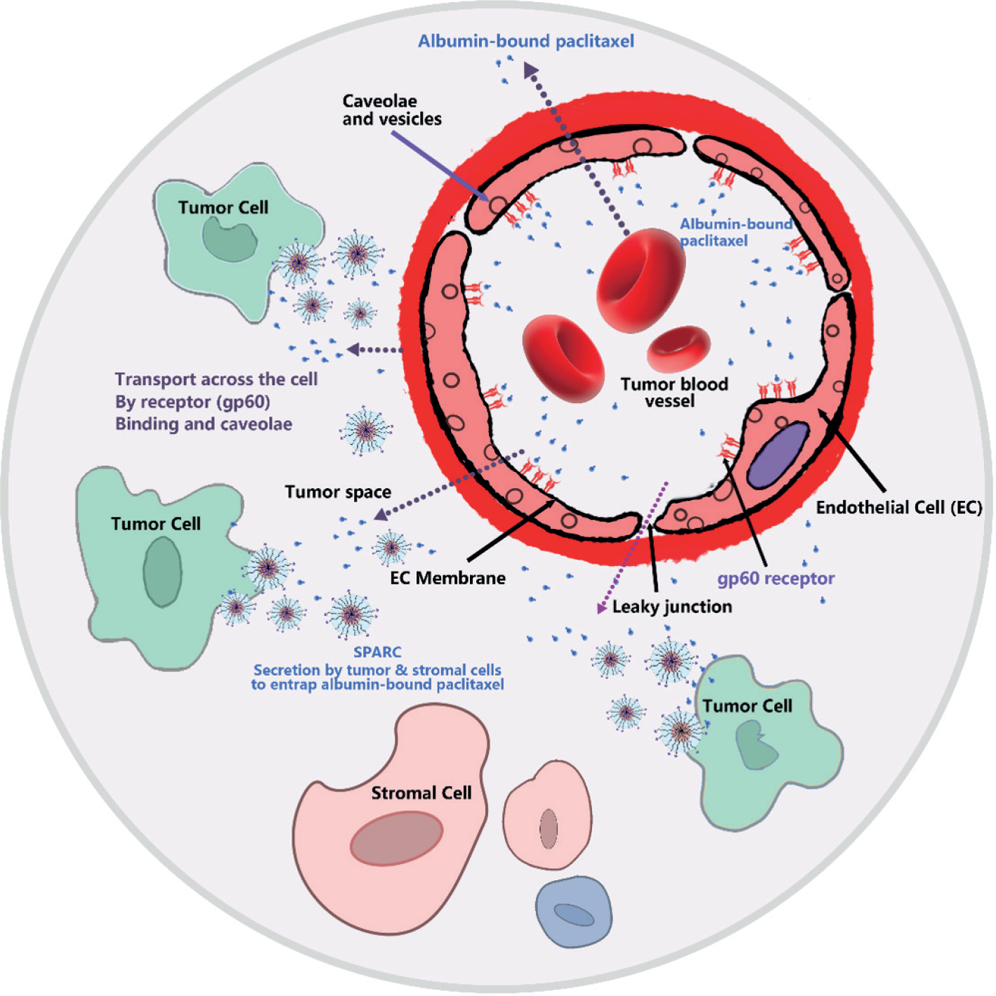
Albumin possesses a distinct endocytosis process, which can be divided into two stages. Following the injection of the albumin drug complex into the human body, it reaches the tumor site via the bloodstream, with a portion of it entering the tumor tissue through the Enhanced Permeability and EPR (Belinskaia, Voronina, Goncharov, 2021). The albumin component activates the specific albumin receptor (gp60) on the membrane surface of the tumor vascular endothelial cells, causing the endothelial cells to form a depression or fossa. Consequently, the nano-albumin-bound PTX is transported into the tumor cell space (Belinskaia et al., 2021). Subsequently, the Secreted Protein Acidic and Rich in Cysteine (SPARC) present in the interstitial space of the tumor cells, which shares sequence homology with gp60, binds to albumin specifically and avidly. This enrichment of Nab-PTX occurs, enabling the achievement of tumor cell endocytosis. As a result, the drug is released in large quantities within the tumor cells, exerting an anti-tumor effect. The specific mechanism is illustrated in Figure 8 (Taguchi et al., 2021).

Mechanism of albumin-bound paclitaxel transport and enrichment in tumors(Alavi, Nokhodchi, 2022).
Moreover, Zhao and his team fabricated bovine serum albumin (BSA) nanoparticles containing human albumin, which were loaded with PTX using a de-solvation method (Esim, Hascicek, 2021). They achieved targeting by surface modification with folic acid. The folic acid-modified nanoparticles successfully targeted human prostate cancer (PC-3) cells due to their enhanced stability and favorable surface properties. Separately, various studies developed a novel octyl-functionalized serum albumin (OSA) to augment the hydrophobicity of albumin. This enabled the facile synthesis of PTX-containing core-shell structured nano micelles. The octyl-modified bovine serum albumin nanoparticles demonstrated improved stability, smaller particle size, and more efficient drug trapping than non-functionalized nanoparticles (Fatima et al., 2023).
Poly (butyl cyanoacrylate) (PBCA) Nanoparticles
Environmentally friendly poly (alkyl cyanoacrylate) (PACA) Nanoparticles have been widely used in the past decade for the delivery of a variety of active substance ingredients (Semyari et al., 2021). PBCA is the most commonly used type of PACA Nanoparticles as a carrier. In 2019, Huang & Chen used microemulsion technology to prepare PTX-loaded PBCA Nanoparticles using pluronic-F127 as a surfactant (Huang, Chen, 2019). It was found that the pluronic-F127-modified Nanoparticles possessed high drug loading capacity and encapsulation rate (EE%) (Li et al., 2011). PBCA Nanoparticles prepared from microemulsions released approximately 82% of the drug within 96 hours. HA-PBCA Nanoparticles coated with PTX showed reduced cytotoxicity and first burst release of the drug compared to PBCA Nanoparticles without hyaluronic acid (HA) modification. More importantly, when administered intravenously to Sarcoma-180 mice, PTX-loaded HA-PBCA Nanoparticles exhibited more potent anti-tumor inhibition than uncoated HA PTX PBCA Nanoparticles, resulting in a 9.5-fold increase in uptake by Sarcoma-180 cells. In 2019, Lin et al. investigated the effect of different surfactants on PTX-encapsulated PBCA Nanoparticles. The study showed that Nanoparticles containing surfactants had smaller particle sizes and better stability than Nanoparticles without surfactants. The surfactants used included dextran 70, cholesterol, PVA, and lecithin. Nanoparticles produced from natural lipids such as cholesterol and lecithin had smaller particle sizes, better drug encapsulation and zeta potential, and more adjustable drug release profiles than the other two surfactants (Li et al., 2011).
Polyethylene glycol Nanoparticles
Hyperbranched poly (ethylene glycol) (HPG) is a less cytotoxic and more water-soluble polymer that can be used to prepare Nanoparticles for drug encapsulation. Mugabe et al. successfully encapsulated PTX in two functionalized HPG Nanoparticles using solvent evaporation (Poellmann et al., 2022). Both Nanoparticles had an average particle size of less than 20 nm, with HPG-C10-PEG releasing up to 80% of PTX, whereas PEI-C18-HPG released only 40% of PTX. The PTX-loaded HPG-C10-PEG Nanoparticles formulation showed better tolerance and anti-tumor effects in in vivo than PTX, but its cytotoxicity in vitro was slightly lower. These findings provide an essential reference for drug delivery using functionalized HPG Nanoparticles (Saadati, Hasanzadeh, Seidi, 2021).
In 2017, Sarisozen et al. prepared micelles made of PE and a diacyl matrix to deliver PTX successfully. PE has two hydrocarbon chains, which make the micelles more hydrophobic and thus increase their stability. The PTX-loade micelles are similar in size to placebo micelles. In the Lewis lung cancer mouse model, In 2018, Upponi et al. concatenated the monoclonal antibody 2C5 with micelles to create immunomicelles. This formulation showed stronger anti-cancer effects in vivo and in vitro than Taxol or non-targeted micelles. The team combined PE-PE, egg phosphatidylcholine or PEG-PE, solid triglycerides, and positively charged Lipofectin to make PTX-loaded hybrid micelles. The hybrid polymer micelles further improved the anti-tumor effect. Rubinstein's lab, on the other hand, made sterically stabilised mixed micelles (SSMM) using egg phosphatidylcholine and polyethylene glycol chilled stearoyl phosphatidylethanolamine (PEG-DSPE) (Saadati, Hasanzadeh, Seidi, 2021). At the same lipid concentration, 1.5 times more PTX was encapsulated in SSMM than in PEG-DSPE. In MCF-7 cells, the cytotoxicity of PTX-loaded SSMM, PEG-DSPE micelles, and free PTX was comparable. By further design, the team fabricated SSMM-VIP, a target-specific vasoactive intestinal peptide (VIP)-gSSMM. against drug-resistant BC19/3 cells, SSMM-VIP-loaded PTX was more effective than SSMM-loaded PTX and free PTX (Wang et al., 2021c). This suggests that SSMM-VIP has a stronger anti-cancer effect.
Genexol®-PM, developed by Samyang, Korea, is mPEG-PDLLA block copolymer micelles (Holder et al., 2023). PTX is dissolved in mPEG-PDLLA block copolymer micelles of approximately 25 nm in diameter. The block copolymer consists of polyethylene glycol, which is used as a non-immunogenic carrier, and biodegradable nucleating poly (D, L-lactic acid), which is required for hydrophobic drug solubilization. The formulation is albumin-free, which avoids albumin donors and reduces the risk of microbial growth. The maximum tolerated dose of Genexol®-PM in nude mice is almost three times higher than that of Taxol®, and the biodistribution of PTX in tissues of the liver, spleen, kidney, and lungs is nearly twoto three times higher than that of Taxol® (Kumar, Garg, Dureja, 2022). In a 3-week Phase I clinical trial, the maximum tolerated dose of Genexol®-PM was 390 mg-m-2, much higher than Taxol® at 175 mg-m-2. Its risk of neutropenia was also lower than that of CrEL-containing PTX (Mu et al., 2020).
Other types of polymer Nanoparticles
In 2021, Gu and his team fabricated gelatin nanoparticles loaded with PTX using a de-solvation method. They discovered that the IC50 values of these PTX-loaded Nanoparticles were highly reminiscent of free PTX in RT4 bladder cancer cells. Subsequent pharmacokinetic and biodistribution investigations revealed that the design of the Nanoparticles influenced the bioavailability, plasma clearance, and biodistribution of the therapeutic agent in rats (Gulsu, Killi, Alper, 2022). Moreover, experiments on dogs with intravesical bladder cancer demonstrated that PTX-encapsulated gelatin Nanoparticles exhibited significantly more potent anticancer effects than free PTX (Verma et al., 2022).
In addition, PEG-poly aspartate micelles (NK 105) were included in the study. It consists of a PEG-p(aspartate) amphiphilic copolymer, in which PEG is a hydrophilic block and polyaspartic acid modified by 4-phenyl-1-butanol esterification is a hydrophobic block. By taking advantage of the hydrophobic interactions between PTX and the polyaspartic acid chains of the block copolymer, NK105 was prepared by introducing PTX into the polymer micelles formed by physical embedding and by facilitating the self-association of the polymer with PTX. The MTD of NK105 was 180 mg-m-2, and the plasma AUC at this dose was approximately 30-fold higher than that of the conventional PTX formulation. An in vivo anti-tumor assay in BALB/c mice bearing HT-29 colon cancer cells showed that the anti-tumor activity of NK105 at a PTX-equivalent dose of 25 mg-kg-1 was comparable to that of free PTX given at 100 mg-kg-1, and mice given NK105 100 mg-kg-1 lost less body weight than those given the same dose of free PTX. NK105 has less neurotoxicity and more radio-sensitizing effect than free PTX. NK105 has been tested in a phase III clinical trial in breast cancer patients in Japan, but failed because it did not meet the endpoints of the clinical trial (Takahashi et al., 2013).
HA (hyaluronic acid) is a naturally occurring polysaccharide, non-toxic and biocompatible, which is the receptor associated with the overexpression of most malignant solid cancers, in particular CD-44 and the receptor for hyaluronic acid-mediated motility (RHAMM). In 2021, Luo et al. reported PTX-loaded HA Nanoparticles, which showed enhanced anticancer efficacy when free PTX 7,12-dimethylated was injected intratumorally into rats with breast cancer tumors in rats, he showed enhanced anticancer efficacy. Even though their in vitro cytotoxicity against MDA/MB-231 and ZR-75-1 cell lines was induced by benzanthracene compared to free PTX. Modifying HA using amine-capped water-soluble auxiliary oligomers increased PTX solubility, increasing drug loading by up to 21%(Shi et al., 2023). Nanoparticles were identified to target overexpressed (CD-44) (SCC-7 cells), which exhibited significant cytotoxicity compared to normal fibroblasts (Liu et al., 2023).
Stimulus-responsive nanoparticles significantly contribute to modern material design and technological breakthroughs (Fang et al., 2023). Their capacity to adjust to changes in their environment makes them ideal for enhancing therapeutic outcomes and drug delivery. The chemical composition of polymers is a crucial aspect of stimulus-responsive Nanoparticles. Minor environmental modifications can swiftly alter the polymers' characteristics, such as morphology, solubility, surface properties, swelling, and dissociation. This responsiveness enables the Nanoparticles to react noticeably to specific stimuli. Stimulus-sensitive polymer Nanoparticles respond to disease-related environmental factors by leveraging internal elements like pH, redox potential, temperature, and particle strength (Inada, Hitotsumatsu, Oshima, 2023). Concurrently, they can be integrated with external factors such as photons and sound waves for controlled drug administration. Combining internal and external elements allows for the precise delivery of therapy to pathological regions in time and space(Kasinski et al., 2023). However, due to the constraints of this article, a detailed explanation cannot be provided. In summary, the advancement of stimulus-responsive Nanoparticles creates new opportunities for material design and technological innovation, enabling more accurate and efficient therapeutic methods (Song et al., 2023).
CONCLUSIONS AND PROSPECTS
Polymer nanoparticles show great potential in PTX-loaded cancer therapy, effectively addressing the challenges of drugstability, specificity, anddistribution. It has been reported that 12 nanocarrier-based PTX-loaded formulations are already on the market or in clinical trials (of which 25% use polymer nanoparticles), which suggests that nanoparticles have a potential advantage over traditional approaches. The current FDA approval of PLA and PLGA suggests that polymer nanoparticles are gradually changing the landscape of cancer therapy. However, the wide variation in synthesis and functional modifications of polymer nanoparticles makes it difficult to predict their pharmacokinetics. Therefore, from the perspective of the composition of polymer nanoparticles and PTX-containing nanoparticles, there is a need to explore in depth the mechanisms driving their toxicity and develop innovative safety assessment methods covering pharmacokinetics, organ toxicity, and cellular interactions to reduce risks. Only through in-depth research in various aspects and collaboration of all parties to overcome the current challenges can we fully release the potential of nanomedicine, promote the development of precision medicine, and ultimately provide cancer patients with more precise, targeted, and safer treatment options, so that nanomedicine can play a more critical role in the field of oncology and even the entire healthcare field for the benefit of the majority of patients.
ACKNOWLEDGMENTS
The authors thank the Beijing University of Technology and Beijing Kang Lisheng Pharmaceutical Technology Development Co., Ltd for providing the database and laboratory facilities.
-
FUNDING
The authors declare no competing interests.
-
DECLARATIONS
Ethics approval and consent to participate Not applicable.
-
CONSENT FOR PUBLICATION
All authors approved the final manuscript and the submission to this journal.
AVAILABILITY OF DATA AND MATERIALS
The datasets used and analyzed during the current study are available from the corresponding author upon reasonable request.
REFERENCES
- Abu Samaan TM, Samec M, Liskova A, Kubatka P, Busselberg D. Paclitaxel's Mechanistic and Clinical Effects on Breast Cancer. Biomolecules. 2019;9(12):1-15.
- Alavi M, Nokhodchi A. Microand nanoformulations of paclitaxel based on micelles, liposomes, cubosomes, and lipid nanoparticles: Recent advances and challenges. Drug Discov Today. 2022;27(2):576-584.
- Amani A, Dustparast M, Noruzpour M, Zakaria RA, Ebrahimi HA. Design and Invitro Characterization of Green Synthesized Magnetic Nanoparticles Conjugated with Multitargeted Poly Lactic Acid Copolymers for Co-delivery of siRNA and Paclitaxel. Eur J Pharm Sci. 2021;167:6007-6017.
- Arruda BR, Mendes MGA, de Freitas PGC, Reis AVF, Lima TS, Eloy JO, et al. Nanocarriers for delivery of taxanes: A review on physicochemical and biological aspects. J Drug Deliv Sci Technol. 2023;80:070-096.
- Asgari S, Pourjavadi A, Setayeshmehr M, Boisen A, Ajalloueian F. Encapsulation of drug-loaded graphene oxide-based nanocarrier into electrospun pullulan nanofibers for potential local chemotherapy of breast cancer. Macromol Chem Phys. 2021;222(15):123-125.
- Ashrafizadeh M, Ahmadi Z, Mohamadi N, Zarrabi A, Abasi S, Dehghannoudeh G, et al. Chitosan-based advanced materials for docetaxel and paclitaxel delivery: Recent advances and future directions in cancer theranostics. Int J Biol Macromol. 2020;145:282-300.
- Barak D, Engelberg S, Assaraf YG, Livney YD. Selective Targeting and Eradication of Various Human Non-Small Cell Lung Cancer Cell Lines Using Self-Assembled Aptamer-Decorated Nanoparticles. Pharmaceutics. 2022;14(8):1650-1663.
- Behroozaghdam M, Dehghani M, Zabolian A, Kamali D, Javanshir S, Sadi FH, et al. Resveratrol in breast cancer treatment: from cellular effects to molecular mechanisms of action. Cell Mol Life Sci. 2022;79(11):539-552.
- Belinskaia DA, Voronina PA, Goncharov NV. Integrative Role of Albumin: Evolutionary, Biochemical and Pathophysiological Aspects. J Evol Biochem Phys. 2021;57(6):1419-1448.
- Belinskaia DA, Voronina PA, Shmurak VI, Jenkins RO, Goncharov NV. Serum Albumin in Health and Disease: Esterase, Antioxidant, Transporting and Signaling Properties. Int J Mol Sci. 2021;22(19):1419-1448.
- Bernabeu E, Cagel M, Lagomarsino E, Moretton M, Chiappetta DA. Paclitaxel: What has been done and the challenges remain ahead. Int J Pharm. 2017;526(1-2):474-495.
- Bhardwaj JK, Bikal P, Sachdeva SN. Chemotherapeutic drugs induced female reproductive toxicity and treatment strategies. J Biochem Mol Toxic. 2023;37(7):119-138.
- Brala CJ, Markovic AK, Kugic A, Toric J, Barbaric M. Combination Chemotherapy with Selected Polyphenols in Preclinical and Clinical Studies-An Update Overview. Molecules. 2023;28(9):3746-3758.
- Buyel JF. Plants as sources of natural and recombinant anti-cancer agents. Biotechnol Adv. 2018;36(2):506-520.
- Cai H, Dai XH, Wang XM, Tan P, Gu L, Luo Q, et al. A nanostrategy for efficient imaging-guided antitumor therapy through a stimuli-responsive branched polymeric prodrug. Adv Sci. 2020;7(6):243-256.
- Cai Q, Wang L, Deng G, Liu JH, Chen QX, Chen ZB. Systemic delivery to central nervous system by engineered PLGA nanoparticles. Am J Transl Res. 2016; 8(2): 749-764.
- Chen Q, Xu S, Liu S, Wang Y, Liu GX. Emerging nanomedicines of paclitaxel for cancer treatment. J Control Release. 2022;342:280-294.
- Chen RY, Pu XQ, Liu RR, Dai XM, Ye FF, Zhao CX, et al. Biocompatible Snowman-like Dimer Nanoparticles for Improved Cellular Uptake in Intrahepatic Cholangiocarcinoma. Pharmaceutics. 2023;15(8):2132-2150.
- Chen TE, Tu LX, Wang G, Qi N, Wu W, Zhang W, et al. Multi-functional chitosan polymeric micelles as oral paclitaxel delivery systems for enhanced bioavailability and anti-tumor efficacy. Int J Pharm. 2020;578:105-115.
- Chen Y, Pan Y, Hu DR, Peng JR, Hao Y, Pan M, et al. Recent progress in nanoformulations of cabazitaxel. Biomed Mater. 2021;16(3):002-015.
- Cheng WT, Ho HO, Lin SY, Liu DZ, Chen LC, Sheu MT. Carfilzomib and Paclitaxel Co-Loaded Protein Nanoparticles an Effective Therapy Against Pancreatic Adenocarcinomas. Int J Nanomed. 2021;16:6825-6841.
- Christina YI, Nafisah W, Widodo W, Rifa’i M, Djati MS. In vitro antioxidant and anticancer activity of crude ethanol extract of mahkota dewa (Phaleria macrocarpa) leaves. J Med Plants. 2021;17:56-57.
- Chubarov AS. Serum Albumin for Magnetic Nanoparticles Coating. Magnetochem. 2022;8(2):13-20.
- Colby AH, Oberlies NH, Pearce CJ, Herrera VLM, Colson YL, Grinstaff MW. Nanoparticle drug-delivery systems for peritoneal cancers: a case study of the design, characterization and development of the expansile nanoparticle. Wires Nanomed Nanobi. 2017;9(3):1451-1471.
- Cui HM, Arnst K, Miller DD, Li W. Recent advances in elucidating paclitaxel resistance mechanisms in non-small cell lung cancer and strategies to overcome drug resistance. Curr Med Chem. 2020;27(39):6573-6595.
- Cunha A, Gaubert A, Latxague L, Dehay B. PLGA-Based Nanoparticles for Neuroprotective Drug Delivery in Neurodegenerative Diseases. Pharmaceutics. 2021;13(7):1042-1053.
- Das T, Anand U, Pandey SK, Ashby CR, Assaraf YG, Chen ZS, et al. Therapeutic strategies to overcome taxane resistance in cancer. Drug Resist Updat. 2021;55:754-774.
- Del Prado-Audelo ML, Caballero-Floran IH, Sharifi-Rad J, Mendoza-Munoz N, Gonzalez-Torres M, Urban-Morlan Z, et al. Chitosan-decorated nanoparticles for drug delivery. J Drug Deliv Sci Tec. 2020;59:896-913.
- Deng CY, Xu C, Zhang XM, Yao J, Zhang YX, Yu B, et al. A Novel Paclitaxel-Loaded Polymeric Micelle System with Favorable Biocompatibility and Superior Antitumor Activity. Anticancer Res. 2018;38(1):219-225.
- Deshpande D, Devalapally H, Amiji M. Enhancement in anti-proliferative effects of paclitaxel in aortic smooth muscle cells upon co-administration with ceramide using biodegradable polymeric nanoparticles. Pharm Res. 2008;25(8):1936-1947.
- Devalapally H, Duan ZF, Seiden MV, Amiji MM. Paclitaxel and ceramide co-administration in biodegradable polymeric nanoparticulate delivery system to overcome drug resistance in ovarian cancer. Int J Cancer. 2007;121(8):1830-1838.
- Ding S, Zhang HJ. Improved antitumor efficacy of paclitaxel with nano-formulation in breast cancer. Nanotechnol Rev. 2017;6(3):291-299.
- Engelberg S, Lin YX, Assaraf YG, Livney YD. Targeted Nanoparticles Harboring Jasmine-Oil-Entrapped Paclitaxel for Elimination of Lung Cancer Cells. Int J Mol Sci. 2021;22(3):1019-1027.
- Esim O, Hascicek C. Albumin-based Nanoparticles as Promising Drug Delivery Systems for Cancer Treatment. Curr Pharm Anal. 2021;17(3):346-359.
- Fang ZX, Lin L, Li ZQ, Gua L, Pan DY, Li YK, et al. Stimuli-responsive heparin-drug conjugates co-assembled into stable nanomedicines for cancer therapy. Acta Biomater. 2023;164:422-434.
- Fatima M, Sheikh A, Almalki WH, Talegaonkar S, Dubey SK, Amin M, et al. Recent advancement on albumin nanoparticles in treating lung carcinoma. J Drug Target. 2023;31(5):486-499.
- Ferrari G, Mello AT, Melo G, Roesler CRD, Salmoria GV, Pinto LPD, et al. Polymeric implants with drug-releasing capabilities: a mapping review of laboratory research. Drug Dev Ind Pharm. 2021;47(10):1535-1545.
- Fridlender M, Kapulnik Y, Koltai H. Plant derived substances with anti-cancer activity: from folklore to practice. Front Plant Sci. 2015;6:799-813.
- Gangannapalle M, Shahnoor H, Sattar L, Nagi TK, Al-Tekreeti M, Khan MW, et al. Nanoparticle albumin-bound paclitaxel and solvent-based paclitaxel as chemotherapy options for patients with advanced gastric cancer: A systematic review and meta-analysis. Cureus J Med Sci. 2023;15(7):711-723.
- Gao YJ, Li YF, Li YS, Yuan L, Zhou YX, Li JW, et al. PSMA-mediated endosome escape-accelerating polymeric micelles for targeted therapy of prostate cancer and the real time tracing of their intracellular trafficking. Nanoscale. 2015a;7(2):597-612.
- Gao YJ, Zhou YX, Zhao L, Zhang C, Li YS, Li JW, et al. Enhanced antitumor efficacy by cyclic RGDyK-conjugated and paclitaxel-loaded pH-responsive polymeric micelles. Acta Biomater, 2015b;23:127-135. <<Please specify 2015a and 2015b here and in citations in table>>
- Gu M, Luan J, Song K, Qiu C, Zhang X, Zhang M. Development of paclitaxel loaded pegylated gelatin targeted nanoparticles for improved treatment efficacy in non-small cell lung cancer (NSCLC): an in vitro and in vivo evaluation study. Acta Biochim Pol. 2021;68(4):583-591.
- Gulsu A, Killi B, Alper M. Paclitaxel Delivery by Cationic Gelatin Nanoparticles. Chem Select. 2022;7(1):495-512.
- Guo BH, Kai Y, Jin HB, Tang KX. Taxol synthesis. Afri J Biotechnol. 2006;5(1):15-20.
- Hao WJ, Zhang J, Wang YX, Fang BY, Jin SS, Yuan J, et al. Immune-related adverse events associated with nab-paclitaxel/paclitaxel combined with immune checkpoint inhibitors: a systematic review and network meta-analysis. Front Immunol. 2023;14:809-823.
- Hashem S, Ali TA, Akhtar S, Nisar S, Sageena G, Ali S, et al. Targeting cancer signaling pathways by natural products: Exploring promising anti-cancer agents. Biomed Pharmacother. 2022;150:054-066.
- Hashemi M, Arani HZ, Orouei S, Fallah S, Ghorbani A, Khaledabadi M, et al. EMT mechanism in breast cancer metastasis and drug resistance: Revisiting molecular interactions and biological functions. Biomed Pharmacother. 2022;155:774-785.
- Hashemi M, Zandieh MA, Talebi Y, Rahmanian P, Shafiee SS, Nejad MM, et al. Paclitaxel and docetaxel resistance in prostate cancer: Molecular mechanisms and possible therapeutic strategies. Biomed Pharmacother. 2023;160:392-407.
- He R, Yin CH. Trimethyl chitosan based conjugates for oral and intravenous delivery of paclitaxel. Acta Biomater. 2017;53:355-366.
- Holder JE, Ferguson C, Oliveira E, Lodeiro C, Trim CM, Byrne LJ, et al. The use of nanoparticles for targeted drug delivery in non-small cell lung cancer. Front Oncol. 2023;13:318-336.
- Hornok V. Serum Albumin Nanoparticles: Problems and Prospects. Polymers. 2021;13(21):3758-3769.
- Hou Y, Jin JZ, Duan HX, Liu C, Chen LQ, Huang W, et al. Targeted therapeutic effects of oral inulin-modified double-layered nanoparticles containing chemotherapeutics on orthotopic colon cancer. Biomaterials. 2022;283:440-459.
- Howat S, Park B, Oh IS, Jin YW, Lee EK, Loake GJ. Paclitaxel: biosynthesis, production and future prospects. New Biotechnol. 2014;31(3):242-245.
- Huang G, Chen J. Preparation and applications of hyaluronic acid and its derivatives. Int J Biol Macromol. 2019;125:478-484.
- Hyun H, Park MH, Jo G, Kim SY, Chun HJ, Yang DH. Photo-Cured Glycol Chitosan Hydrogel for Ovarian Cancer Drug Delivery. Mar Drugs. 2019;17(1).
- Ignatiadis M, Zardavas D, Lemort M, Wilke C, Vanderbeeken MC, D'Hondt V, et al. Feasibility Study of EndoTAG-1, a Tumor Endothelial Targeting Agent, in Combination with Paclitaxel followed by FEC as Induction Therapy in HER2-Negative Breast Cancer. Plos One. 2016;11(7):5009-5021.
- Inada A, Hitotsumatsu M, Oshima T. Development of Amphiphilic Short Peptides to Control Drug Inclusivity through Metal Complexation Reactions. J Chem Eng Jpn. 2023;56(1):291-309.
- Jain A, Tiwari A, Verma A, Saraf S, Jain SK. Combination Cancer Therapy Using Multifunctional Liposomes. Crit Rev Ther Drug Carrier Syst. 2020; 37(2): 105-134.
- Ji BY, Xuan LS, Zhang YX, Mu WR, Paek KY, Park SY, et al. Application of Data Modeling, Instrument Engineering and Nanomaterials in Selected Medid the Scientific Recinal Plant Tissue Culture. Plants. 2023;12(7):1505-1523.
- Ji YH, Liu CK, Landis JB, Deng M, Chen JH. Plastome phylogenomics of Cephalotaxus (Cephalotaxaceae) and allied genera. Ann Bot. 2021;127(5):697-708.
- Jiang MX, Zeng J, Zhao LP, Zhang MG, Ma JL, Guan XW, et al. Chemotherapeutic drug-induced immunogenic cell death for nanomedicine-based cancer chemo-immunotherapy. Nanoscale. 2021;13(41):218-235.
- Kasinski A, Swierczek A, Zielinska-Pisklak M, Kowalczyk S, Plichta A, Zgadzaj A, et al. Dual-Stimuli-Sensitive Smart Hydrogels Containing Magnetic Nanoparticles as Antitumor Local Drug Delivery Systems-Synthesis and Characterization. Int J Mol Sci. 2023;24(8):6906-6915.
- Ke WD, Zha ZS, Mukerabigwi JF, Chen WJ, Wang YH, He CX, et al. Matrix Metalloproteinase-Responsive Multifunctional Peptide-Linked Amphiphilic Block Copolymers for Intelligent Systemic Anticancer Drug Delivery. Bioconjugate Chem. 2017;28(8):2190-2198.
- Khoury W, Trus R, Chen XY, Baghaie L, Clark M, Szewczuk MR, et al. Parsimonious Effect of Pentoxifylline on Angiogenesis: A Novel Pentoxifylline-Biased Adenosine G Protein-Coupled Receptor Signaling Platform. Cells. 2023;2(8):1199-1203.
- Kim H-R, Jang J-W, Park J-W. Carboxymethyl chitosan-modified magnetic-cored dendrimer as an amphoteric adsorbent. J Hazard Mater. 2016;317:608-616.
- Kornel A, Nadile M, Tsiani E. Evidence of the Beneficial Effects of Ursolic Acid against Lung Cancer. Molecules. 2022;27(21):7466-7479.
- Kosaka Y, Saeki T, Takano T, Aruga T, Yamashita T, Masuda N, et al. Multicenter Randomized Open-Label Phase II Clinical Study Comparing Outcomes of NK105 and Paclitaxel in Advanced or Recurrent Breast Cancer. Int J Nanomed. 2022;17:4567-4578.
- Kumar V, Garg V, Dureja H. Nanomedicine-based approaches for delivery of herbal compounds. Tradit Med Res. 2022;7(5):48-59.
- Lee SW, Yun MH, Jeong SW, In CH, Kim JY, Seo MH, et al. Development of docetaxel-loaded intravenous formulation, Nanoxel-PM (TM) using polymer-based delivery system. J Control Release. 2011;155(2):262-271.
- Li BX, Chen XJ, Ding TJ, Liu YH, Ma TT, Zhang GL, et al. Potentially Overestimated Efficacy of Nanoparticle Albumin-bound Paclitaxel compared with Solvent-based Paclitaxel in Breast Cancer: A Systemic Review and Meta-analysis. J Cancer. 2021;12(17):5164-5172.
- Li N, Wang JL, Yang XG, Li LB. Novel nanogels as drug delivery systems for poorly soluble anticancer drugs. Colloid Surface B. 2011;83(2):237-244.
- Lin MH-C, Chung C-Y, Chen K-T, Yeh J-C, Lee T-H, Lee M-H, et al. Comparison between polybutylcyanoacrylate nanoparticles with either surface-adsorbed or encapsulated brain-derived neurotrophic factor on the neural differentiation of iPSCs. Int J Mol Sci. 2019;20(1):182.
- Liu ZB, Chen XJ, Jin Q, Li M, Zhu SQ, Zhang Y, et al. Dual functionalized hyaluronic acid micelles loading paclitaxel for the therapy of breast cancer. Front Bioeng Biotech. 2023;11:6906-6918.
- Lohan-Codeco M, Barambo-Wagner ML, Nasciutti LE, Ribeiro LF, Da Costa NM, et al. Molecular mechanisms associated with chemoresistance in esophageal cancer. Cell Mol Life Sci. 2022;79(2):1571-1582.
- Lu Y, Su H, Zhang JZ, Wang YT, Li HJ. Treatment of Poor Sperm Quality and Erectile Dysfunction With Oral Pentoxifylline: A Systematic Review. Front Pharmacol. 2022;12:787-792.
- Luo K, Gao Y, Yin S, Yao Y, Yu H, Wang G, et al. Co-delivery of paclitaxel and STAT3 siRNA by a multifunctional nanocomplex for targeted treatment of metastatic breast cancer. Acta Biomater. 2021;134:649-663.
- Mahabady MK, Mirzaei S, Saebfar H, Gholami MH, Zabolian A, Hushmandi K, et al. Noncoding RNAs and their therapeutics in paclitaxel chemotherapy: Mechanisms of initiation, progression, and drug sensitivity. J Cell Physiol. 2022;237(5):2309-2344.
- Maharati A, Moghbeli M. Role of microRNAs in regulation of doxorubicin and paclitaxel responses in lung tumor cells. Cell Div. 2023;18(1):11-24.
- Manthalkar L, Ajazuddin, Bhattacharya S. Evidence-based capacity of natural cytochrome enzyme inhibitors to increase the effectivity of antineoplastic drugs. Discov Oncol. 2022;13(1):142-164.
- McKinlay CJ, Waymouth RM, Wender PA. Cell-Penetrating, Guanidinium-Rich Oligophosphoesters: Effective and Versatile Molecular Transporters for Drug and Probe Delivery. J Am Chem Soc. 2016;138(10):3510-3517.
- Mendoza JMF, Ibarra D. Technology-enabled circular business models for the hybridisation of wind farms: Integrated wind and solar energy, power-to-gas and power-to-liquid systems. Sustain Prod Consump. 2023;36:308-327.
- Meng X, Lou QY, Yang WY, Wang YR, Chen R, Wang L, et al. The role of non-coding RNAs in drug resistance of oral squamous cell carcinoma and therapeutic potential. Cancer Commun. 2021;41(10):981-1006.
- Miguel RD, Hirata AS, Jimenez PC, Lopes LB, Costa-Lotufo LV. Beyond Formulation: Contributions of Nanotechnology for Translation of Anticancer Natural Products into New Drugs. Pharmaceutics. 2022a;14(8):1722-1735.
- Miguel RD, Hirata AS, Jimenez PC, Lopes LB, Costa-Lotufo LV. Beyond Formulation: Contributions of Nanotechnology for Translation of Anticancer Natural Products into New Drugs. Pharmaceutics. 2022b;14(8):1722-1751.
- Min L, Han JC, Zhang W, Gu CC, Zou YP, Li CC. Strategies and Lessons Learned from Total Synthesis of Taxol. Chem Rev. 2023;123(8):4934-4971.
- Moghbeli M, Taghehchian N, Akhlaghipour I, Samsami Y, Maharati A. Role of forkhead box proteins in regulation of doxorubicin and paclitaxel responses in tumor cells: A comprehensive review. Int J Biol Macromol. 2023;248:5995-6001.
- Mostafa-Hedeab G, Al-Kuraishy HM, Al-Gareeb AI, Jeandet P, Saad HM, Batiha GE. A raising dawn of pentoxifylline in management of inflammatory disorders in Covid-19. Inflam Pharmacol. 2022;30(3):799-809.
- Mu WW, Chu QH, Liu YJ, Zhang N. A Review on Nano-Based Drug Delivery System for Cancer Chemoimmunotherapy. Nano-Micro Lett. 2020;12(1):142-153.
- Mugabe C, Liggins RT, Guan D, Manisali I, Chafeeva I, Brooks DE, et al. Development and in vitro characterization of paclitaxel and docetaxel loaded into hydrophobically derivatized hyperbranched polyglycerols. Int J Pharmaceut. 2011;404(1-2):238-249.
- Mustafa G, Hassan D, Ruiz-Pulido G, Pourmadadi M, Eshaghi MM, Behzadmehr R, et al. Nanoscale drug delivery systems for cancer therapy using paclitaxel-A review of challenges and latest progressions. J Drug Deliv Sci Tec. 2023;84:4494-4520.
- Nakao M, Fujita K, Suzuki Y, Arakawa S, Sakai Y, Sato H, et al. Nab-paclitaxel Monotherapy for Relapsed Small Cell Lung Cancer: Retrospective Analysis and Review. Anticancer Res. 2020;40(3):1579-1585.
- Narmani A, Jahedi R, Bakhshian-Dehkordi E, Ganji S, Nemati M, Ghahramani-Asl R, et al. Biomedical applications of PLGA nanoparticles in nanomedicine: advances in drug delivery systems and cancer therapy. Expert Opin Drug Del. 2023;20(7):937-954.
- Nawara HM, Afify SM, Hassan G, Zahra MH, Seno A, Seno M. Paclitaxel-Based Chemotherapy Targeting Cancer Stem Cells from Monoto Combination Therapy. Biomid. 2021;9(5):500-514.
- Niznansky L, Osinova D, Kuruc R, Szabo AH, Szoradova A, Masar M, et al. Natural Taxanes: From Plant Composition to Human Pharmacology and Toxicity. Int J Mol Sci. 2022;23(24):619-627.
- Oktay B, Eroglu GO, Demir S, Kuruca SE, Apohan NK. Poly(lactic acid) nanofibers containing phosphorylcholine grafts for transdermal drug delivery systems. Mater Today Sustain. 2022a;18:132-150.
- Oktay B, Eroglu GO, Demir S, Kuruca SE, Apohan NK. Poly(lactic acid) nanofibers containing phosphorylcholine grafts for transdermal drug delivery systems. Mater Today Sustain. 2022b;18:132-143.
- Ortiz M, Wabel E, Mitchell K, Horibata S. Mechanisms of chemotherapy resistance in ovarian cancer. Cancer Drug Resist. 2022;5(2):304-316.
- Patel P, Raval M, Manvar A, Airao V, Bhatt V, Shah PA. Lung cancer targeting efficiency of Silibinin loaded Poly Caprolactone /Pluronic F68 Inhalable nanoparticles: In vitro and In vivo study. Plos One. 2022;17(5):257-267.
- Patel S, Patel V, Yadav M, Panjwani D, Ahlawat P, Dharamsi A, et al. Development of surface conjugated block co polymeric micelles as targeted therapeutics: characterization and in-vitro cell viability. J Polym Res. 2023;30(1):1191-1203.
- Perez-Matas E, Hidalgo-Martinez D, Escrich A, Alcalde MA, Moyano E, Bonfill M, et al. Genetic approaches in improving biotechnological production of taxanes: An update. Front Plant Sci. 2023;14:228-235.
- Petrignet J, Boudhar A, Blond G, Suffert J. Step-Economical Synthesis of Taxol-like Tricycles through a Palladium-Catalyzed Domino Reaction. Angew Chem Int Ed. 2011;50(14):3285-3289.
- Petrova SL, Vragovic M, Pavlova E, Cernochova Z, Jager A, Jager E, et al. Smart Poly(lactide)-b-poly(triethylene glycol methyl ether methacrylate) (PLA-b-PTEGMA) Block Copolymers: One-Pot Synthesis, Temperature Behavior, and Controlled Release of Paclitaxel. Pharmaceutics. 2023;15(4):1191-1213.
- Poellmann MJ, Rawding P, Kim D, Bu J, Kim Y, Hong S. Branched, dendritic, and hyperbranched polymers in liquid biopsy device design. Wires Nanomed Nanobi. 2022;14(3):1770-1784.
- Qu D, Jiao MY, Lin HJ, Tian CL, Qu GW, Xue JW, et al. Anisamide-functionalized pH-responsive amphiphilic chitosan-based paclitaxel micelles for sigma-1 receptor targeted prostate cancer treatment. Carbohyd Polym. 2020;229.
- Ramezani M, Ebrahimian M, Hashemi M. Current Strategies in the Modification of PLGA-based Gene Delivery System. Curr Med Chem. 2017;24(7):728-739.
- Ramezanpour A, Ansari L, Rahimkhoei V, Sharifi S, Bigham A, Lighvan ZM, et al. Recent advances in carbohydrate-based paclitaxel delivery systems. Polym Bull. 2023;166-178.
- Ramirez-Estrada K, Altabella T, Onrubia M, Moyano E, Notredame C, Osuna L, et al. Transcript profiling of jasmonate-elicited Taxus cells reveals a beta-phenylalanine-CoA ligase. Plant Biotechnol J. 2016;14(1):85-96.
- Rehman S, Ullah H, Nagarajan JS, Sukaina M, Ghafoor B, Masthan SS, et al. Pneumothorax as a complication of bevacizumab-containing chemotherapy: A systematic review of case reports. Cureus J Med Sci. 2022;14(7):338-346.
- Rezvantalab S, Drude NI, Moraveji MK, Guvener N, Koons EK, Shi Y, et al. PLGA-based nanoparticles in cancer treatment. Front Pharmacol. 2018;9:2018-2025.
- Saadati A, Hasanzadeh M, Seidi F. Biomedical application of hyperbranched polymers: Recent Advances and challenges. TrAC. 2021;142:308-319.
- Sanchez-Munoz R, Perez-Matas E, Almagro L, Cusido RM, Bonfill M, Palazon J, et al. A Novel Hydroxylation Step in the Taxane Biosynthetic Pathway: A New Approach to Paclitaxel Production by Synthetic Biology. Front Bioeng Biotechnol. 2020;8:410-423.
- Sarisozen C, Pan J, Dutta I, Torchilin VP. Polymers in the co-delivery of siRNA and anticancer drugs to treat multidrug-resistant tumors. J Pharm Investig. 2017;47:37-49.
- Semyari S, Azizi S, Kundu D, Boroumandmoghaddam A, Moniri M, Ebrahimifar M, et al. A Review of Poly Butyl Cyanoacrylate Nanoparticles as a Cancer Drug Delivery and Targeting. J Nanostruct. 2021;11(4):754-771.
- Shazly T, Torres WM, Secemsky EA, Chitalia VC, Jaffer FA, Kolachalama VB. Understudied factors in drug-coated balloon design and evaluation: A biophysical perspective. Bioeng Transl Med, 2023;8(1):370-381.
- Shi GY, Li ZT, Zhang ZC, Yin QQ, Li N, Wang SC, et al. Functionalized europium-doped hollow mesoporous silica nanospheres as a cell imaging and drug delivery agents. Biochem Biophys Res Commun. 2023;674:1-9.
- Shimada Y, Nakamura M, Suzuka T, Matsui J, Tatsumi R, Tsutsumi K, et al. A new route for the construction of the AB-ring core of Taxol. Tetrahedron Lett. 2003;44(7):1401-1403.
- Simon LC, Sabliov CM. The effect of nanoparticle properties, detection method, delivery route and animal model on poly(lactic-co-glycolic) acid nanoparticles biodistribution in mice and rats. Drug Metab Rev. 2014;46(2):128-141.
- Slingerland M, Guchelaar HJ, Rosing H, Scheulen ME, van Warmerdam LJC, Beijnen JH, et al. Bioequivalence of liposome-entrapped paclitaxel Easy-To-Use (LEP-ETU) formulation and paclitaxel in polyethoxylated castor oil: A randomized, two-period crossover study in patients with advanced cancer. Clin Ther. 2013;35(12):1946-1954.
- Sodeyama MK, Ikeda H, Nagamatsu Y, Masaki C, Hosokawa R, Shimizu H. Printable PICN Composite Mechanically Compatible with Human Teeth. J Dent Res. 2021;100(13):1475-1481.
- Sofias AM, Dunne M, Storm G, Allen C. The battle of "nano" paclitaxel. Adv Drug Deliver Rev. 2017;122:20-30.
- Song JX, Cheng MY, Xie Y, Li KK, Zang XL. Efficient tumor synergistic chemoimmunotherapy by self-augmented ROS-responsive immunomodulatory polymeric nanodrug. J Nanobiotechnol. 2023;21(1):1423-1437.
- Sousa-Pimenta M, Estevinho LM, Szopa A, Basit M, Khan K, Armaghan M, et al. Chemotherapeutic properties and side-effects associated with the clinical practice of terpene alkaloids: paclitaxel, docetaxel, and cabazitaxel. Front Pharmacol. 2023;14:306-313.
- Soyata A, Hasanah AN, Rusdiana T. Isoflavones in Soybean as a Daily Nutrient: The Mechanisms of Action and How They Alter the Pharmacokinetics of Drugs. Turk J Pharm Sci. 2021;18(6):799-810.
- Spada A, Emami J, Tuszynski JA, Lavasanifar A. The Uniqueness of Albumin as a Carrier in Nanodrug Delivery. Mol Pharmaceutics. 2021;18(5):1862-1894.
- Stirland DL, Nichols JW, Miura S, Bae YH. Mind the gap: A survey of how cancer drug carriers are susceptible to the gap between research and practice. J Control Release. 2013;172(3):1045-1064.
- Sun HT, Yan LY, Zhang RS, Lovell JF, Wu Y, Cheng C. A sulfobetaine zwitterionic polymer-drug conjugate for multivalent paclitaxel and gemcitabine co-delivery. Biomater Sci. 2021;9(14):5000-5010.
- Taguchi K, Okamoto Y, Matsumoto K, Otagiri M, Chuang VTG. When Albumin Meets Liposomes: A Feasible Drug Carrier for Biomedical Applications. Pharmaceuticals. 2021;14(4):296-307.
- Takahashi A, Yamamoto Y, Yasunaga M, Koga Y, Kuroda J, Takigahira M, et al. NC-6300, an epirubicin-incorporating micelle, extends the antitumor effect and reduces the cardiotoxicity of epirubicin. Cancer Sci. 2013;104(7):920-925.
- Tang BQ, Zaro JL, Shen Y, Chen Q, Yu YL, Sun PP, et al. Acid-sensitive hybrid polymeric micelles containing a reversibly activatable cell-penetrating peptide for tumor-specific cytoplasm targeting. J Control Release. 2018;279:147-156.
- Tian H, Ma DD, Tan XN, Yan WT, Wu XJ, He C, et al. Platinum and Taxane Based Adjuvant and Neoadjuvant Chemotherapy in Early Triple-Negative Breast Cancer: A Narrative Review. Front Pharmacol. 2021;12:663-675.
- Upponi JR, Jerajani K, Nagesha DK, Kulkarni P, Sridhar S, Ferris C, et al. Polymeric micelles: theranostic co-delivery system for poorly water-soluble drugs and contrast agents. Biomaterials. 2018;170:26-36.
- Utsugi M, Kamada Y, Nakada M. Synthetic studies on the taxane skeleton: effective construction of eight-membered carbocyclic ring by palladium-catalyzed intramolecular alpha-alkenylation of a methyl ketone. Tetrahedron Lett. 2008;49(32):4754-4757.
- Vale N, Duarte D, Silva S, Correia AS, Costa B, Gouveia MJ, et al. Cell-penetrating peptides in oncologic pharmacotherapy: A review. Pharmacol Res. 2020;162:231-264.
- Velhal K, Barage S, Roy A, Lakkakula J, Yamgar R, Alqahtani MS, et al. A Promising review on cyclodextrin conjugated paclitaxel nanoparticles for cancer treatment. Polymers. 2022;14(15):3162-3169.
- Verma N, Tiwari A, Bajpai J, Bajpai AK. Swelling triggered release of cisplatin from gelatin coated gold nanoparticles. Inorg Nano-Met Chem. 2022;52(7):961-973.
- Villegas C, Gonzalez-Chavarria I, Burgos V, Iturra-Beiza H, Ulrich H, et al. Epothilones as Natural Compounds for Novel Anticancer Drugs Development. Int J Mol Sci. 2023a;24(7):6063-6077.
- Villegas C, Gonzalez-Chavarria I, Burgos V, Iturra-Beiza H, Ulrich H, et al. Epothilones as Natural Compounds for Novel Anticancer Drugs Development. Int J Mol Sci. 2023b;24(7):6063-6072.
- Vitiello A, Ferrara F, Lasala R, Zovi A. Precision medicine in the treatment of locally advanced or metastatic Urothelial Cancer: New molecular targets and pharmacological therapies. Cancers. 2022;14(20):5267-5173.
- Voci S, Gagliardi A, Molinaro R, Fresta M, Cosco D. Recent Advances of Taxol-Loaded Biocompatible Nanocarriers Embedded in Natural Polymer-Based Hydrogels. Gels. 2021;7(2):33-45.
- Wang C, Wang Y, Wang Y, Fan M, Luo F, Qian Z. Characterization, pharmacokinetics and disposition of novel nanoscale preparations of paclitaxel. Int J Pharm. 2011;414(1-2):251-259.
- Wang CW, Aguilar A, Ojima I. Strategies for the drug discovery and development of taxane anticancer therapeutics. Expert Opin Drug Discov. 2022;17(11):1193-1207.
- Wang G, Uludag H. Recent developments in nanoparticle-based drug delivery and targeting systems with emphasis on protein-based nanoparticles. Expert Opin Drug Discov. 2008;5(5):499-515.
- Wang JY, Li XQ, Wu WN, Xu XM, Xu H, Zhang T. Recent progress of paclitaxel delivery systems: Covalent and noncovalent approaches. Adv Ther. 2023:281-292.
- Wang L, Li P, Feng K. EGCG adjuvant chemotherapy: Current status and future perspectives. Eur J Med Chem. 2023;250:197-209.
- Wang LK, Chen HY, Wang FS, Zhang XK. The development of peptide-drug conjugates (PDCs) strategies for paclitaxel. Expert Opin Drug Del. 2022;19(2):147-161.
- Wang T, Li LY, Zhuang WB, Zhang FJ, Shu XC, Wang N, et al. Recent research progress in taxol biosynthetic pathway and acylation reactions mediated by taxus acyltransferases. Molecules. 2021a;26(10):2855-2867.
- Wang YM, Cui SY, Wu B, Zhang QX, Jiang W. PLA-based core-shell structure stereocomplexed nanoparticles with enhanced loading and release profile of paclitaxel. Front Biosci-Landmark. 2021b;26(9):517-532.
- Wang Y, Zhou L, Xiao M, Sun ZL, Zhang CY. Nanomedicine-based paclitaxel induced apoptotic signaling pathways in A562 leukemia cancer cells. Colloids Surf B-Biointer. 2017;149:16-22.
- Wang XW, Li YF, Wang D, Wang XL, Yuan W, Zhao WL, et al. Evaluation of antitumor efficacy of folate-poly(2-ethyl-2-oxazoline)-distearoyl phosphatidyl ethanolamine based liposome. Pharm Dev Technol. 2021c;26(1):110-118.
- Windmeier C, Gressner AM. Pharmacological aspects of pentoxifylline with emphasis on its inhibitory actions on hepatic fibrogenesis. Gen Pharmacol-Vasc Syst. 1997;29(2):181-196.
- Wu X, Wang XY, Zhang HY, Chen H, He HS, Lu Y, et al. Enhanced in vivo Stability and Antitumor Efficacy of PEGylated Liposomes of Paclitaxel Palmitate Prodrug. Int J Nanomed. 2024;19:11539-11560.
- Wu YL, Yang Y, Lv XL, Gao MH, Gong XJ, Yao QQ, et al. Nanoparticle-Based Combination Therapy for Ovarian Cancer. Int J Nanomed. 2023;18:1965-1987.
- Xiao Y, Zhang T, Ma XB, Yang QC, Yang LL, Yang SC, et al. Microenvironment-responsive prodrug-induced pyroptosis boosts cancer immunotherapy. Adv Sci. 2021;8(24):840-849.
- Xin HL, Chen LC, Gu JJ, Ren XQ, Wei Z, Luo JQ, et al. Enhanced anti-glioblastoma efficacy by PTX-loaded PEGylated poly(ɛ-caprolactone) nanoparticles: In vitro and in vivo evaluation. Int J Pharm. 2010;402(1-2):238-247.
- Xin HL, Jiang XY, Gu JJ, Sha XY, Chen LC, Law K, et al. Angiopep-conjugated poly(ethylene glycol)-co-poly(ɛ-caprolactone) nanoparticles as dual-targeting drug delivery system for brain glioma. Biomater. 2011;32(18):4293-4305.
- Xu JH, Hu SL, Shen GD, Shen G. Tumor suppressor genes and their underlying interactions in paclitaxel resistance in cancer therapy. Cancer Cell Int. 2016;16:5569-5578.
- Xu Z, Wang T, Lou J, Wei S. Study on optimization of cell suspension culture system and kinetics of Taxus chinensis var. J Pharm Sci. 2019;2:321-335
- Yadav S, van Vlerken LE, Little SR, Amiji MM. Evaluations of combination MDR-1 gene silencing and paclitaxel administration in biodegradable polymeric nanoparticle formulations to overcome multidrug resistance in cancer cells. Cancer Chemother Pharmacol. 2009;63(4):711-722.
- Yan JJ, Fei WD, Song QQ, Zhu Y, Bu N, Wang L, et al. Cell membrane-camouflaged PLGA biomimetic system for diverse biomedical application. Drug Deliv. 2022;29(1):2296-2319.
- Yang JI, Jin BW, Kim SY, Li Q, Nam A, Ryu MO, et al. Antitumour effects of Liporaxel (oral paclitaxel) for canine melanoma in a mouse xenograft model. Vet Comp Oncol. 2020;18(2):152-160.
- Yang JJ, Song DK, Li BQ, Gao XX, Wang YT, Li XH, et al. Replacing the tropolonic methoxyl group of colchicine with methylamino increases tubulin binding affinity with improved therapeutic index and overcomes paclitaxel cross-resistance. Drug Resist Updat. 2023;68:951-960.
- Yang YH, Mao JW, Tan XL. Research progress on the source, production, and anti-cancer mechanisms of paclitaxel. Chin J Nat Med. 2020;18(12):890-897.
- Yao Q, Dai Z, Choi JH, Kim D, Zhu L. Building Stable MMP2-Responsive Multifunctional Polymeric Micelles by an All-in-One Polymer-Lipid Conjugate for Tumor-Targeted Intracellular Drug Delivery. ACS Appl. Mater Interfaces. 2017;9(38): 32520-32533.
- Yi XQ, Hu JJ, Dai J, Lou XD, Zhao ZJ, Xia F, et al. Self-Guiding Polymeric Prodrug Micelles with two aggregation-induced emission photosensitizers for enhanced chemo-photodynamic therapy. Acs Nano. 2021;15(2):3026-3037.
- Zare H, Ahmadi S, Ghasemi A, Ghanbari M, Rabiee N, Bagherzadeh M, et al. Carbon Nanotubes: Smart Drug/ GeneDelivery Carriers. Int J Nanomed. 2021;16:1681-1706.
- Zhang B, Maiti A, Shively S, Lakhani F, McDonald-Jones G, Bruce J, et al. Microtubule-binding drugs offset tau sequestration by stabilizing microtubules and reversing fast axonal transport deficits in a tauopathy model. Proc Natl Acad Sci. 2005;102(1):227-231.
- Zhang XJ, Zhang XG, Yu PE, Han YC, Li YG, Li CX. Hydrotropic polymeric mixed micelles based on functional hyperbranched polyglycerol copolymers as hepatoma-targeting drug delivery system. J Pharm Sci. 2013;102(1):145-153.
- Zhang Y, Sun C, Zhang QT, Deng YB, Hu X, Chen P. Intranasal delivery of Paclitaxel encapsulated nanoparticles for brain injury due to Glioblastoma. J Appl Biomater Func. 2020;18:423-451.
- Zhao S, Tang YF, Wang RH, Najafi M. Mechanisms of cancer cell death induction by paclitaxel: an updated review. Apoptosis. 2022;27(9-10):647-667.
- Zheng Y, Li Z, Wang Y, Chen WJ, Lin YF, Guo JM, et al. CircRNA: A new class of targets for gastric cancer drug resistance therapy. Pathol Oncol Res. 2023;29:1033-1040.
- Zhi KN, Raji B, Nookala AR, Khan MM, Nguyen XH, Sakshi S, et al. PLGA Nanoparticle-Based Formulations to Cross the Blood-Brain Barrier for Drug Delivery: From R&D to cGMP. Pharmaceutics. 2021;13(4):500-513.
- Zhou M, Wen LJ, Wang C, Lei Q, Li YX, Yi XQ. Recent Advances in Stimuli-Sensitive Amphiphilic Polymer-Paclitaxel Prodrugs. Front Bioeng Biotechnol. 2022;10:5034-5041.
- Zhou P, Xu J, Zhuang DY, Li XL, Yue T, Hu HQ, et al. Postoperative cerebral hemorrhage death in a patient with secondary hyperparathyroidism: a report of one case and literature review. Front Neurosci. 2023;17:3453-3459.
- Zhou Y, Wen HY, Gu L, Fu JJ, Guo JY, Du LR, et al. Aminoglucose-functionalized, redox-responsive polymer nanomicelles for overcoming chemoresistance in lung cancer cells. J Nanobiotechnol. 2017;15.
- Zhu CQ, Zhang HB, Li W, Luo LH, Guo XM, Wang ZH, et al. Suppress orthotopic colon cancer and its metastasis through exact targeting and highly selective drug release by a smart nanomicelle. Biomater. 2018;161:144-153.
- Zhu L, Wang T, Perche F, Taigind A, Torchilin VP. Enhanced anticancer activity of nanopreparation containing an MMP2-sensitive PEG-drug conjugate and cell-penetrating moiety. Proc Natl Acad Sci. 2013;110(42):17047-17052.
- Zhu LY, Chen LQ. Progress in research on paclitaxel and tumor immunotherapy. Cell Moll Biol Lett. 2019;24:40-51.
- Zhu ZS, Li Y, Li XL, Li RT, Jia ZJ, Liu BR, et al. Paclitaxel-loaded poly(N-vinylpyrrolidone)-b-poly(epsilon-caprolactone) nanoparticles: Preparation and antitumor activity in vivo. J Control Release. 2010;142(3):438-446.
Edited by
-
Associate Editor:
Marcos Bruschi