 Biosafety of milk-derived extracellular vesicles and the mechanism of their cellular uptake
Abstract
The biosafety of cow’s milk-derived extracellular vesicles (mEVs) has not been fully explored, particularly regarding its potential as an in vivo drug carrier. This study systematically evaluated the biosafety of mEVs by assessing their in vitro cytotoxicity as well as in vivo toxicity, immunogenicity, and potential for irritation in mice. The results showed that mEVs exhibited minimal cytotoxicity in vitro, with no significant effect on cell viability. Furthermore, repeated injections of mEVs did not induce aberrant immune responses, irritation or toxicity in mice, indicating that mEVs are a highly biocompatible and safe drug carrier with promising biomedical applications. However, further research is needed to fully understand their long-term effects and optimize their use.
Keywords:
mEV; Biosafety; Immunogenicity; Irritation; Cellular uptake mechanism.
INTRODUCTION
Extracellular vesicles (EVs) are phospholipid bilayer vesicles, approximately 30 to 150 nm in diameter, that are secreted by cells and are ubiquitously present in nearly all body fluids (Chen et al., 2024; Lang et al., 2023; Senesi et al., 2024). These EVs play a crucial and multifaceted role in intercellular communication and are intricately involved in tumor metastasis processes (Juodeikis, Carding, 2022). The importance of EVs gained further recognition in 2013, when the Nobel Prize in Medicine was awarded for the discovery of the machinery regulating vesicle traffic, laying the foundation for modern exosome biology. This milestone has since stimulated extensive research into the biomedical applications of EVs across a wide range of disciplines (Patel, Gaikwad, Prasad, 2024).
Compared to other synthetic nanocarriers, EVs possess distinct advantages, notably their enhanced stability for long-range drug delivery and reduced susceptibility to rapid clearance by the body (Wang et al., 2023). These characteristics render EVs as highly promising candidates for next-generation drug delivery systems, as supported by a growing body of research (Hendrix, De Wever, 2022; Liu et al., 2021; Su et al., 2024).
The current methods for extracting EVs from cell culture systems are hindered by several challenges, including high costs, complex procedures, and limited efficiency. These obstacles have significantly hampered the widespread application and clinical translation of EV-based technologies. To overcome these limitations, scientists have turned their attention to alternative sources and successfully extracted milk-derived EVs (mEVs). Emerging evidence indicates that mEVs can encapsulate and transport bioactive substances, exhibiting comparable information-transmitting functions to EVs derived from other sources. Additionally, milk is an abundant, affordable, and readily available resource, making it ideal for large-scale extraction. With targeted modification strategies, mEVs hold great promise as highly effective drug delivery carriers (Heusermann et al., 2016).
Despite the considerable potentials of mEVs, a comprehensive understanding of their biosafety following in vivo administration remains lacking, and the precise mechanisms underlying their cellular uptake are yet to be elucidated. These critical gaps in knowledge represent significant barriers to the advancement and application of mEVs in the biomedical field. In this context, the present study investigated the biosafety and cellular uptake mechanisms of mEVs extracted from cow’s milk, providing support for their future clinical application and addressing current gaps in the scientific understanding of their properties and mechanisms.
MATERIAL AND METHODS
Reagents
Skim milk (Inner Mongolia Yili Industrial Group Co., Ltd.); glacial acetic acid (analytical grade; Tianjin Damao Chemical Reagent Co., Ltd.); PBS (Beijing Solarbio Technology Co., Ltd.); RIPA lysis buffer and 5× protein loading buffer (Beyotime Biotechnology Co., Ltd.); BCA protein assay kit, Omni-Easy One-Step gel preparation kit, and protein-free rapid blocking buffer (Shanghai Yase Biotechnology Co., Ltd.); protein marker (Wuhan Sevier Biotechnology Co., Ltd.); skim milk powder (Beijing Langeco Technology Co., Ltd.); Rabbit anti -TSG101 antibody (Beijing Boosen Biotechnology Co., Ltd.); HRP-conjugated goat anti-rabbit IgG (Hangzhou Hua'an Biotechnology Co., Ltd.); and Tween 20 (Wuxi Yatai United Chemical Co., Ltd.).
Instruments
The JEM 1200EX transmission electron microscope (JEOL, Japan) was utilized for morphological characterization. A TGL-16WS high-speed centrifuge (Xiangyi Centrifuge Instrument Co., LTD.) and a TGL-16G low-speed benchtop centrifuge (Shanghai Anting Scientific Instrument Factory) were employed for sample centrifugation. A hundred-thousand electronic balance and an AR124CN electronic balance (USA Ohaus, US) were used for precise weighing. The Optima XPN-100 ultra-fast centrifuge (Beckman Coulter LTD., USA) was used for high-speed centrifugation during sample preparation. A Nano-ZS90 laser particle size analyzer (Malvern Company, UK) was used to measure the particle size and potential of mEVs. A ZD-85 dual-function gas-bath constant-temperature oscillator (Jintan Analytical Instrument Co., Ltd., Jiangsu Province) was used for sample oscillation. Gel electrophoresis apparatuses (Beijing Liuyi Biotechnology Co., Ltd. and Bio-Rad Corporation) were utilized for protein separation. A DZF- 6021 vacuum drying chamber (Shanghai Jinghong Experimental Equipment Co., Ltd.) was used for sample drying. The Spectra MRTM (China Thermo Fisher Technology Co., Ltd.) was employed for relevant measurements. An automatic gel chemiluminescence imaging system (UVP) was used for protein detection, and a high-precision automatic AC voltage regulator (Zhejiang Changnan Electric Co., Ltd.) was used to ensure stable power supply.
Cell Culture
Human umbilical vein endothelial cells (HUVECs) were cultured in DMEM complete medium supplemented with 10% fetal bovine serum (containing 100 U/mL penicillin and 100 μg/mL streptomycin) at 37°C with 5% CO2.
Experimental animals
SPF female BALB/c mice (6-8 weeks old, 15-25 g) were purchased from SPV (Beijing) Biotechnology Co., Ltd (animal production license No.: SCXK (Beijing) 2019-0010; quality certificate No.: SYXK (Henan) 2018-0004). The animals were housed in a barrier facility at 20-25°C, with 40%-70% relative humidity and 12h/12h light/dark cycle. Animals had free access to food and drinking water, and the bedding was replaced daily.
Extraction and purification of mEVs
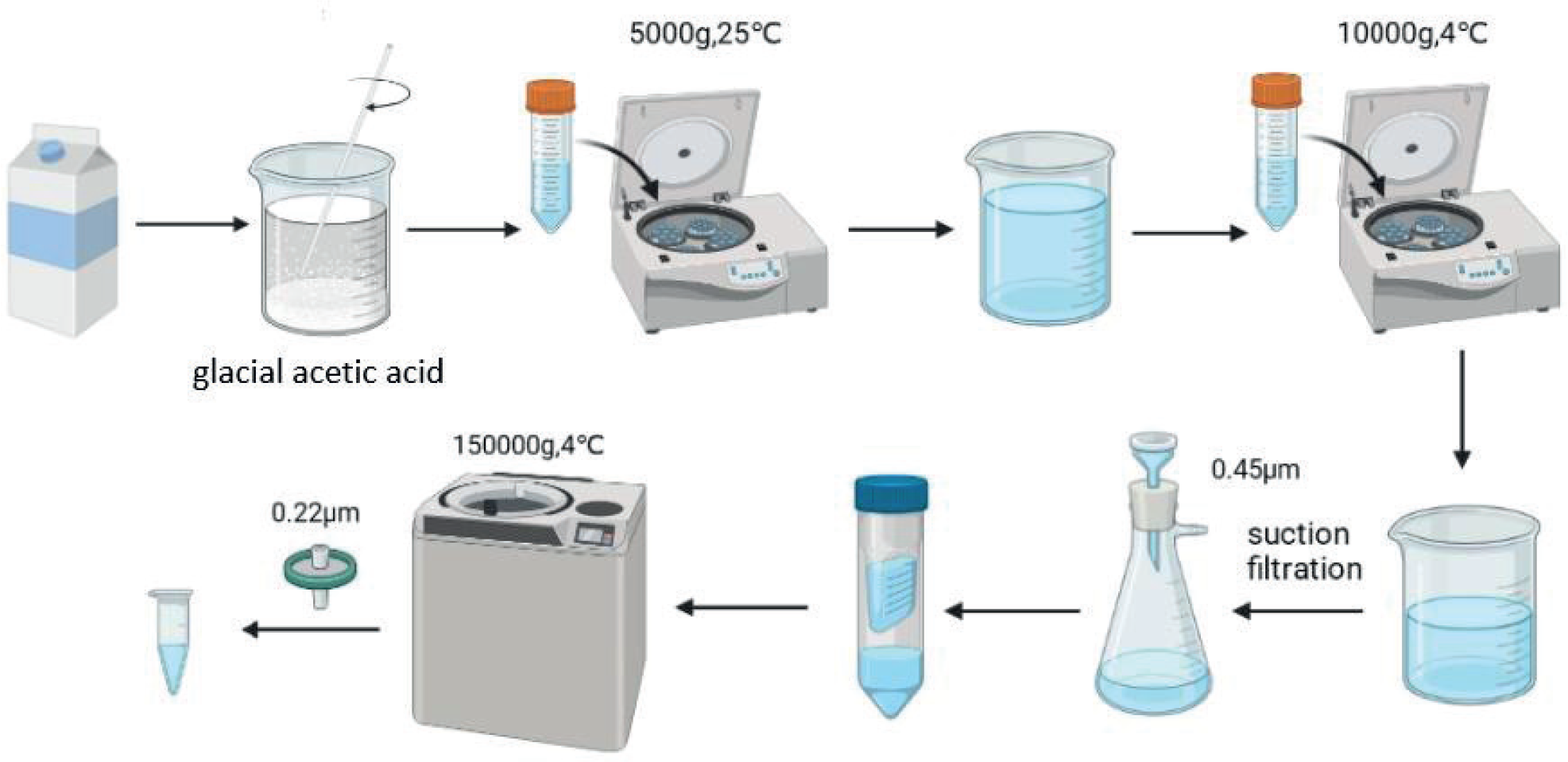
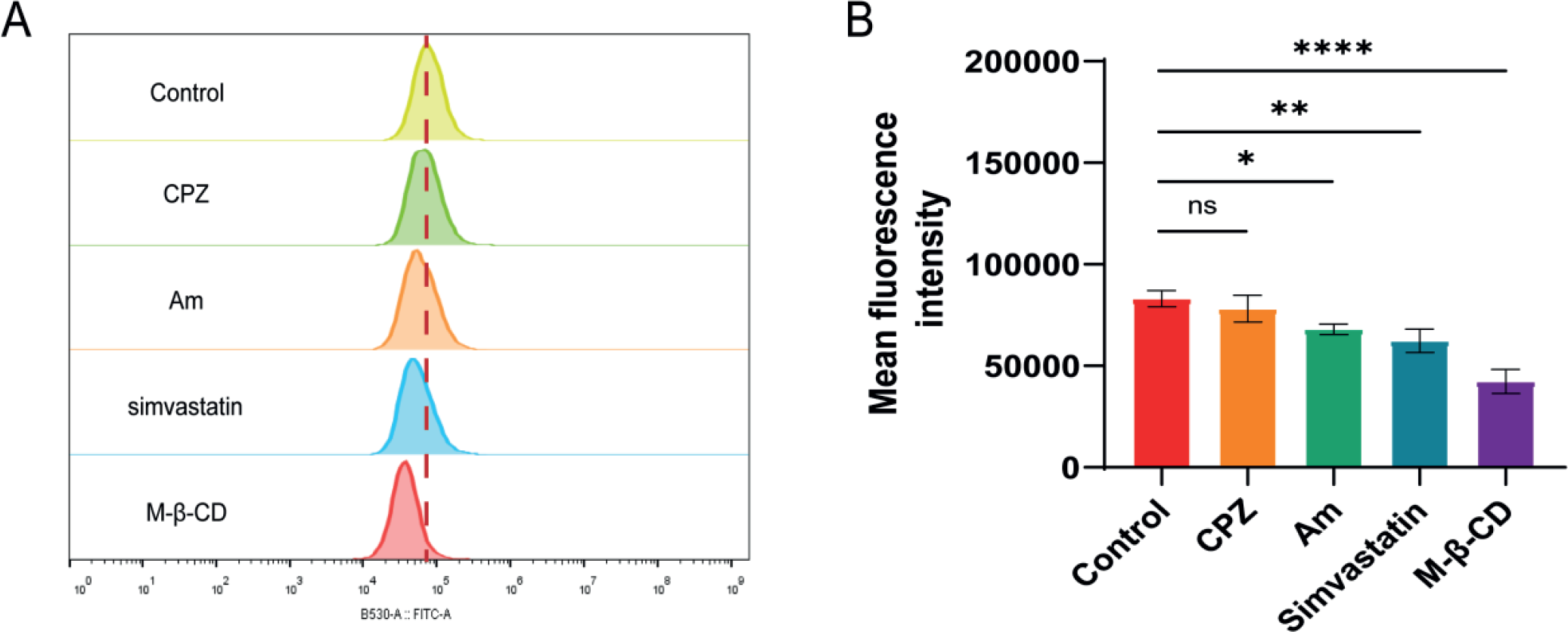
Skim milk was mixed with glacial acetic acid at a volume ratio of 100:1. The mixture was stirred at room temperature for 5 minutes and subsequently centrifuged at 5000×g for 20 minutes at 25°C. The upper aqueous phase was collected and subsequently centrifuged at 10000×g for 40 minutes at 4°C. The middle layer was carefully extracted, while the upper and lower layers were discarded. The collected liquid was filtered through a 0.45-μm membrane, and the filtrate was concentrated using an ultrafiltration tube with a molecular weight cut-off (MWCO) of 100 kDa. The concentrated liquid was centrifuged at 150,000×g for 90 minutes at 4°C. The precipitate was collected, resuspended in phosphate-buffered saline (PBS), and centrifuged at 150,000×g for 90 minutes. This washing process was repeated three times. Finally, the precipitate was resuspended in PBS and filtered through a 0.22-μm membrane to obtain purified mEVs (Figure 1).
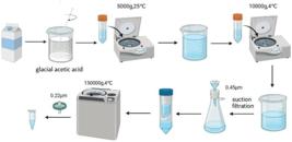
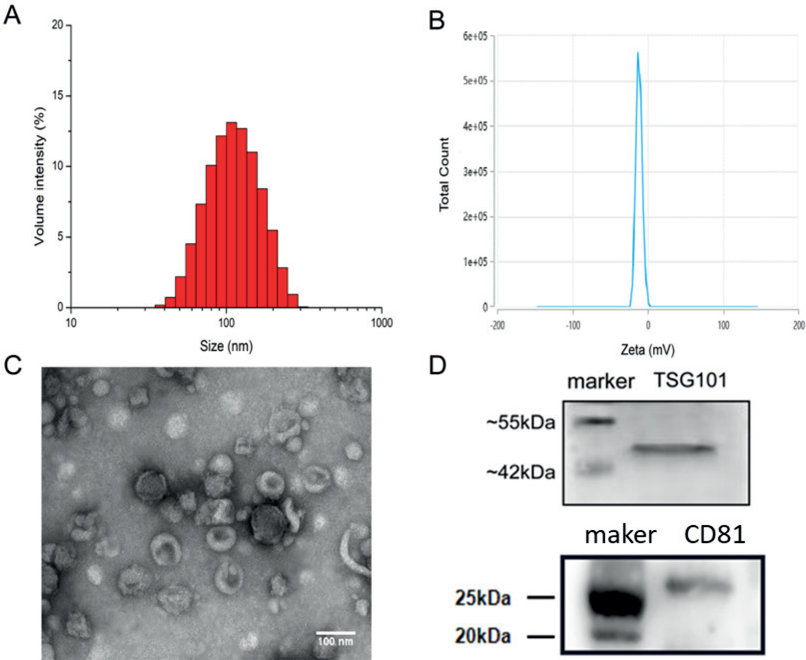
mEV characterization. A: Particle size of mEV; B: Electric potential of mEV; C: TEM of mEV (100 nm); D: Characteristic protein bands.
Characterization of mEVs
The morphology of the extracted mEVs was characterized using transmission electron microscopy (TEM). The particle size distribution and zeta potential of mEVs were determined using a nano-laser particle size analyzer. Characteristic mEV proteins were analyzed by Western blotting, and protein content within mEVs was quantified using a BCA protein assay kit.
In brief, an aliquot of mEVs was mixed with RIPA lysis buffer (containing RIPA and PMSF at a ratio of 100:1) at an appropriate ratio, incubated on ice for 20 minutes, and centrifuged at 14,000 ×g for 5 minutes. The supernatant was carefully transferred to a new tube for subsequent protein concentration determination. The protein sample was mixed with 5× loading buffer at a volume ratio of 4:1, heated at 95°C for 5 minutes, and loaded for Western blot analysis.
Stability of mEVs
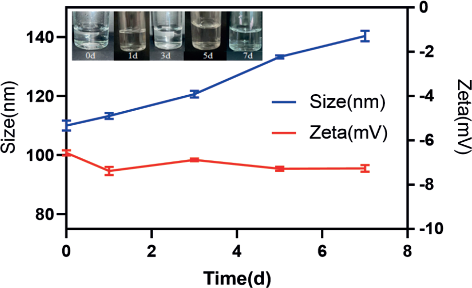
Freshly extracted mEVs were resuspended in PBS (pH 7.2) and stored at 4°C. To evaluate their stability, mEV particle size distribution and zeta potential were measured at day 0, 1, 3, 5, and 7 after preparation. This time-series analysis of key physicochemical properties provides critical insights into the long-term integrity and functionality of mEVs under refrigerated conditions, which is essential for their potential applications in biomedical research and therapeutic development.
In vitro cytotoxicity of mEVs
The effect of mEVs on the proliferation of HUVECs was determined using the MTT assay. HUVECs were cultured in a 10-cm petri dish until 80%-90% confluency, then digested with trypsin, counted, and seeded into a 96-well plate at a density of 1.5×10⁴ cells/ well. The cells were cultured overnight at 37°C with 5% CO2.
Once cell density reached 80%, the original culture medium was removed, and freshly prepared mEV solutions (100, 200, 300, 400, 500, and 1000 μg/ mL; experimental group), vehicle (control group), and culture medium (blank group) were added to the cells, with six replicate wells per condition. After 24 hours of incubation, 20 μL of MTT solution (5 mg/mL) was added to each well and incubated in the dark for 4-6 hours. The solution was then discarded, and 150 μL of dimethyl sulfoxide (DMSO) was added to dissolve the formazan crystals. Absorbance was measured at a wavelength of 490 nm using a microplate reader within 10 minutes. Cell viability was calculated according to formula (1) based on the ratio of the absorbance values of the experimental groups to those of the control group.
In vivo toxicity of mEVs in mice
Twenty-five SPF female BALB/c mice (6-8 weeks old) were randomly assigned into the normal saline control group, low-dose mEVs group (10 μg/20 g body weight), medium-dose mEVs group (100 μg/20 g), high-dose mEVs group (500 μg/20 g), and lipopolysaccharide (LPS) positive control group (10 μg/20 g), with five mice per group. All substances were administered via tail vein injection at a volume of 200 μL per 20 g of body weight, once every two days for a total of five doses. Mice were observed daily for changes in behavior, fur condition, or respiratory patterns. Body weight was measured and recorded every two days to monitor the potential effects of mEVs on growth and metabolism.
At 48 hours after the final dose, the mice were humanely euthanized according to the Institutional Animal Care and Use Committee guidelines. Immediately after euthanasia, the heart, liver, spleen, lungs, and kidneys were harvested, gently rinsed with pre-warmed normal saline to remove surface blood and contaminants, and promptly immersed in 4% paraformaldehyde solution for 24 hours to ensure proper tissue fixation. Subsequently, the fixed tissues were paraffin embedded, cut into 4 μm-thick sections, dewaxed, stained with hematoxylin and eosin (H&E), and examined under a light microscope by experienced pathologists.
Irritation potential of mEVs in mice
BALB/c mice were grouped, administered, and euthanized as previously described. Blood samples were collected via cardiac puncture and centrifuged at 3000 rpm for 15 minutes at 4°C to separate the serum for hematology and liver and renal function tests. Specifically, aspartate transaminase (AST) and alanine aminotransferase (ALT) levels were measured to assess hepatocellular integrity and potential liver damage, while creatinine (Cr) and urea nitrogen (UREA) concentrations were determined to evaluate kidney health and excretory function. All biochemical assays were performed using validated commercial kits, and the analyses were completed on an automated clinical chemistry analyzer. Stringent quality control measures, including the use of appropriate calibration standards and control samples, were implemented to ensure the accuracy, precision, and reliability of the obtained results.
Immunogenicity of mEVs in mice
The immunogenicity of mEVs in BALB/c mice was systematically evaluated using a multi-parameter approach. This involved calculating the spleen index, assessing the proliferation of splenic immune cells, quantifying the number of peritoneal macrophages, and measuring the levels of key inflammatory cytokines within the peritoneal fluid. In brief, BALB/c mice were grouped and treated as previously described. After the final dose of treatment and euthanasia, the body weight of each mouse was measured and recorded as BM, and the spleen was harvested and weighed to obtain the spleen weight (SW). The spleen index, a key biomarker reflecting the overall immune status and potential immunomodulatory effects of mEVs, was then calculated using formula (2). This parameter serves as a standardized metric for comparing immune responses across treatment groups, enabling a better understanding of the immunogenicity of mEVs in vivo.
The spleens of mice were carefully excised under aseptic conditions and immediately transferred to a sterile petri dish placed on ice. The spleens were gently dissociated into single-cell suspensions by grinding them through 70 μm cell strainers. Hank's balanced salt solution was added dropwise to maintain cell viability and facilitate efficient filtration. The resulting cell suspensions were centrifuged at 4°C and 2000 rpm for 15 minutes, and the cell pellets were resuspended in 2-3 mL of red blood cell lysis buffer and incubated at room temperature for 5 minutes. After lysis, 10-15 mL of D-Hank's solution was added to the cell suspensions, which were then centrifuged at 2000 rpm for 15 minutes at 4°C. The supernatant was removed, and the cell pellet was resuspended in Dulbecco's Hank's balanced salt (D-Hank's) solution.
This washing procedure was repeated three times to ensure the removal of residual lysing buffer and cell debris. After the final wash, the cell concentration was determined using a hemocytometer. Subsequently, 1×10⁶ cells were transferred to an Eppendorf tube. The cells were incubated with fluorochrome-conjugated CD19 and CD3 antibodies (diluted according to the manufacturer's recommendations) for 30 minutes at 4°C in the dark, washed three times with PBS to remove unbound antibodies, and resuspended in 500 μL of PBS for flow cytometry. Data acquisition and analysis were performed using appropriate software to accurately quantify the percentages of splenic B cells (CD19⁺) and T cells (CD3⁺).
Following euthanasia, mice were placed in a supine position, and the abdominal area was disinfected with 75% ethanol. A defined volume of sterile physiological saline was then injected into the peritoneal cavity using a sterile syringe. The abdomen was gently massaged for two minutes to ensure thorough dispersion of the saline and facilitate the collection of peritoneal-derived components. The peritoneal fluid was carefully aspirated with a sterile syringe and transferred to a conical centrifuge tube. The sample was centrifuged at 2000 rpm for 15 minutes at 4°C. The supernatant, rich in soluble factors, was carefully aliquoted and stored at -80°C until analysis. Inflammatory cytokines present in the supernatant were quantitatively detected using highly sensitive enzyme-linked immunosorbent assay (ELISA) kits. Each ELISA was performed according to the manufacturer's instructions, including standard curve preparation, sample dilution, and incubation time, to ensure result accuracy and reproducibility. The cell pellets from centrifugation were resuspended in D-Hank's solution, and 1×10⁶ cells were transferred to a sterile Eppendorf tube. The cells were incubated with fluorochrome-conjugated F4/80 antibody (well-established marker for macrophages) at 4°C for 30 minutes in the dark, washed three times with PBS to remove any unbound antibodies, and resuspended for flow cytometry analysis. Data acquisition was performed using a high-performance flow cytometer, and subsequent analysis was carried out with specialized software to determine the percentage of F4/80-positive macrophages in the samples.
Cellular uptake of mEVs
It has been well-documented that endocytosis is the primary pathway through which mEVs enter cells (Costa et al., 2017). To investigate the underlying mechanism of endocytosis, a panel of specific endocytosis inhibitors was utilized to block distinct endocytic pathways.
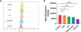
MCF-7 cells were seeded into 12-well plates at a density of 1.5×10⁵ cells/well and cultured at 37°C with 5% CO2 for 24 hours. Cells were treated with 20 μM chlorpromazine (CPZ, a selective inhibitor of clathrin-mediated endocytosis), 2 μM simvastatin (inhibitor of raft-mediated endocytosis), 10 mg/mL methyl-β-cyclodextrin (M-β-CD, a dual inhibitor of raftand caveolin-mediated endocytosis), or 100 μM amiloride (Am, inhibitor of pinocytosis) for 1 hour, then washed three times with pre-warmed PBS to remove any unbound inhibitors, and incubated with 50 μg/mL of FITC-labeled mEVs (FITC-mEVs) for 4 hours under standard cell culture conditions to allow mEV uptake. At the end of the incubation, the cells were washed three times with PBS to remove non-internalized FITC-mEVs, digested using trypsin-EDTA, resuspended in 1 mL of PBS, and analyzed by flow cytometry.
Statistical analysis
The experimental data were processed using GraphPad Prism 8.0 and presented as mean ± standard deviation. A P < 0.05 was considered statistically significant.
Stability of mEVs
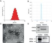
The mEVs were uniformly dispersed in PBS (pH7.2), resulting in a clear and translucent solution that remained stable for 7 days. Particle size, but not zeta potential, moderately increased over time. These results indicated that mEVs are stable at pH 7.2 and 4°C for at least 7 days (Table I and Figure 3).
In vitro cytotoxicity of mEVs
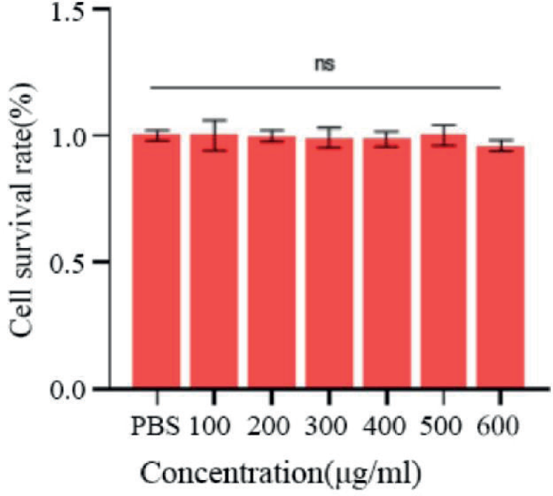
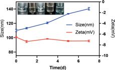
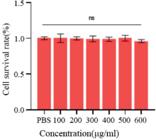
HUVEC viability after treatment with different concentrations of mEVs was determined using the MTT assay. There was no significant difference in cell viability between the control group and mEV groups. Notably, cell viability remained above 95% in the presence of 1000 μg/mL mEVs, which demonstrates that the mEVs exhibit minimal cytotoxicity in vitro (Figure 4).
In vivo toxicity of mEVs in mice
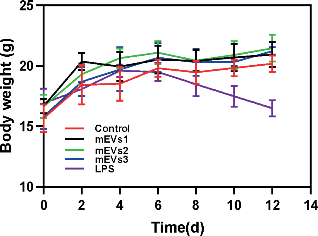
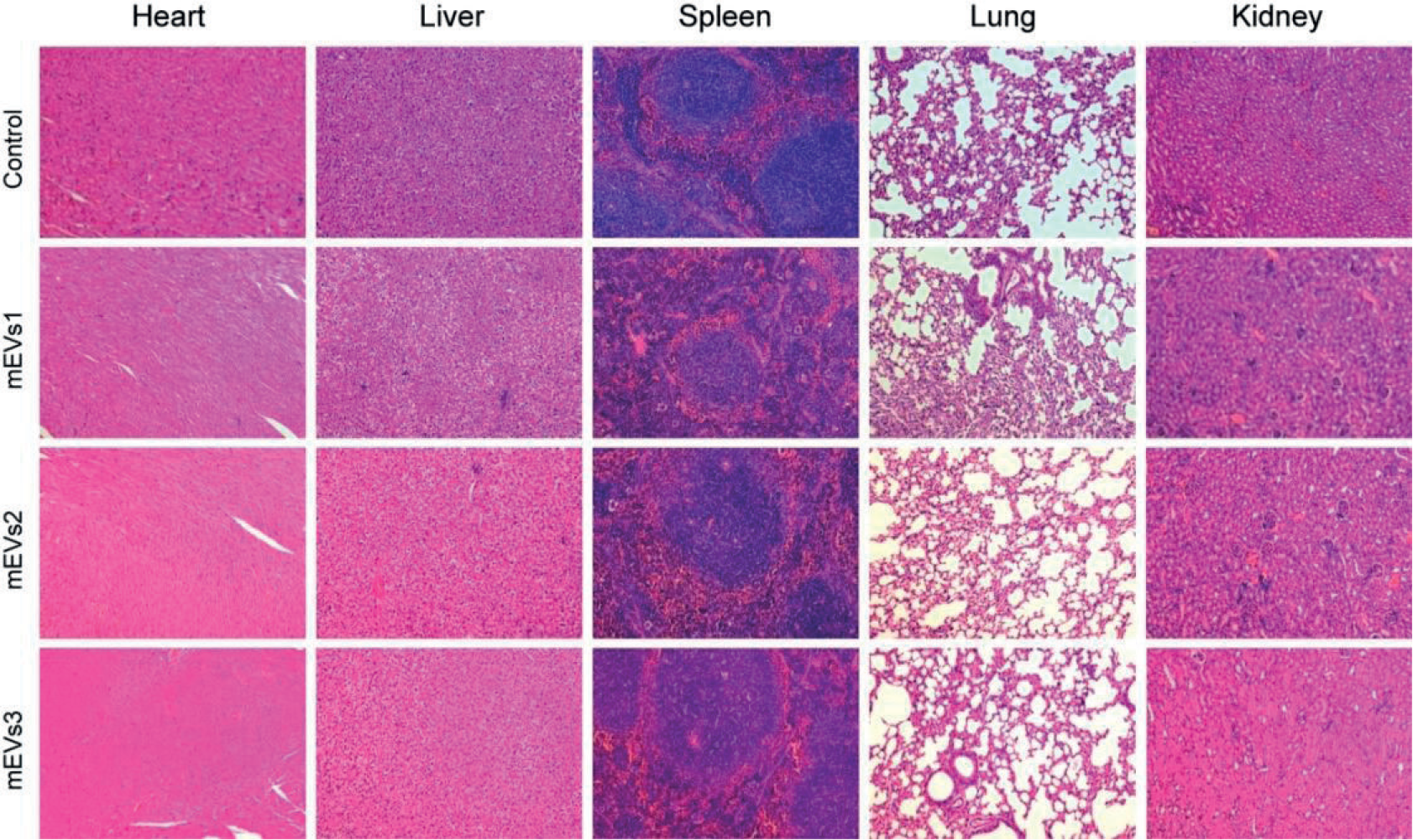
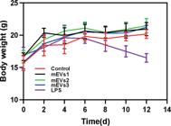
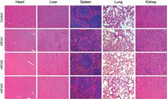
Weight loss is a common manifestation of toxicity in mice. As shown in Figure 5, weight gain was com-parable between the saline group and mEV groups but significantly decreased in the LPS group. No abnormal behavior or death was observed throughout the study. H&E staining of the hearts, livers, spleens, lungs, and kidneys of mEVs-treated mice revealed no significant histopathological changes compared to those observed in the normal saline group (Figure 6), indicating mini-mally toxicity in mice.
Irritation potential of mEVs in mice
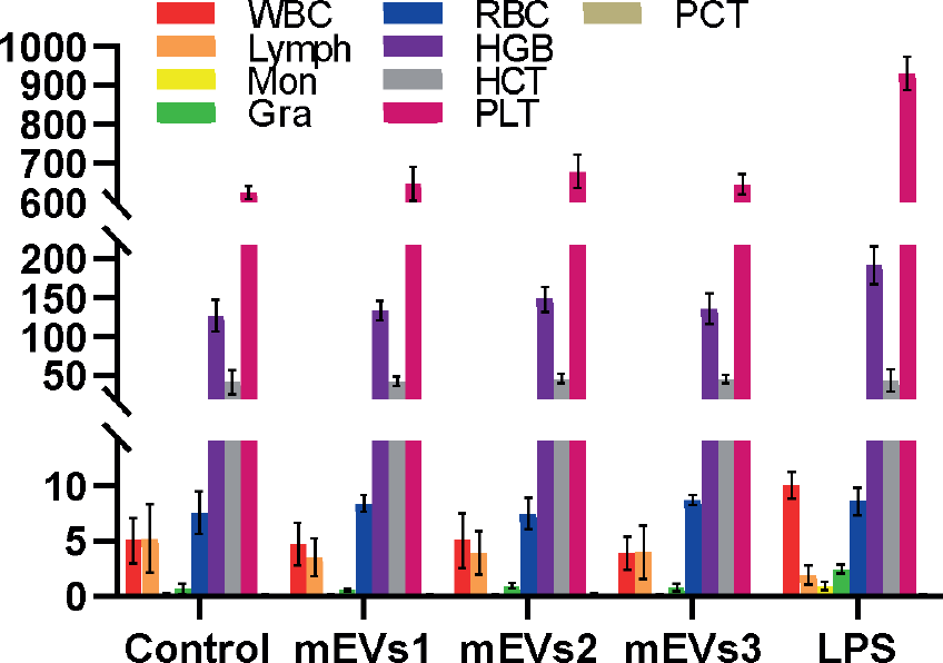
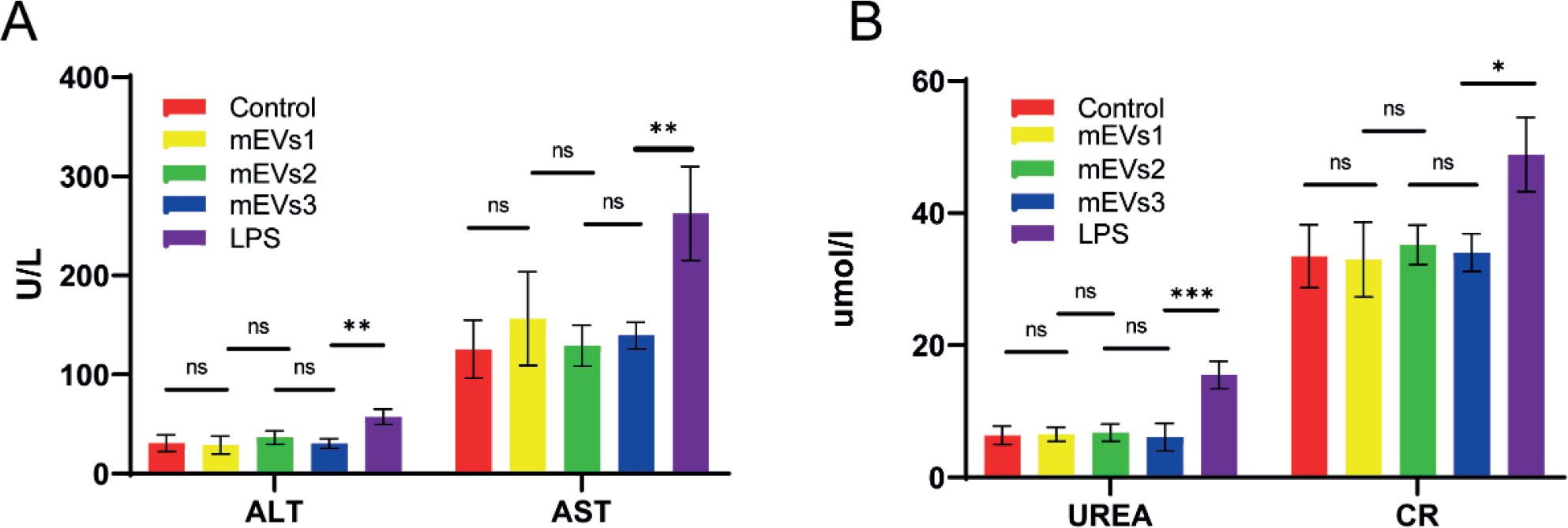
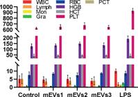
Hematology test revealed significantly higher white blood cell (WBC), neutrophil (Gra), and monocyte (Mon) counts and markedly lower lymphocyte (Lymph) counts in the LPS group compared to the control group (Figure 7). In contrast, hematology parameters were comparable between the control and mEVs groups. Additionally, ALT, AST, UREA, and CR levels were comparable between the mEVs groups and normal saline group (all P > 0.05), but significantly higher in the LPS group (all P < 0.05) (Figure 8). Collectively, these results indicate that mEVs do not induce irritation in mice.
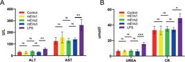
Liver and kidney parameters of mice in each group (n=5). (A) ALT and AST; (B) UREA and CR. N = 5, *P < 0.05, **P < 0.01, ***P < 0.001, ****P < 0.0001.
Immunogenicity of mEVs in mice
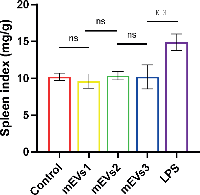
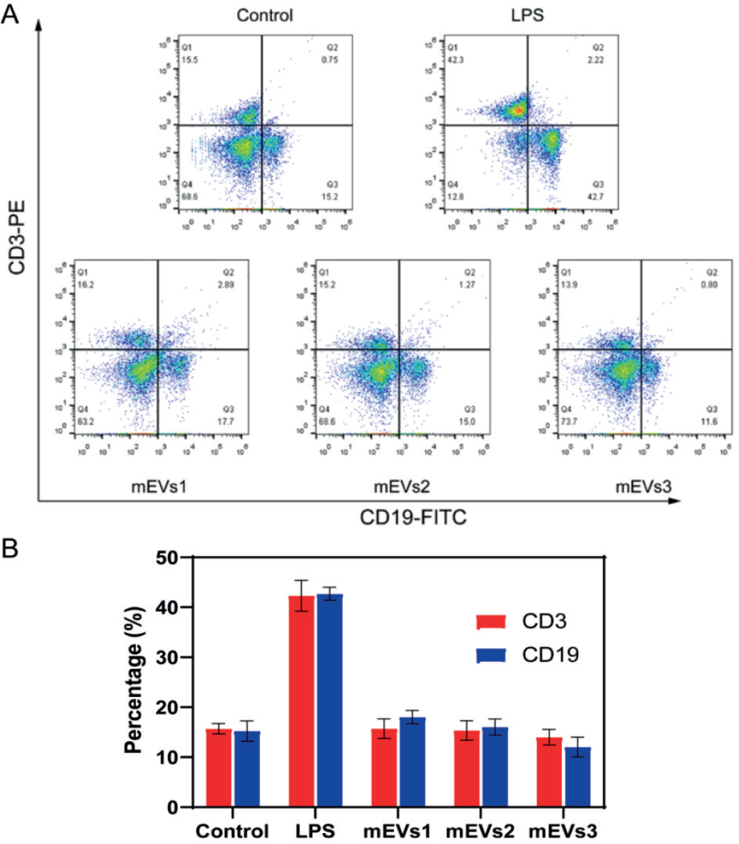
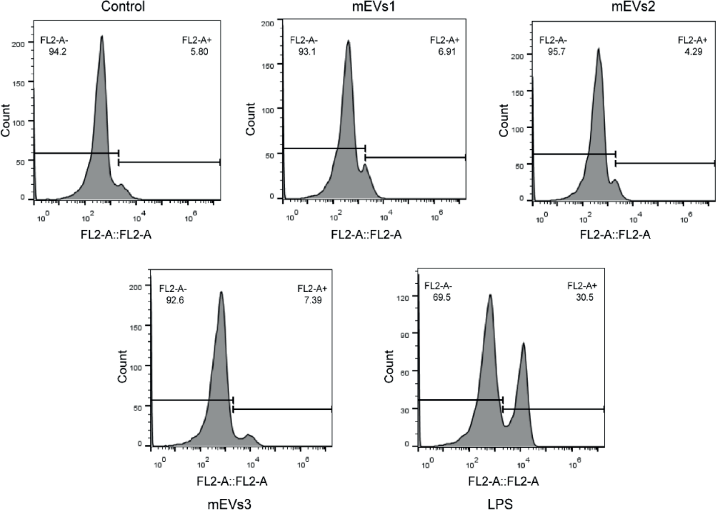
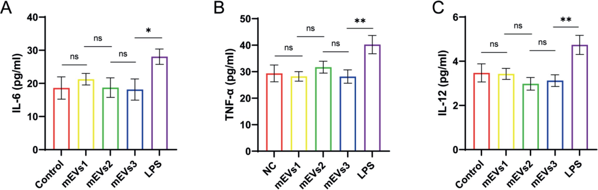
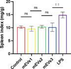
The spleen index of mice was similar between the mEV groups and normal saline group, but significantly higher in the LPS group (P < 0.01, Figure 9). Furthermore, the LPS group had higher percentages of splenic CD3+ T cells (42.3 ± 2.4% vs. 15.2 ± 3.8%) and CD19+ B cells (42.3 ± 2.4% vs. 15.5 ± 1.5%) than the normal saline control group (Figure 10), indicating that LPS stimulated splenic B and T cell proliferation. On the other hand, the percentages of splenic B and T cells were similar between the mEVs group and normal saline group, demonstrating that the mEVs does not induce splenic immune cell proliferation. Similarly, the percentage of peritoneal macrophages was markedly higher in the LPS group but similar between the mEVs groups and normal saline group (Figure 11). Peritoneal levels of IL-6, IL-12 and TNF-α were significantly upregulated in the LPS group (all P < 0.05) but were comparable between the mEVs and normal saline groups (all P > 0.05) (Figure 12). Altogether, these data suggest that mEVs do not elicit robust immune responses in vivo.
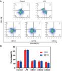
Percentage of splenic immune cells in each group. A: Flow cytometry graph, B: Quantitative analysis graph.

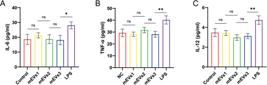
Cellular uptake of mEVs
M-β-CD, a dual inhibitor of raftand caveolin-mediated endocytosis, had the strongest inhibitory effect on mEV uptake, as evidenced by significantly lower mean fluorescence intensity compared to other groups (P < 0.0001; Figure 13). Simvastatin (inhibitor of raft-mediated endocytosis) and amiloride (inhibitor of pinocytosis) also inhibited mEV uptake, with simvastatin exhibiting a stronger effect than amiloride. The clathrin-mediated endocytosis inhibitor chlorpromazine had the weakest inhibitory effect (P > 0.05). These results demonstrated that mEVs enter cells through multiple mechanisms rather than a single pathway, predominantly via raft-mediated endocytosis and pinocytosis.
DISCUSSION
Milk sources can vary considerably based on factors such as cow breed, feeding environment, and lactation stage, all of which may influence the consistency of mEVs. For example, different cow breeds may secrete mEVs with distinct protein and nucleic acid profiles. A study on bovine milk-derived components has shown that genetic factors can influence in milk protein composition, which may in turn be reflected in the mEVs (Ou et al., 2024). Lactation stage is another crucial determinant of mEV characteristics. During early lactation, the colostrum contains a higher concentration of immune-related components. mEVs isolated from colostrum may thus possess enhanced immunomodulatory properties compared to those from mature milk (Rahman et al., 2021). As lactation progresses, the composition of milk and mEVs gradually changes, potentially impacting their suitability for different therapeutic applications.
Regarding the long-term effects of repeated mEV administration, although our short-term study in mice showed no significant adverse effects, prolonged exposure could potentially disrupt metabolic balance or elicit cumulative immune responses (Gutierrez et al., 2022). In translational research aimed at human applications, numerous ethical considerations and technical challenges must be addressed. The source of milk is a critical ethical concern, as ensuring the welfare of cows and maintaining high standards in milk production are essential. Regarding potential allergenicity, milk is a common allergen, and although mEVs are a sub-component of milk, they may carry allergenic proteins. Additionally, designing appropriate clinical trials to determine optimal dosage and treatment duration presents a significant challenge. Similar challenges have been encountered in nanomedicine, as discussed in a review, where factors such as biodistribution, clearance rate, and toxicity of nanocarriers must be carefully considered when formulating clinical trial protocols (Metselaar, Lammers, 2020).
We selected BALB/c mice as the animal model due to their well-characterized immune system and homogeneous genetic background, making them ideal for evaluating the immunogenicity of mEVs and minimizing variability in experimental results (Liang et al., 2021). A power analysis was conducted using data from preliminary experiments and estimates from similar studies. Based on the expected effect size, significance level (α = 0.05), and power (1-β = 0.8), a sample size of 5 mice per group was determined to be sufficient for detecting significant differences between groups. This sample size also considered practical limitations, such as resource availability and animal housing capacity, while ensuring reliable statistical results.
All animal experiments were conducted in strict accordance with ethical guidelines. To minimize animal suffering during surgical procedures and sample collection, mice were anesthetized using an appropriate anesthetic agent, with close monitoring of the depth of anesthesia. The experimental procedures were optimized to reduce the duration of stress on the animals. In accordance with the 3Rs principles, although no alternative non-animal models were available for this specific study, we made efforts to minimize animal use. The sample size was determined through a rigorous power analysis to ensure that the minimum number of animals was used while still obtaining reliable results. In terms of refinement, the handling and housing conditions of the animals were continuously optimized. The cages were furnished with comfortable bedding, and environmental factors such as temperature, humidity, and light-dark cycle, were tightly controlled to provide a stress-free living environment for the mice. This approach aligns with the recommendations of animal welfare organizations, including the National Institutes of Health's Guide for the Care and Use of Laboratory Animals, which emphasizes the importance of adhering to the 3Rs principles in animal research.
CONCLUSION
This study systematically evaluated the biosafety of mEVs and discerned the mechanisms of their cellular uptake. mEVs extracted from cow's milk had no cytotoxic effect in vitro and did not induce toxicity, irritation, or immune responses in mice following repeated intravenous injections. mEVs are taken up by cells through several mechanisms, predominantly via lipid raft-mediated endocytosis and pinocytosis. Collectively, these findings provide strong evidence for the favorable biosafety of mEVs, highlighting their potential as promising candidates for biomedical applications. Their efficient uptake by cells through specific mechanisms further supports their development as novel drug delivery systems. However, further research is warranted to address the variability in milk sources, evaluate the long-term effects of repeated mEV administration, and explore the translation of these findings to human applications. Such studies will be crucial for fully realizing the therapeutic potential of mEVs in clinical settings.
ETHICAL COMPLIANCE
All animal studies were approved by the Ethical Committee and responsible authorities of our research organization(s), in accordance with applicable guidelines, regulations, and ethical standards for humans or animals.
ACKNOWLEDGMENTS
This study was supported by grants from the Key Scientific Research Project Plan of Colleges and Universities in Henan Province (24A350020), Key Research and Development and Promotion Projects of Henan Province (242102311233), Science andTechnique Foundation of Henan Province (No. 212102310778 for J.Z), and Medical Science and Technique Foundation of Henan Province (No. LHGJ20210201 for J.Z).
DATA AVAILABILITY STATEMENT
Use of data not disclosed.
REFERENCES
- Chen W, Wu P, Jin C, Chen Y, Li C, Qian H. Advances in the application of extracellular vesicles derived from three-dimensional culture of stem cells. J Nanobiotechnol. 2024;22(1):215.
- Costa Verdera H, Gitz-Francois JJ, Schiffelers RM, Vader P. Cellular uptake of extracellular vesicles is mediated by clathrin-independent endocytosis and macropinocytosis. J Control Release. 2017; 266:100-108.
- Gutierrez A, Harvey EL, Creehan KM, Taffe MA. The long-term effects of repeated heroin vapor inhalation during adolescence on measures of nociception and anxiety-like behavior in adult Wistar rats. Psychopharmacology (Berl). 2022;239(12):3939-3952.
- Hendrix A, De Wever O. Systemically circulating bacterial extracellular vesicles: origin, fate, and function. Trends Microbiol. 2022;30(3):213-216.
- Heusermann W, Hean J, Trojer D, Steib E, von Bueren S, Graff-Meyer A, et al. Exosomes surf on filopodia to enter cells at endocytic hot spots, traffic within endosomes, and are targeted to the ER. J Cell Biol. 2016;213(2):173-84.
- Juodeikis R, Carding SR. Outer Membrane Vesicles: Biogenesis, Functions, and Issues. Microbiol Mol Biol Rev. 2022;86(4):e0003222.
- Lang HL, Zhao YZ, Xiao RJ, Sun J, Chen Y, Hu GW, et al. Small extracellular vesicles secreted by induced pluripotent stem cell-derived mesenchymal stem cells improve postoperative cognitive dysfunction in mice with diabetes. Neural Regen Res. 2023;18(3):609-617.
- Liang YJ, Duan L, Lu JP, Xia J. Engineering exosomes for targeted drug delivery. Theranostics. 2021;11(7):3183-3195.
- Liu H, Zhang Q, Wang S, Weng W, Jing Y, Su J. Bacterial extracellular vesicles as bioactive nanocarriers for drug delivery: Advances and perspectives. Bioact Mater. 2021;14:169-181.
- Metselaar JM, Lammers T. Challenges in nanomedicine clinical translation. Drug Deliv Transl Res. 2020, 10(3):721-725.
- Ou HR, Csuth TI, Czompoly T, Krisztian K. Dairy: Friend or Foe? Bovine Milk - Derived Extracellular Vesicles and Autoimmune Diseases. Int J Mol Sci. 2024;25(21):11499.
- Patel B, Gaikwad S, Prasad S. Exploring the significance of extracellular vesicles: Key players in advancing cancer and possible theranostic tools. cancer pathogenesis&therapy. 2024;02(00):E01-E44.
- Rahman MM, Takashima S, Kamatari YO, Shimizu K, Okada A, Inoshima Y. Comprehensive Proteomic Analysis Revealed a Large Number of Newly Identified Proteins in the Small Extracellular Vesicles of Milk from Late-Stage Lactating Cows. Animals (Basel). 2021;11(9):2506.
- Senesi G, Lodrini A, Mohammed S, Mosole S, Ceresa D, Bolis S, et al. Human cardiomyocyte derived extracellular vesicles regulate cardiac fibroblast activation through miR-24. Cardiovasc Res. 2024;120(Supplement 1):i44.
- Su LY, Tian Y, Zheng Q, Cao Y, Yao M, Wang S, et al. Anti-tumor immunotherapy using engineered bacterial outer membrane vesicles fused to lysosome-targeting chimeras mediated by transferrin receptor. Cell Chem Biol. 2024;31(6):1219-1230.e5.
- Wang L, Wang G, Mao W, Chen Y, Rahman MM, Zhu C, et al. Bioinspired engineering of fusogen and targeting moiety equipped nanovesicles. Nat Commun. 2023;14(1):3366.
Edited by
-
Associate Editor:
Guilherme Martins Gelfuso