 Neuroprotective activity of fluoxetine in the retina of rats subjected to experimental light exposure
Abstract
This study evaluated the neuroprotective effects of fluoxetine in the retina of 45 Wistar rats exposed to intense light (3000 LUX for 12 hours). The animals were assigned to prevention (CG, PhotoG, and FG) and regeneration (RCG, RPhotoG, FG 7, FG 14, FG 21, and FG 30) groups, receiving daily fluoxetine treatment (10 mg/kg/day, intraperitoneally [i.p.]) either before or after photoexposure. Retinal analysis was performed using histology and histomorphometry (hematoxylin and eosin staining), detection of apoptosis (TUNEL assay), and electroretinography (ERG). Rats in the PhotoG group demonstrated reduced outer nuclear layer (ONL) thickness (~25 µm), an increased apoptotic index, and decreased b-wave amplitudes in the ERG (~60 µV), compared to controls. The FG group maintained ONL thickness (~36 µm), exhibited reduced apoptosis, and demonstrated improved electrophysiological responses. In the FG 30 group, ONL thickness increased to approximately 39 µm, apoptosis decreased, and the b-wave amplitude increased to ~120 µV, accompanied by reduced implicit times. These findings indicate that fluoxetine protects the retina against light-induced damage through antioxidant, anti-inflammatory, and antiapoptotic mechanisms, with enhanced efficacy following prolonged administration, suggesting its potential as a therapeutic agent for retinal degenerative diseases.
Keywords:
Fluoxetine; Retina; Retinal degeneration; Neuroprotective agent.
INTRODUCTION
The process by which light is the causative agent of injury is called phototoxicity or light-induced retinal degeneration (Cingolani et al., 2006). Photoreceptors are also directly affected by the byproducts of excessive phototransduction. The large number of free radicals in the intercellular environment induces the formation of damaged membrane proteins and lipids, leading to an imbalance in the cell that can collapse and initiate the cascade of events that will culminate in death by apoptosis. Phototoxicity mechanisms may vary depending on the intensity, duration, and spectrum of light (Cingolani et al., 2006).
There is currently no treatment that restores photoreceptors lost in retinal degeneration. Fluoxetine, a selective serotonin reuptake inhibitor (SSRI), is commonly used in the treatment of depression. Several studies have found important neurobiological roles of fluoxetine related to the central nervous system (CNS). Novio et al. (2011) demonstrated a positive effect of fluoxetine against oxidative cellular damage resulting from stress. Zafir and Banu (2007) also demonstrated the antioxidant potential of fluoxetine, which represents an important intracellular mechanism underlying the protective pharmacological effects observed clinically in the treatment of several stress-related diseases.
The anti-inflammatory action of fluoxetine was also demonstrated by Abdel-Salam, Baiuomy, and Arbid (2004), who induced inflammatory responses in rats using carrageenan and observed effects comparable to those of standard anti-inflammatory drugs. Kolla et al. (2005) demonstrated greater neuronal survival and a reduction in the levels of apoptosis mediators and oxidative substances such as superoxide dismutase and hydrogen peroxide (H2O2). Makkonen et al. (2011) demonstrated an increase in the concentration of insulin-like growth factor 1 (IGF-1) following fluoxetine administration. Caiaffo et al. (2016) demonstrated the anti-inflammatory, antioxidant, and antiapoptotic activities of fluoxetine in their review.
Fluoxetine has demonstrated significant neuroprotective potential in different models of retinal injury. In the study by Romano et al. (2023), mice subjected to retinal ischemia/reperfusion and treated with topical fluoxetine showed functional preservation of retinal ganglion cells, with significant improvement in the amplitude of the pattern electroretinogram (PERG) and reduction in latency (p < 0.05). In addition, there was a significant reduction in the expression of inflammatory markers such as IL-6, TNF-α, IL-1β, and Iba-1, indicating modulation of the inflammatory response by fluoxetine.
Furthermore, Ambati et al. (2021) identified that fluoxetine is a direct inhibitor of the NLRP3 inflammasome, an intracellular structure involved in the pathogenesis of age-related macular degeneration (AMD). The drug binds to the NACHT domain of NLRP3, inhibiting its activation and preventing Alu RNA-induced retinal pigment epithelium (RPE) degeneration. In a murine model, fluoxetine treatment significantly reduced RPE cell loss. Moreover, analyses of large population-based databases showed that patients with depression treated with fluoxetine had a lower risk of developing dry AMD (HR: 0.814; 95% CI: 0.676-0.98).
The incessant search for therapies that can promote the protection and/or regeneration of retinal cells, especially photoreceptors, has been the focus of several studies, both in vitro and in vivo. Therefore, the present study evaluated the neuroprotective activity of fluoxetine in the retinas of rats subjected to experimental light exposure.
MATERIAL AND METHODS
Sample
45 male rats (Rattus norvergicus albinus), 60 days, obtained from Federal Rural University of Pernambuco, Brazil. All experiments were conducted in accordance with the Ethics Committee for the Use of Animals of the University under process number 054/2014 as well as the Statute for the Use of Animals of the Association for Research in Vision and Ophthalmology.
Experimental design
This experiment was divided into two: first, called prevention, the animals were randomly divided into three groups: control group (CG), which was not submitted to any intervention; photo-exposed group (PhotoG), which was submitted to light exposure (3000 LUX); and fluoxetine group (FG), which was treated with fluoxetine administered with an intraperitoneal dose of 10 mg/kg for seven consecutive days prior to light exposure. The animals were euthanized seven days after exposure to light. In the second experiment, regeneration, the animals were divided into 6 experimental groups: RCG (Regeneration Control Group) (the animals in this group were not subjected to the stress of photo-exposure; they only received doses of 0.9% NaCl, being 1ml/100g intraperitoneally), RPhotoG (subjected to photoexposure and also only received doses of 0.9% NaCl, being 1ml/100g intraperitoneally), FG 7, FG 14 FG 21 FG 30 (subjected to photoexposure and treated with fluoxetine 10mg/kg, intraperitoneally, for 7, 14, 21 and 30 consecutive days). In all groups, treatments always started at the same time (at 12 pm). All animals were euthanized immediately after the end of fluoxetine treatment.
Exposure to light
The animals were submitted to adaptation to the dark for 24 hours in individualized cages in the interior of the light exposure chamber with a mean temperature of 22 ± 2 ºC. Next, the pupils were dilated with the topical application of Tropicamide 1% and the animals were submitted to 3000 LUX (measured by Instrutherm LD-240) of white light for 12 hours beginning at 6:00 am (Montalbán-Soler et al., 2012). The light exposure chamber was made of wood and had white inner walls, an adequate ventilation system and two 40W fluorescent bulbs. After exposure, the animals were returned to the laboratory and kept again in a cyclic light regimen until euthanasia.
Electroretinogram (ERG)
The protocol described by Montalbán-Soler et al. (2012) was used, with modifications. Twelve hours after photoexposure, three animals per group were anesthetized (ketamine 60 mg/kg and xylazine 20 mg/kg) and then adapted to the dark for 30 minutes under a red light. The left pupils were dilated with tropicamide 1% and the animals were positioned on a cushion heated to 37°C to maintain the core body temperature. A diode emitter with luminous flashes was positioned 1 cm from the eye. The light intensity was calibrated by a dual biosignal generator device specifically adapted for ERG recordings. The electroretinographic signals were acquired with the use of a DTL fiber electrode positioned on the cornea. A reference electrode was placed approximately 3 cm from the ipsilateral eyelid commissure and a ground electrode was positioned on the tail.
The electrical signals generated on the retina were amplified (x 1000) and filtered (bandpass filter of 1 to 1000 Hz) using the Nihon Kohdem system, Neuropack 2. MEB-7102A/k. The light stimuli were calibrated prior to ensure standardization across animals.
The retina was stimulated using different intensities depending on the type of ERG: 0.01 cd.s/m2 at 0.2 Hz for two flashes was used for the scotopic ERG: 3 cd.s/m2 at 0.1 Hz for two flashes was used for the mixed scotopic ERG; 3 cd.s/m2 at 2 Hz for five flashes was used for the photopic ERG; and 3 cd.s/m2 at 30 Hz for 20 flashes was used for the flicker ERG. The amplitude of the a-wave was measured as the difference in voltage between the mean of the 10 μs baseline recorded prior to the flashes and the trough of the a-wave. The amplitude of the b-wave was measured as the voltage difference between the peak of the b-wave and the trough of the a-wave.
Euthanasia, collection of material and histological analysis
After the ERG, the animals were anesthetized (ketamine 60 mg/Kg and xylazine 20 mg/Kg), followed by euthanasia using sodium pentobarbital. Perfusion of the left ventricle was then performed with sodium chloride 0.9%, followed by paraformaldehyde in 0.1 M phosphate buffer (pH 7.4). The left eye was enucleated and immersed in paraformaldehyde 4% in a phosphate buffer 0.1 M (pH 7.4) for 48 hours. The material was then processed according to standard protocols and embedded in Paraplast Plus (Sigma-Aldrich). The blocks were sliced into 5 μm-thick sections. Histological slides were prepared and stained with hematoxylin and eosin.
For the histological analysis, the slides were photographed using the the LAEZ image capture system (Leica). Total thickness and the thickness of the outer nuclear layer (ONL) were measured using ImageJ® for Windows. Eight measurements were made per slide (15 slides per group): four in the portion above the optical disc (superior or dorsal retina) and four in the portion below the optical disc (inferior or ventral retina). The measurements were separated by a distance of 200 μm beginning with the optic disc.
TdT-mediated DUTP nick-end labeling (TUNEL) procedure
After being cleared with xylol, the slices were rehydrated and submitted to the TUNEL procedure using the Apoptag® Red in Situ kit (Merk Millipore), following the manufacturer’s protocol. Immunostaining was performed on paraffin sections according to the manufacturer’s instructions, and DNA was also extracted from frozen tissue by homogenization for 5 minutes at 37°C, followed by DNA isolation as described by Distelhorst, Lam & McCormick (1996). An AxioPlan epifluorescence microscope (Carl Zeiss, Göttingen, Germany) was used to capture photomicrographs with a 40 x objective, using a G 365 nm excitation filter and LP 420 nm emission filter. Nucleus undergoing apoptosis emits a red or purple fluorescence while those with intact DNA emit blue fluorescence.
Cells in apoptosis were counted using 15 fields per slide with the aid of a grid with 441 points. The mean number of apoptotic cells per animal in each group was compared.
Statistical analysis
The data obtained from the morphometric evaluations, the apoptosis assay (TUNEL) and the electroretinography (ERG) were expressed as means ± standard deviations, reflecting the intraand intergroup variability.
For comparison between the experimental groups, one-way analysis of variance (ANOVA) test was used, followed by Tukey’s post hoc test, when applicable. This statistical approach allowed us to verify significant differences between multiple groups simultaneously, controlling The type I error, and identifying which groups presented specific differences between them. The significance level of p < 0.05 was adopted, indicating that the observed results have, at most, a 5% probability of being due to chance.
RESULTS AND DISCUSSION
The histomorphometric analysis demonstrated reduced outer nuclear layer (ONL) thickness values as well as increased degeneration and structural disorganization of the outer segment layer of photoreceptors in the PhotoG and RphotoG groups. These animals exhibited pronounced vacuolization in the intracellular matrix, loss of the elongated, fusiform morphology of the outer segment of the photoreceptors, and absence of the inner segment.
The administration of fluoxetine preserved ONL thickness, attenuating the loss of photoreceptor nuclei, and protecting both the inner and outer segments of these cells. This neuroprotective effect was observed in the FG group and showed progressive enhancement in the FG 7, FG 14, FG 21, and FG 30 groups, demonstrating that longer durations of fluoxetine treatment promoted a greater regenerative effect against light-induced damage (Figures 1 and 2).

A - CG. B - PhotoG. Note retina damage: reduced thickness, with loss of nuclei of photoreceptors on ONL; disorganization and degeneration of OSPL, with presence of vacuoles and the loss of elongated, fusiform morphology. C - FG. Preservation of thickness of ONL and better maintenance of elongated, forsiform shape of OSPL. Total thickness of retina (D) and outer nuclear layer (E) of rats submitted to light exposure and the administration of fluoxetine. Note the increase in thickness in animals of FG. *p < 0.05 between PhotoG and FG. RGCL - retinal ganglion cell lay. IPL - inner plexiform layer. INL - inner nuclear layer. OPL - outer plexiform layer. ONL - outer nuclear layer. OSPL - outer segment photoreceptor layer. H.E. staining.

A - RCG. B - RPhotoG. Note retina damage: reduced thickness, with loss of nuclei of photoreceptors on ONL; disorganization and degeneration of OSPL, with presence of vacuoles and the loss of elongated, fusiform morphology. C - FG 7, D - FG 14, E - FG 21 and F - FG 30. Preservation of thickness of ONL and better maintenance of elongated, forsiform shape of OSPL. Total thickness of retina (G) and outer nuclear layer (H) of rats submitted to light exposure and the administration of fluoxetine. Note the increase in thickness in animals of FG. *p<0,05 entre os grupos FG 30 and RPhotoG.
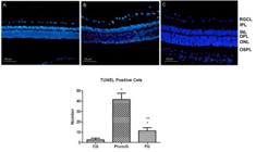
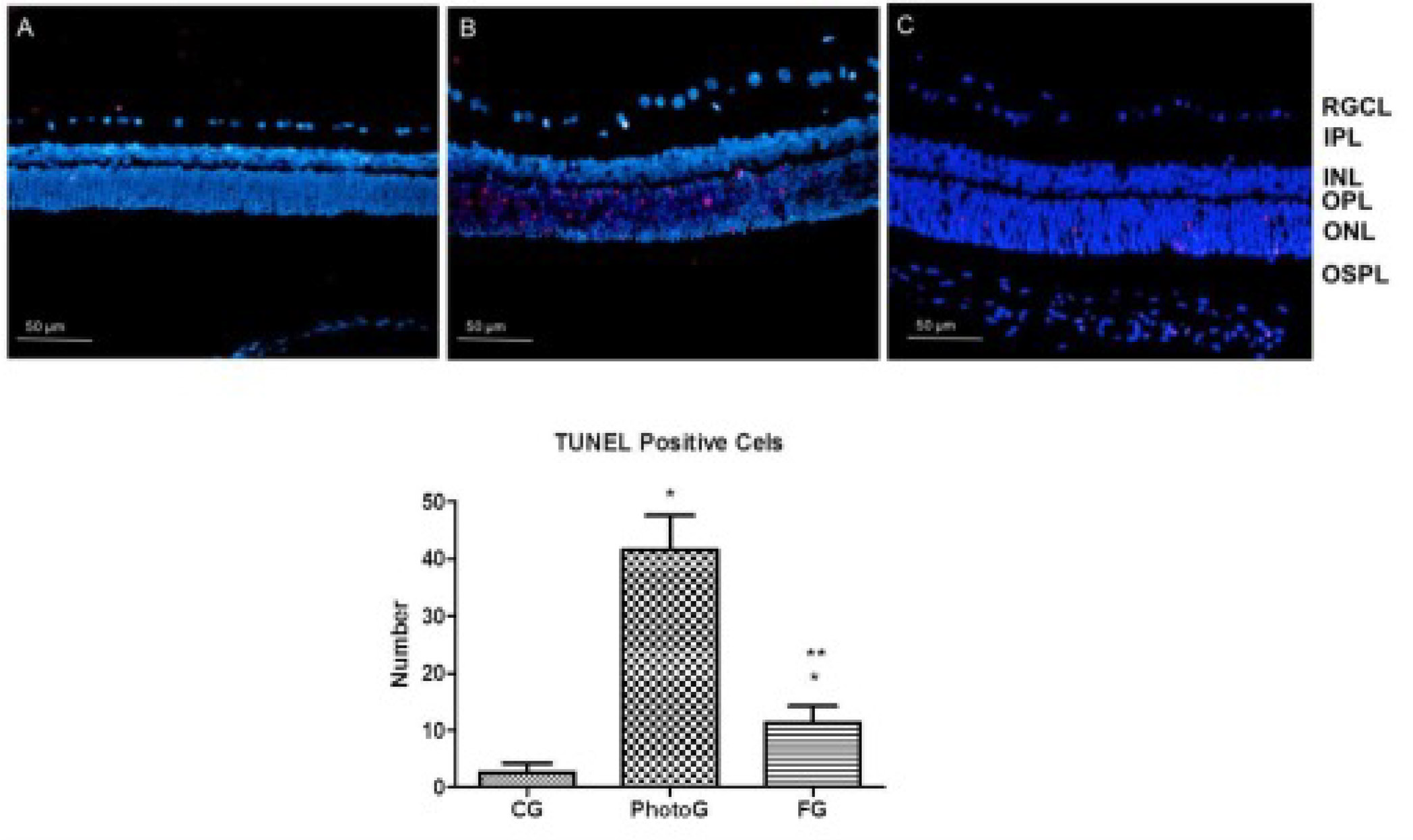
Photomicrographs with fluorescence of the retina of rats submitted to light exposure and the administration of fluoxetine, prevention experiment, demonstrating labeling of cells in apoptosis using TUNEL method in red color. And Number of positive TUNEL cells of the outer nuclear layer of rats submitted to light exposure and the administration of fluoxetine. *p < 0.05, PhotoG x CG and FG x CG. **p < 0.05, FG x PhotoG.

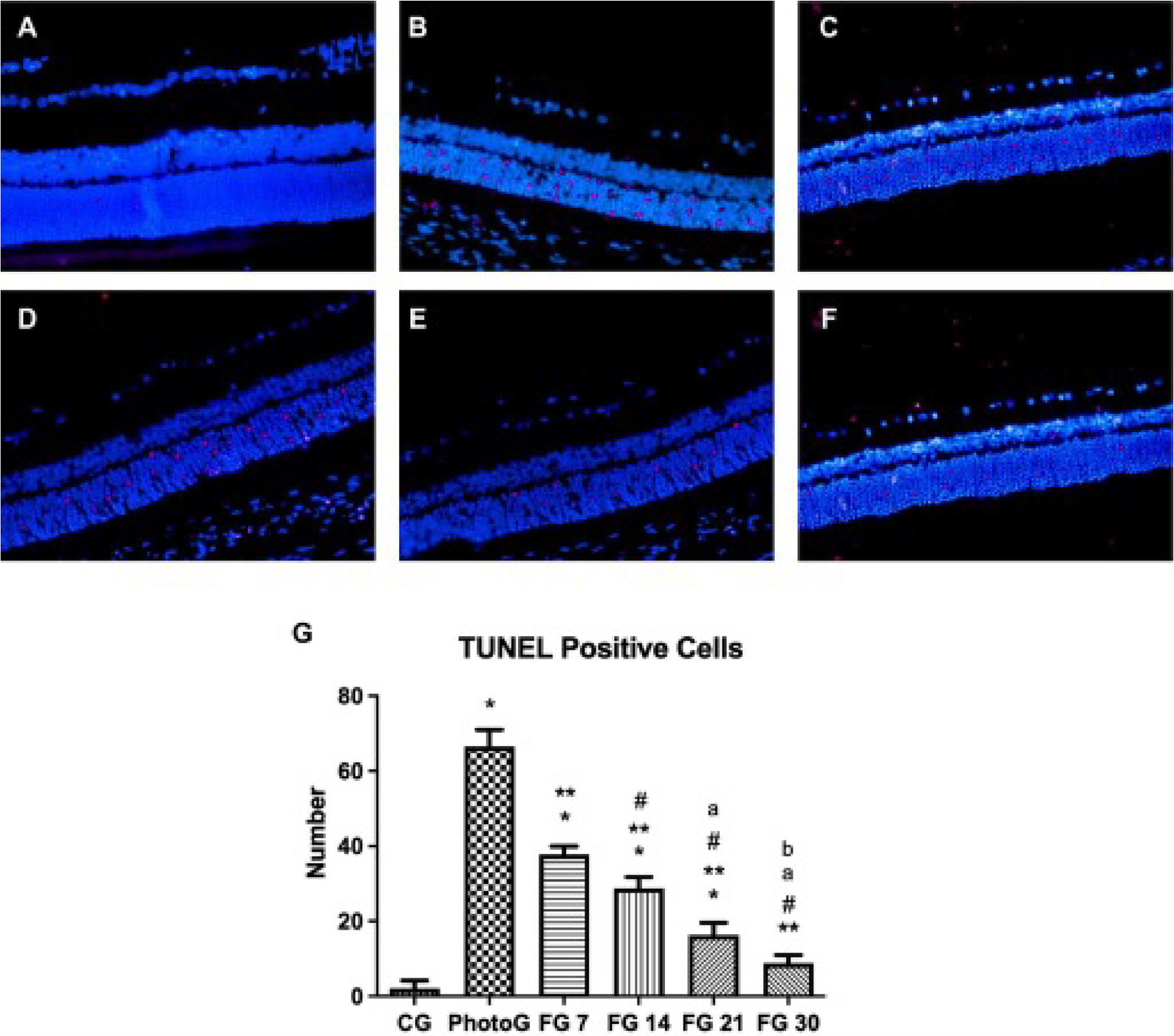
Photomicrographs with fluorescence of the retina of rats submitted to light exposure and the administration of fluoxetine, regeneration experiment, demonstrating labeling of cells in apoptosis using TUNEL method in red color. And Number of positive TUNEL cells of the outer nuclear layer of rats submitted to light exposure and the administration of fluoxetine. A - RCG, B - RPhotoG, C - FG 7, D - FG 14, E - FG 21 and F - FG 30. *p<0.05, RPotoG x RCG, FG 7 x RCG, FG 14 x RCG, FG 21 x RCG; **p<0,05, FG 7 x RPotoG, FG 14 x RPotoG, FG 21 x RPotoG, FG 30 x RPotoG; # p<0,05, FG 14 x FG 7, FG 21 x FG 7, FG 30 x FG 7; a p<0,05, FG 21 x FG 14, FG 30 x FG 14; b p<0,05, FG 30 x FG 21.
Montalbán-Soler et al. (2012) demonstrated more pronounced photoreceptor degeneration in the dorsocentral region of the retina in mice submitted to light exposure. Some authors attribute the reduced damage in the ventral retina to a lower concentration of reactive oxygen species, the shorter outer segment length of band cells, and a lower rhodopsin content comparte to the dorsal region (Penn, Anderson, 1987). According to some studies (Li, Cao, Anderson, 2003; Liu et al., 2011) and Stone et al. (1999), the direction and localization of the light source in the environment where animals are housed may also influence the regional distribution of retinal damage. In the present study, the light source was positioned on the ceiling of the exposure chamber, and upon striking the convex curvature of the cornea, the light beams were refracted and redirected, resulting in predominant exposure of the dorsocentral region of the retina.
The outer segment layer of photoreceptors consists of membranous discs derived from the plasma membrane, which are impregnated with photopigments responsible for phototransduction (Cohen, 1972). Thus, photoreceptors are directly affected by the byproducts of excessive phototransduction. The large number of free radicals in the intracellular environment induces the formation of proteins and lipids with damaged membranes, leading to cellular imbalance that can result in cell collapse and initiate a cascade of events culminating in apoptotic cell death (Costa et al., 2008).
In the present study, light exposure induced a higher rate of apoptosis in animals from the PhotoG and RPhotoG groups. In contrast, animals subjected to fluoxetine treatment, in both the prevention and regeneration protocols, exhibited a progressive reduction in apoptotic cell numbers (Figures 3 and 4).
Several in vivo studies have demonstrated that fluoxetine (FLX) can act as a neuroprotective agent in various models of neuronal injury (Ludka et al., 2017; Peric et al., 2017; Khodanovich et al., 2018). It has been shown that FLX protects the brain following ischemic injury by reducing both early and long-term neuronal loss and inflammation, improving survival and functional recovery, and enhancing neurogenesis (Sun et al., 2015). FLX has also demonstrated significant immunomodulatory and anti-inflammatory effects in neurodegenerative disease models, mitigating oxidative damage by reducing lipid peroxidation, suppressing the production of reactive oxygen and nitrogen species, and increasing the activity of antioxidant enzymes (Ludka et al., 2017; Peric et al., 2017). Moreover, protection against Aβ-induced neurotoxicity has been shown to be mediated by TGF-β1 (Caraci et al., 2016).
Ambati et al. (2021) demonstrated that fluoxetine exerts protective effects against retinal pigment epithelium (RPE) degeneration in experimental models of macular degeneration. The authors reported a dose-dependent inhibitory effect of fluoxetine on Alu RNA-induced RPE cell loss. These findings reveal that fluoxetine exhibits a specific protective activity in a disease-relevant animal model, an effect not observed with several other FDA-approved antidepressants.
Réus et al. (2012) found that fluoxetine can increase the expression of Akt, CREB, BDNF, and Bcl-2, all of which are involved in anti-apoptotic signaling pathways. Other studies have also shown that antidepressants such as fluoxetine upregulate Bcl-2 expression (Montalbán-Soler et al., 2012; Chiou et al., 2006). Furthermore, BDNF stimulation, in conjunction with the Akt signaling pathway, enhances the expression of Bcl-2, which is regarded as a key inhibitor of neuronal cell death.
The anti-apoptotic protein Bcl-2 and the pro-apoptotic protein BAD regulate the release of cytochrome c from the mitochondria, which plays a central role in apoptosis initiation (Li et al., 1997). Agostinho et al. (2011a) evaluated the effects of fluoxetine on mitochondrial respiratory chain complexes and reported alterations in enzymatic activity in the brains of treated rats. Additionally, acute fluoxetine treatment was found to alter citrate synthase activity, while both acute and chronic administration affected the activity of creatine kinase (Agostinho et al., 2011b). These enzymes are involved in cellular energy metabolism, and it has been clearly established that fluoxetine is associated with modulation of metabolic pathways, which correlates with reduced cell death in neuropsychiatric conditions (Ben-Shachar and Karry, 2008).
Ex vivo studies have shown that antidepressants inhibit the secretion of IL-1β, IL-2, tumor necrosis factor-α (TNF-α), and IFN-γ, as well as the proliferative activity of T cells and the cytotoxic function of natural killer (NK) cells (Ghosh et al., 2015). Liu et al. (2003) demonstrated that fluoxetine reduces the production of IL-6, TNF-α, and nitric oxide. These effects are attributed to suppression of gene expression, as evidenced by reduced transcriptional levels of IL-6 and TNF-α mRNA. Furthermore, fluoxetine may inhibit phosphorylation of the mitogen-activated protein kinase (MAPK) pathway, a major signaling cascade for pro-inflammatory cytokines, and may also suppress nuclear factor kappa B (NF-κB) activation, a central regulator of inflammatory responses.
Ghosh et al. (2015) observed changes in the levels of IL-4, IL-6, IL-10, and IL-12 in macrophages cultured in the presence of tumor fluid. These cytokine shifts indicated an altered macrophage phenotype, suggestive of a polarization switch from M1 (classically activated) to M2 (alternatively activated). Upon fluoxetine treatment, a significant reversal in IL-4, IL-6, and IL-10 levels was observed. The authors proposed that fluoxetine may promote macrophage repolarization toward a host-protective phenotype, likely via NF-κB inhibition, supporting its potential role in modulating immune responses in inflammatory conditions.
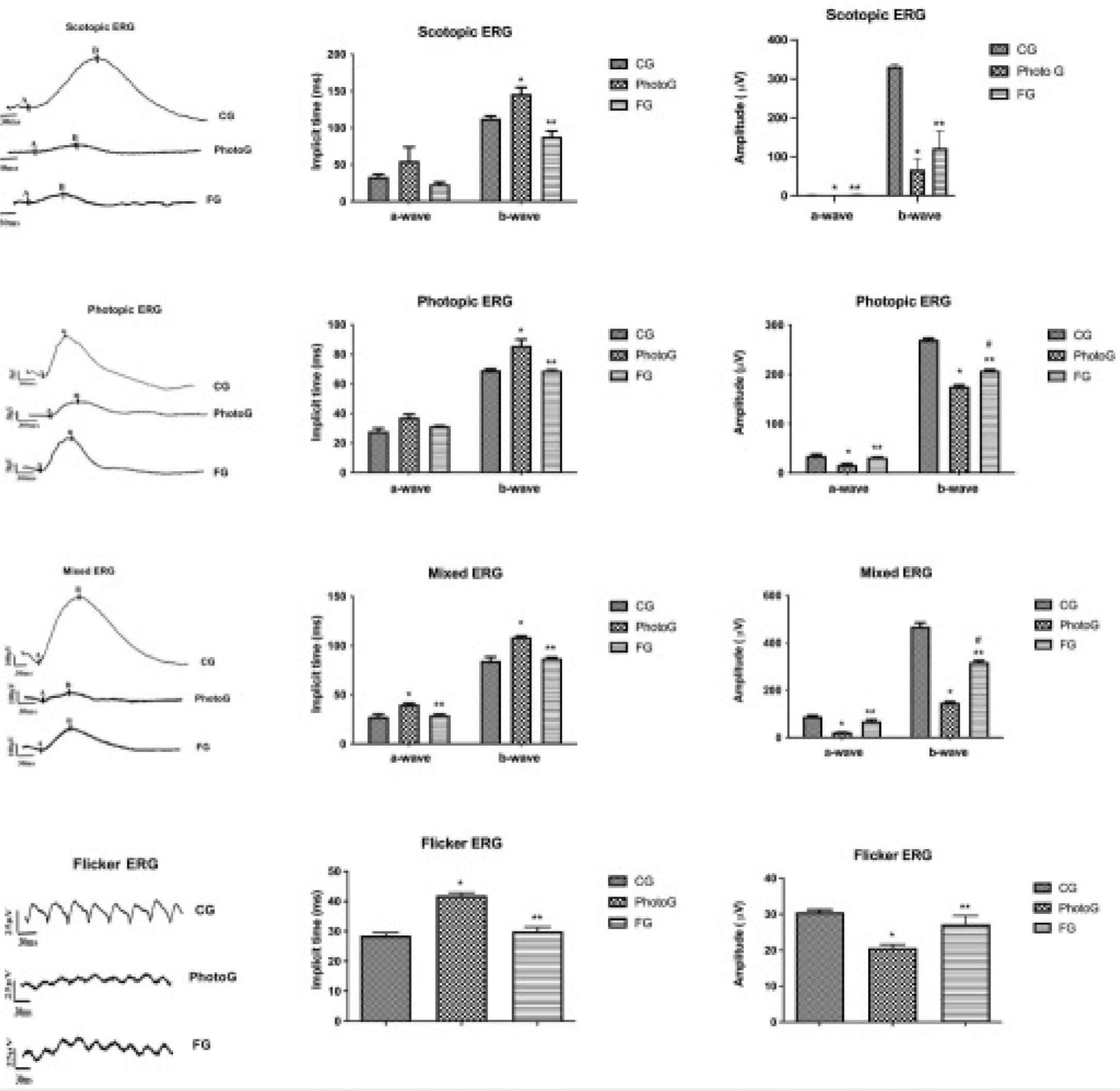
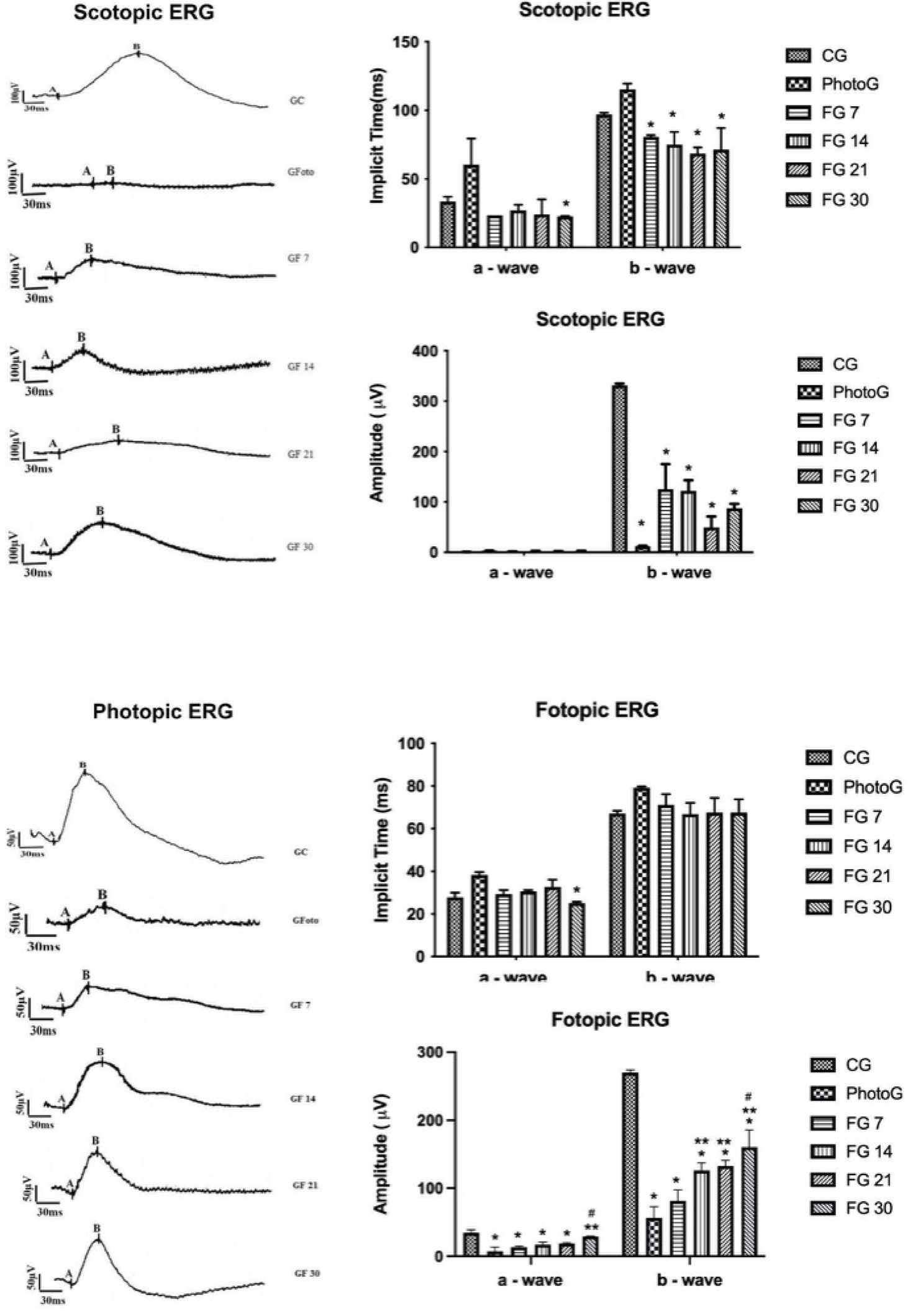
In the present study, all ERG modalities (scotopic, photopic, mixed, and flicker) demonstrated shorter implicit times (latencies) in the retinal responses of animals from the PhotoG and RPhotoG groups. Notably, a significant improvement in ERG parameters was observed in the FG and FG 30 groups, indicating a more pronounced effect of fluoxetine when administered for extended periods (Figures 5, 6, and 7).
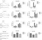
Scotopic ERG: Implicit response time (ms) of retinal cells from the emission of the flash of light to the triggering of the action potential. *p < 0.05, PhotoG x CG; **p < 0.05, FG x PhotoG. Amplitude of a-wave in μV. **p < 0.05, PhotoG x CG; **p < 0.05, FG x PhotoG. Amplitude of b-wave in μV. *p < 0.05, PhotoG x CG; **p < 0.05, FG x PhotoG. Photopic ERG: Implicit Time (ms) *p < 0.05, PhotoG x CG; **p < 0.05, FG x PhotoG. Amplitude of a-wave in μV. *p < 0.05, PhotoG x CG; **p < 0.05, FG x PhotoG. Amplitude of b-wave in μV. *p < 0.05, PhotoG x CG; **p < 0.05, FG x PhotoG; # p< 0.05, FG x CG. Mixed ERG: Implicit Time (ms) *p < 0.05, PhotoG x CG; **p < 0.05, FG x PhotoG. Amplitude of a and b-waves in μV. *p < 0.05, PhotoG x CG; **p < 0.05, FG x PhotoG.; #p < 0.05, FG x CG. Flicker ERG: Implicit Time (ms) *p <0.05, PhotoG x CG. Amplitude in μV.; **p < 0.05, FG x PhotoG.
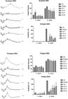
Scotopic ERG: Implicit time (ms) a-wave: *p<0,05, RPhotoG x FG 30. b-wave: *p<0,05, RPhotoG x FG 7; RPhotoG x FG 14; RPhotoG x FG 21; RPhotoG x FG 30. Amplitude (μV) a-wave: *p<0,05, RPhotoG x RCG, FG 7 x RCG, FG 14 x RCG, FG 21 x RCG and FG 30 x RCG; **p<0,05, FG 14 x RPhotoG, FG 21 x RPhotoG, FG 30 x RPhotoG. # p<0,05, FG 30 x FG 7. Amplitude b-wave *p<0,05, RPhotoG x RCG, FG 7 x RCG, FG 14 x RCG, FG 21 x RCG e FG 30 x RCG; **p<0,05, FG 7 x RPhotoG, FG 14 x RPhotoG, FG 30 x RPhotoG. Photopic ERG: Implicit time (ms) a-wave: *p<0,05, RPhotoG x FG 30. Amplitude (μV) a-wave: *p<0,05, RPhotoG x RCG, FG 7 x RCG, FG 14 x RCG, FG 21 x RCG; **p<0,05, FG 30 x RPhotoG. # p<0,05, FG 30 x FG 7. Amplitude b-wave *p<0,05, RPhotoG x RCG, FG 7 x RCG, FG 14 x RCG, FG 21 x RCG and FG 30 x RCG; **p<0,05, FG 14 x RPhotoG, FG 21 x RPhotoG, FG 30 x RPhotoG. # p<0,05, FG 30 x FG 7.

Mixed ERG: Implicit time (ms) a-wave: *p<0,05, RPhotoG x RCG. **p<0,05, FG 7 x RPhotoG, FG 14 x RPhotoG, FG 21 x RPhotoG, FG 30 x RPhotoG. b-wave: *p<0,05, RPhotoG x RCG, FG 21 x RCG, FG 30 x RCG. **p<0,05, FG 7 x RPhotoG, FG 14 x RPhotoG, FG 21 x RPhotoG, FG 30 x RPhotoG. Amplitude (μV) a-wave: *p<0,05, RPhotoG x RCG, FG 7 x RCG, FG 14 x RCG, FG 21 x RCG, FG 30 x RCG. **p<0,05, FG 14 x RPhotoG, FG 30 x RPhotoG. Amplitude (μV) a-wave: *p<0,05, RPhotoG x RCG, FG 7 x RCG, FG 14 x RCG, FG 21 x RCG and FG 30 x RCG; **p<0,05, FG 7 x RPhotoG, FG 14 x RPhotoG, FG 21 x RPhotoG, FG 30 x RPhotoG. Flicker ERG: Implicit time (ms) *p<0,05, RPhotoG x RCG. **p<0,05, FG 7 x RPhotoG, FG 14 x RPhotoG, FG 21 x RPhotoG, FG 30 x RPhotoG. Amplitude (μV): *p<0,05, RPhotoG x RCG. **p<0,05, FG 7 x RPhotoG, FG 14 x RPhotoG, FG 21 x RPhotoG, FG 30 x RPhotoG.
Similar to the present study, Romano et al. (2023) reported an improvement in the electroretinographic (ERG) responses of rats subjected to retinal ischemic injury and treated with fluoxetine. Their data show a significant increase in ERG amplitude and a reduction in implicit time (latency) in rats treated with fluoxetine, suggesting preservation of retinal ganglion cell function.
Montalbán-Soler et al. (2012) stated that the retina possesses compensatory mechanisms that may attempt to reverse the damage caused by light-induced degeneration, evidenced by a gradual recovery of the b-wave in the ERG. However, the same authors emphasized that such mechanisms do not fully account for the rapid recovery of the b-wave observed in their experiments, and that protein regulation is only one component of the complex array of compensatory processes involved in retinal remodeling. Likewise, the present findings demonstrate enhanced amplitudes of both aand b-waves, along with shorter implicit times. This accelerated recovery of ERG responses may be influenced by fluoxetine treatment.
Collier et al. (2011) demonstrated that light-induced retinal degeneration can be prevented or attenuated through treatment with serotonin receptor (5-HT1A) agonists. Although the mechanisms of action of 5-HT1A agonists are not yet fully elucidated, some proposed pathways include neuronal membrane hyperpolarization mediated by G protein-coupled inwardly rectifying potassium channels (GIRKs), which reduce glutamate release and block calcium (Ca2+) or sodium (Na+) channels.
Several studies suggest that the generation of free radicals and the accumulation of intracellular calcium (Ca2+) are closely associated with retinal injury caused by excessive light exposure (Hensler, 2003). In addition to being a selective serotonin reuptake inhibitor (SSRI) with antioxidant, anti-inflammatory, and neuroprotective properties (Caiaffo et al., 2016), fluoxetine may also act as an ion channel inhibitor.
Hahn et al. (1999) found that fluoxetine exerts a potent inhibitory effect on potassium (K+), sodium (Na+), and calcium (Ca2+) channels. Using hippocampal and prefrontal cortex neurons from rat embryos, Deák et al. (2000) demonstrated fluoxetine’s ability to inhibit L-, N-, and T-type voltage-dependent calcium channels. Similarly, Choi et al. (2004), also using hippocampal neurons, showed that fluoxetine reduces the frequency of action potentials and the amplitude of membrane depolarization. The authors attributed this modulation of membrane potential to the inhibition of voltage-dependent K+ currents, as well as other ion channels, such as Na+ and Ca2+, since fluoxetine is not a selective ion channel blocker.
Ionic currents play a critical role in shaping neuronal action potentials, and modulation of any of these currents can significantly alter neuronal excitability. In this context, Kim et al. (2005) demonstrated that fluoxetine inhibits ATP-induced currents that promote increases in intracellular Ca2+ levels. This inhibitory action reduces both the influx of extracellular Ca2+ and the release of Ca2+ from intracellular stores.
This attenuation of calcium dynamics was also observed by Steele, Chen, and MacLeish (2005). As previously noted, an elevated intracellular Ca2+ concentration is directly associated with photoreceptor damage induced by light overstimulation. Therefore, fluoxetine’s inhibitory effects on Ca2+ channels may facilitate membrane depolarization of photoreceptors, resulting in reduced phototransduction activity and, consequently, lower production of reactive oxygen species and free radicals, thereby providing neuroprotection to retinal cells.
Montalbán-Soler et al. (2012) report that the retina possesses compensatory mechanisms, which could attempt to reverse the harm caused by light-induced degeneration, with a slow recovery of the b-wave on the ERG. However, the same authors state that such mechanisms do not explain the rapid recovery of the b-wave in their studies and that such proteins are only part of the diverse compensatory mechanisms involved in retinal alterations. Likewise, the present findings demonstrate better amplitude of the a and b-waves as well as a shorter implicit time. The fast recovery of the b-wave may have been influenced by fluoxetine. Collier et al. (2011) demonstrated that light-induced degeneration can be prevented or suppressed by treatment with agonists of serotonin receptors (5-HT1A). The mechanisms by which agonists of 5-HT1A, work have not yet been fully clarified, but some isolated mechanisms have been identified, such as the hyperpolarization of neuronal membranes mediated by the G protein coupled to K+ channels which diminishes the release of glutamate [and blocks either Ca++ or Na+ channels.
Some studies indicate that the formation of free radicals and excess of intracellular Ca++ are closely related to damage to the retina stemming from an excess of light (Hensler, 2003). Besides being a selective serotonin reuptake inhibitor with various functions (antioxidant, anti-inflammatory and neuroprotective) (Caiaffo et al., 2016), fluoxetine may also act as an ionic channel inhibitor. Hahn et al. (1999) found that fluoxetine has a potent inhibiting effect on K+, Na+ and Ca++ channels. Using neurons from the hippocampus and prefrontal cortex of rat embryos, Dea´k et al. (2000) demonstrated the inhibitory power of fluoxetine on the voltage-dependent L, N and T calcium channels. Also using hippocampus neurons, Choi et al. (2004) demonstrated the effect of fluoxetine on the reduction in the frequency of the action potential and the amplitude of the depolarization of the membrane potential. The authors state that this modulation of the membrane potential may be due to an inhibition of voltage-dependent K+ currents and different ionic currents, such as Na+ and Ca++, since fluoxetine is not a selective ionic blocker. Ionic currents affect the modulation of the neuronal action potential and changes in any of these currents could alter the activity of the action potential. Likewise, Kim et al. (2005) demonstrated that fluoxetine clearly inhibits an ATP-induced current that increases the concentration of intracellular Ca++ ions. This inhibitory effect diminishes both the inflow of Ca++ from the extracellular environment to the intracellular environment and the release of Ca++ from internal cell reserves. This reduction in the dynamics of Ca++ ions was also demonstrated by Steele, Chen, MacLeish (2005). As stated previously, the increase in the intracellular Ca++ concentration is directly related to damage in the retina stemming from excessive light. Thus, such inhibitory effects of fluoxetine may favor the depolarization of photoreceptor membranes, leading to a lower rate of phototransduction and, consequently, less production of reactive oxygen species and free radicals, thereby offing neuroprotection to retinal cells.
CONCLUSIONS
The results of this study demonstrate that fluoxetine exhibits significant neuroprotective effects in the retina of rats subjected to intense light exposure. Administration of the drug preserved outer nuclear layer (ONL) thickness, significantly reduced photoreceptor apoptosis, and enhanced retinal function, as evidenced by increased ERG wave amplitudes and shortened implicit times. These effects were more pronounced with prolonged treatment, particularly in the group treated for 30 consecutive days, suggesting a time-dependent mechanism of action.
Among the principal advantages of fluoxetine are its antioxidant, anti-inflammatory, and antiapoptotic properties, as well as its ability to modulate ion channels, which directly influence neuronal excitability and oxidative stress. As a drug already approved for clinical use in humans, its potential for therapeutic repurposing in retinal degenerative diseases is particularly noteworthy. However, fluoxetine also presents limitations, including the possibility of systemic side effects associated with long-term use, and the need for additional studies to assess its safety and efficacy in broader populations, including female subjects and models with comorbidities.
Finally, this study has some important limitations, such as the exclusive use of male animals, the absence of detailed molecular characterization of the underlying mechanisms, and the potential overinterpretation of translational applicability to humans, which should be approached with caution. Further research, including studies with both sexes, more comprehensive biochemical analyses, and clinical trials, is essential to validate the use of fluoxetine as a therapeutic strategy for retinal diseases in humans.
ACKNOWLEDGMENTS
To students Marta Sarinho, Kalyne Monique, Bruno Daby and Elton Hugo for their support and help in carrying out the experiments
DATA AVAILABILITY STATEMENT
Use of data not disclosed.
REFERENCES
- Abdel-Salam OME, Baiuomy AR, Arbid MS. Studies on the anti-inflammatory effect of fluoxetine in the rat. Pharmacol Res. 2004;49:119-131.
- Agostinho FR, Réus GZ, Stringari RB, Ribeiro KF, Ferraro AK, Benedet J, et al. Treatment with olanzapine, fluoxetine and olanzapine/fluoxetine alters citrate synthase activity in rat brain. Neurosci Lett. 2011b;487:278-281.
- Agostinho FR, Réus GZ, Stringari RB, Ribeiro KF, Ferreira GK, Jeremias IC, et al. Olanzapine plus fluoxetine treatment alters mitochondrial respiratory chain activity in the rat brain. Acta Neuropsychiatr. 2011a;23:282-291.
- Ambati M, Apicella I, Wang SB, Narendran S, Leung H, Pereira F, et al. Identification of fluoxetine as a direct NLRP3 inhibitor to treat atrophic macular degeneration. Proc Natl Acad Sci U S A. 2021;118(41).
- Ben-Shachar D, Karry R. Neuroanatomical pattern of mitochondrial complex I pathology varies between schizophrenia, bipolar disorder and major depression. PLoS One. 2008;3(11):1-13.
- Caraci F, Tascedda F, Merlo S, Benatti C, Spampinato SF, Munafo A, et al. Fluoxetine Prevents Abeta1-42-Induced Toxicity via a Paracrine Signaling Mediated by Transforming-Growth-Factor-beta1. Front Pharmacol. 2016;7:389.
- Caiaffo V, Oliveira BDR, Sá FB, Evêncio Neto J. Anti-inflammatory, antiapoptotic, and antioxidant activity of fluoxetine. Pharmacol Res Perspec. 2016;4(3):1-9.
- Chiou SH, Ku HH, Tsai TH, Lin HL, Chen LH, Chien CS, et al. Moclobemide upregulated Bcl-2 expression and induced neural stem cell differentiation into serotoninergic neuron via extracellular regulated kinase pathway. Br J Pharmacol. 2006;148:587-598.
- Choi JS, Choi BH, Ahn HS, Kim MJ, Han TH, Rhie DJ, et al. Fluoxetine inhibits A-type potassium currents in primary cultured rat hippocampal neurons. Brain Res. 2004;1018:201-207.
- Cingolani C, Rogers B, Lu L, Kachi S, Shen J, Campochiaro PA. Retinal degeneration from oxidative damage. Free Radic Biol Med. 2006;40(4):660-669.
- Cohen AI. Rods and cones, in Fuortes M.G.F. (Eds), Handbook of sensory physiology. Springer- Verlag, Berlin, 1972.
- Collier RI, Patel Y, Martin EA, Dembinska O, Hellberg M, Krueger DS, et al. Agonists at the serotonin receptor (5-HT1a) protect the retina from severe photo-oxidative stress. Invest Ophthalmol Vis Sci. 2011;52(5): 2118-2126.
- Costa BL, Fawcett R, Li GY, Safa R, Osborne NN. Orally administered epigallocatechin gallate attenuates light-induced photoreceptor damage. Brain Res Bull. 2008;76(4):412-423.
- Distelhorst CW, Lam M, McCormick TS. Bcl-2 inhibits hydrogen peroxide-induced ER Ca2+ pool depletion. Oncogene. 1996;12(10):2051-2055.
- Ghosh S, Mukherjee S, Choudhury S, Gupta P, Adhikary A, Baral R, et al. Reactive oxygen species in the tumor niche triggers altered activation of macrophages and immunosuppression: Role of fluoxetine. Cell Signal. 2015;27:1398-1412.
- Hahn SJ, Choi JS, Rhie DJ, Oh CS, Jo YH, Kim MS. Inhibition by fluoxetine of voltage-activated ion channels in rat PC12 cells. Eur J Pharmacol. 1999;367:113-118.
- Hensler JG. Regulation of 5-HT1A receptor function in brain following agonist or antidepressant administration. Life Sci. 2003;72:1665-1682.
- Kim JK, Choi JS, Lee YM, Shim EY, Hong SH, Kim MJ, et al. Fluoxetine inhibits ATP-induced [Ca2C]i increase in PC12 cells by inhibiting both extracellular Ca2C influx and Ca2C release from intracellular stores. Neuropharmacology. 2005;49:265-274.
- Khodanovich M, Kisel A, Kudabaeva M, Chernysheva G, Smolyakova V, Krutenkova E, et al. Effects of Fluoxetine on Hippocampal Neurogenesis and Neuroprotection in the Model of Global Cerebral Ischemia in Rats. Int J Mol Sci. 2018;19:162.
- Kolla N, Wei Z, Richardson JS, Li XM. Amitriptyline and fluoxetine protect PC2 cells from cell death induced by hydrogen peroxide. J Psychiatr Neursosci. 2005;30(3):196-201.
- Li F, Cao W, Anderson RE. Alleviation of constant-light-induced photoreceptor degeneration by adaptation of adult albino rat to bright cyclic light. Invest Ophthalmol Vis Sci. 2003;44:4968-4975.
- Li P, Nijhawan D, Budihardio I, Srinivasula SM, Ahmad M, Alnemri ES, et al. Cytochrome c and dATP dependent formation of Apaf-1/caspase-9 complex initiates an apoptotic protease cascade. Cell. 1997;91:479-89.
- Liu D, Wang Z, Liu S, Wang F, Zhao S, Hao A. Anti-inflamatory effects of fluoxetine in lipopolysaccharide(LPS)-stimulated microglial cells. Neuropharmacology. 2011;61:592-599.
- Ludka FK, Dal-Cim T, Binder LB, Constantino LC, Massari C, Tasca CI. Atorvastatin and Fluoxetine Prevent Oxidative Stress and Mitochondrial Dysfunction Evoked by Glutamate Toxicity in Hippocampal Slices. Mol Neurobiol. 2017;54:3149-3161.
- Makkonen L, Kokki H, Kuikka J, Turpeinen U, Riikonen R. Effects of fluoxetine treatment on striatal dopamine transporter binding and cerebrospinal fluid insulin-like growth factor-1 in children with autism. Neuropediatrics. 2011;42:207-209.
- Montalbán-Soler L, Alarcón-Martinez L, Jimenez-Lopez M, Salinas-Navarro M, Galindo-Romero C, Sá FB, et al. Retinal compensatory changes after light damage in albino mice. Mol Vis. 2012;18:675-693.
- Novio S, Nunez MJ, Amigo G, Freire-Garabal M. Effects of fluoxetine on the oxidative status of peripheral blood leucocytes of restraint-stressed mice. Basic Clin Pharmacol Toxicol. 2011;109:365-371.
- Penn JS, Anderson RE Effect of light history on rod outer segment membrane composition in the rat. Exp Eye Res. 1987;44:767-778.
- Peric I, Stanisavljevic A, Gass P, Filipovic D. Fluoxetine reverses behavior changes in socially isolated rats: Role of the hippocampal GSH-dependent defense system and proinflammatory cytokines. Eur Arch Psychiatry Clin Neurosci. 2017;267:737-749.
- Réus GZ, Abelaria HM, Agostinho FR, Ribeiro KF, Vitto MF, Luciano TF, et al. The administration of olanzapine and fluoxetine has synergistic effects on intracellular survival pathways in the rat brain. J Psychiat Res. 2012;46:1029-1035.
- Romano GL, Gozzo L, Maurel OM, Di Martino S, Riolo V, Micale V, et al. Fluoxetine Protects Retinal Ischemic Damage in Mice. Pharmaceutics. 2023;15(5):1370.
- Steele Jr EC, Chen X, MacLeish PR. Fluoxetine inhibits calcium-activated currents of salamander rod photoreceptor somata and presynaptic terminals via modulation of intracellular calcium dynamics. Mol Vis. 2005;11:1200-1210.
- Stone J, Maslim J, Valter-kocsi K, Mervin K, Bowers F, Chu Y, et al. Mechanisms of photoreceptor death and survival in mammalian retina. Prog Ret Eye Res. 1999;18(6):689-735.
- Sun X, Sun X, Liu T, Zhao M, Zhao S, Xiao T, et al. Fluoxetine enhanced neurogenesis is not translated to functional outcome in stroke rats. Neurosci Lett. 2015;603:31-36.
- Zafir A, Banu N. Antioxidant potencial of fluoxetine in comparison to Curcuma longa in restraint-stressed rats. Eur J Pharmacol. 2007;572:23-31.