 Unveiling the binding ballet: A molecular dynamics study on caffeic acid interaction with Ilhéus virus NS2B-NS3 protease complex
Abstract
Ilhéus virus (ILHV), an arbovirus belonging to the Flaviviridae family, poses a significant health threat with limited knowledge about its protein structures. This study aims to fill this gap by using structural analysis and molecular dynamics-based pharmacophore modeling. These advanced molecular tools enable us to develop a robust virtual screening model for identifying potential antiviral drugs against ILHV. The present study used the Robetta protein molecular modeling tool (https://robetta.bakerlab.org), molecular dynamics simulations using GROMACS, molecular docking using AutoDock Vina, and the nAPOLI server for analysis of protein-ligand interactions. Our molecular docking results reveal the remarkable affinity of caffeic acid to act like an allosteric inhibitor by binding the NS2B- NS3 protease. This finding is important as the NS2B-NS3 protease has highly conserved domains among Flavivirus, suggesting that caffeic acid could be a pan-flavivirus antiviral drug. Although this research is in silico, it provides insights into essential molecular interactions, paving the way for targeted drug discovery in the fight against ILHV and related arboviruses.
Keywords:
Ilhéus virus; Caffeic acid; Molecular docking; Molecular dynamics; Drug discovery
INTRODUCTION
The Flavivirus genus comprises clinically important arboviruses, including West Nile (WNV), Yellow Fever (YFV), Zika (ZIKV), Dengue (DENV), and Ilhéus (ILHV) viruses. ILHV, a neurotropic virus capable of affecting the Central Nervous System (CNS), causes Ilhéus fever, an emerging zoonotic disease primarily transmitted to humans via mosquito bites from the Psorophora and Ochlerotatus genera (Saivish et al., 2023b; Smith, 2017). First isolated in 1944 from mosquitoes in Ilhéus, Bahia, Brazil, (Laemmert, Hughes, 1947), ILHV has since been detected sporadically in the Amazon rainforest and other regions of Central and South America (Johnson et al., 2007; Venegas et al., 2012). Humans, horses, and other mammals are considered dead-end hosts due to their short and low viremia levels, which terminate the transmission cycle (Pierson, Diamond, 2020).
The clinical presentation of Ilhéus fever is acute, with nonspecific symptoms resembling dengue, such as headache, myalgia, retro-ocular pain, nausea, vomiting, jaundice, and abdominal discomfort (Causey et al., 1961; Johnson et al., 2007; Spence, Anderson, Downs, 1962; Venegas et al., 2012). Severe cases can progress to Central Nervous System (CNS) involvement, including encephalitis and meningoencephalitis, which may be fatal (Milhim et al., 2020). The high degree of genetic and structural similarity among flaviviruses, coupled with their co-circulation in endemic regions, complicates the accurate laboratory diagnosis of ILHV. Limited access to viral isolates further exacerbates this diagnostic challenge, resulting in the underreporting of cases and a lack of public health interest (Musso, Desprès, 2020). Consequently, the limited reporting of ILHV cases has primarily stemmed from academic investigations rather than systematic surveillance efforts. This background underscores the need for a deeper exploration of ILHV’s molecular features, particularly those related to its replication machinery.
ILHV encodes seven nonstructural proteins (NS1– NS5), essential for viral replication and pathogenesis. However, structural data for these proteins remain sparse, with the NS3 protease being the only experimentally resolved structure to date (RCSB Protein Data Bank: 7WD4). Studies on well-characterized flaviviruses such as DENV, ZIKV, and YFV suggest that ILHV shares structural similarities with these viruses, allowing for comparative analyses to infer its molecular features. Among these, the NS2B-NS3 protease complex is critical for processing the viral polyprotein into functional units, a key step in the replication cycle. NS3 provides the catalytic domain, while NS2B acts as a cofactor, aiding substrate recognition and cleavage specificity for sequences rich in arginine or lysine residues (Chappell et al., 2005; Li et al., 2005). As the NS2B-NS3 complex is indispensable for viral replication, it represents an attractive target for antiviral drug development.
Currently, no approved vaccines or antiviral treatments are available to combat ILHV infections in humans, emphasizing the urgent need for novel therapeutic strategies. To address this gap, we developed an in silico model of the NS2B-NS3 protease complex using molecular dynamics simulations. This model provides a robust foundation for structure-based virtual screening (SBVS), a computational approach widely used to identify potential drug candidates with pharmacological activity against specific molecular targets (Rifaioglu et al., 2019). Using this model, we evaluated the antiviral potential of caffeic acid, a naturally occurring compound with documented antioxidant, anti-inflammatory, and antiviral properties (Gulcin, 2006; Wu et al., 2017). Our findings highlight the utility of SBVS in accelerating the identification of promising candidates for ILHV therapeutics, offering a stepping stone toward mitigating the public health risks posed by this neglected virus.
METHODS
Molecular Modeling
The molecular modeling of the NS2B-NS3 protein was done through the Robetta server (https://robetta.bakerlab.org). This server was ranked as one of the best protein prediction servers in the last Critical Assessment of Methods Structure Prediction (CASP14), a biennial community experiment to assess the state of the art in modeling protein. The sequences of NS2B and the NS3 protease were obtained from the genomic sequence of the viral polyprotein available in GenBank (accession number MH932545).
The model quality was evaluated using the MolProbity server (Williams et al., 2018). The transmembrane region of NS2B was omitted post- modeling, as it wasn’t identified as a potential target region. After model validation, the online PropKa server (Jurrus et al., 2018) was used to predict the protonation states of histidines at physiological pH (7.4).
Molecular Dynamics Simulations
The initial steps of Molecular Dynamics (MD) were performed using GROMACS version 5.1.2 (Abraham et al., 2015), applying the AMBER99SB-ILDN force field (Case et al., 2023) for calculations. This force field has been specially optimized to improve the accuracy of side-chain torsional potentials. It effectively simulates both folded and intrinsically disordered proteins, making it versatile for various biological systems (Lindorff⎕Larsen et al., 2010). The initial structure was placed in a cubic box, ensuring a minimum distance of 10 angstroms (Å) between any edge of the box and a protein atom. The box was then solvated with TIP3P water, and sodium ions were added to neutralize the system.
To preserve the rigid internal structure of the solvent and constrain the hydrogen bonds of the solute, the system temperature and pressure were set to 310 K (36.85ºC) and 1 atm. The maximum distance for electrostatic calculations between non-bonded atoms was set to 1.0 nm, employing the Particle Mesh Ewald (PME) summation method. The leap-frog method was utilized with a time step of 2 femtoseconds (fs) to integrate the equations of motion.
The solvated system underwent two rounds of Energy Minimization (EM). The first round involved a maximum of 500 steps, with the protein in its constrained positions, focusing on solvent adjustment. The second EM consisted of up to 10,000 steps, allowing the protein to move freely while keeping the water flexible. In both EM steps, the steepest descent algorithm was employed.
After the two EM steps, the system had its thermodynamic variables (temperature and pressure) adjusted in two different stages: NVT (temperature adjustment) and NPT (pressure adjustment) ensembles, with the protein constrained in its positions. Before the production step, a short NVT ensemble of 1 nanosecond (ns) was executed with the protein without protein position restrain. Finally, the production step was conducted as a long NPT ensemble for 150 ns, which is an accurate time to study protein stability. Trajectory analysis was performed using the tools from the GROMACS 5.1.2 package. Cluster analysis was carried out using the single linkage method, where a structure is added to a cluster when its distance to any element of the cluster is less than the specified cutoff value. This method has been accurately performed in previous studies with NS2B-NS3 and E flavivirus proteins (Saivish et al., 2023a; Saivish et al., 2023b).
MD simulations were carried out using protein and Caffeic Acid (CA). An extra step was necessary to acquire the bond properties of the CA atoms compatible with GROMACS. The ACPYPE tool, accessible at https://www.bio2byte.be/acpype/, was utilized for this purpose. The charging mechanism was configured to “gasteiger” and the atom type to “GAFF2”. The necessary parameter files for GROMACS were acquired.
Molecular Docking
Based on the cluster analysis, two NS2B-NS3pro structures from molecular dynamics were selected for Caffeic Acid docking. A blind docking was performed, comprising the entire protein. The protein and ligand docking preparation was done in MGLTools (https://ccsb.scripps.edu/mgltools/). Moreover, the ligand pH was set to 7.4 through OpenBabel software (O’Boyle et al., 2011). The AutoDock Vina program (Huey, Morris, Forli, 2012) was used for blind docking. Ten runs were performed for each protein conformation, for a total of 20 docking runs. Each run generated ten ligand binding conformations (a total of 100 ligand binding conformations for each protein structure).
All 100 protein-ligand complexes for each protein were submitted to the nAPOLI server (Fassio et al., 2019) for protein-ligand interaction analysis. In nAPOLI, protein-ligand interactions are analyzed in three steps: first, atomic-level contacts are computed using Delaunay Tessellation (DT); next, atoms are classified by their physicochemical properties; and finally, interactions are identified based on distance, angle, and property criteria. nAPOLI detects aromatic stacking, hydrogen bonds (including water-mediated), hydrophobic, and electrostatic interactions while filtering results to include only target chain-ligand pairs. Hydrogen bonds are calculated with HBPlus, refined using HBAdd, and labeled accordingly, including water- mediated hydrogen bonds (Fassio et al., 2019). This analysis is important to identify the main residues of the protein-ligand interactions and significant conserved interacting residues.
Finally, to evaluate the potential binding of Caffeic Acid, the PRODIGY server (https://rascar.science.uu.nl/prodigy/) was used to predict the binding affinity of experimental data from the Protein Data Bank where Caffeic Acid is an inhibitor. The complexes were Caffeic Acid with ERK2 (PDB ID: 4N0S) and Caffeic Acid with HCAII (PDB ID: 6YRI). The top ten complexes for each protein from the AutoDock Vina docking results were submitted to PRODIGY to standardize the predicted binding affinity energy and compare it with the experimental data.
RESULTS
Molecular Modeling
The tridimensional structure of the Robetta NS2B-NS3pro output model (Figure 1A) showed 96.5 % of all residues in the favored region and 100% of all residues in allowed regions, as can be seen in the Ramachandran plot (Figure 1B). According to the MolProbity server, Clashscore (which represents the number of serious steric overlaps per 1000 atoms) was 0.33 and MolProbity score (which combines clashscore, rotamer, and Ramachandran evaluations into a single score) was 0.86. These values corresponded to the 99th and 100th percentile, respectively, where the 100th is the best value among experimentally resolved structures of comparable resolution. There were no outliers, and all these results together indicated a high-quality protein model.
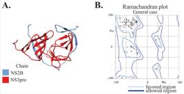
Protein modeled by Robetta server. (A) Tridimensional structure of NS2B-NS3pro model protein represented in cartoon. (B) Ramachandran plot of NS2B-NS3pro Robetta output model. The plot shows 96.5 % of all residues in favored region (light blue line) and 100 % of all residues in allowed regions (dark blue line).
Molecular Dynamic Simulations
For the molecular dynamics simulations, the Root Mean Square Deviation (RMSD) analysis (Figure 2A) was performed to evaluate the structural stability of the protein over the simulation time. The results showed that the RMSD values initially fluctuated during the equilibration phase but began to stabilize after approximately 75 ns, indicating that the protein reached a relatively stable conformation for the remainder of the simulation.

(A) Root Mean Square Deviation of 150 ns trajectory. It is observed RMSD starts to stabilize near 75 ns. (B) Two main conformational groups (clusters) were observed.
To further analyze the conformational space sampled during the simulation, a cluster analysis was performed using a cutoff of 0.25 nm (Figure 2B). This analysis revealed two main clusters, which represent the predominant conformations of the protein throughout the trajectory. The central structure of each cluster, defined as the structure with the smallest average RMSD compared to all other structures within the same cluster, was extracted and used for subsequent analyses. Figure 2B illustrates the distribution of the two clusters and highlights the central structures that were selected as representative conformations.
Molecular Docking
The primary interactions identified in the AutoDock Vina docking results by nAPOLI server are detailed in Figure 3. Specifically, Figure 3A shows the docking outcomes between the NS2B-NS3pro cluster 1 and the CA molecule. For all docking runs, interactions were observed with both subunits of the NS2B-NS3pro protein (labeled as A for the NS2B subunit and B for the NS3pro subunit), while no interactions occurred within the catalytic triad (HIS51, ASP75, and SER135). Instead, conserved interactions primarily involved residues PHE81A, ILE107B, and ARG57B. These findings highlight key binding regions outside the active site, providing insights into potential alternative binding modes. Figure 3B focuses on the interaction analysis of the top 10 docking poses. In these poses, conserved interactions were identified with residues PHE81A, THR150B, ALA148B, ASN136B, ILE107B, and ARG57B. Together, these figures demonstrate the consistent engagement of specific residues across the best docking conformations.
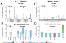
Interactions from AutoDock Vina docking results according to nAPOLI server analysis. In the left panel (A-B), results from NS2B-NS3pro cluster 1 structure and in the right panel (C-D) results from NS2B-NS3pro cluster 2 structure. (A-C) Results from all docking runs – 100 protein-ligand complexes. (B-D) Results from top ten Auto-Dock Vina scores.
Such as the NS2B-NS3pro cluster 1 structure, the docking results between the NS2B-NS3pro cluster 2 and the CA molecule are presented in Figure 3C. Across all docking runs, interactions were observed with both the NS2B (Chain A) and NS3pro (Chain B) subunits. Compared to cluster 1, a broader range of interactions was detected across different regions of the protein. However, no specific residues emerged as consistently conserved in these interactions, suggesting more variable binding patterns.
In contrast, the top 10 docking poses with the lowest AutoDock Vina scores (Figure 3D) showed interactions predominantly concentrated on the NS3pro Chain (Chain B). Notably, the main conserved residue across these best-ranked docking results was TYR18B, which may play a key role in stabilizing the CA molecule within the NS3pro subunit. This figure highlights a shift in binding preference toward specific residues on the NS3pro chain when focusing on the most favorable docking poses.
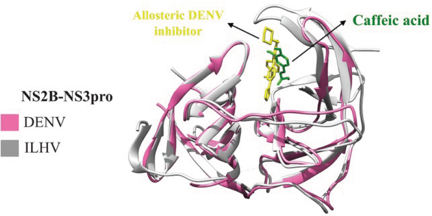
For the best 10 AutoDock Vina scores, the main conservation interactions involved PHE81A, THR150B, ALA148B, ASN136B, ILE107B, and ARG57B. Figure 4 shows the binding site of the 10 best poses of NS2B- NS3 cluster 1, highlighting the interactions of the best conformation. This binding site is defined as an allosteric binding site, which is the same as the one observed in the DENV structure of NS2B-NS3pro (PDB ID: 6MO0) complexed with an allosteric inhibitor that blocks viral replication. The comparison of the binding site of caffeic acid and the allosteric DENV inhibitor is shown in Figure 5.

Best ten ligand binding conformations from AutoDock Vina results for NS2B-NS3pro cluster 1 structure. It has been highlighted the interactions between ligand and protein residues showing -7.1 kcal/mol predicted binding energy by PRODIGY server. Residue color name refers to protein chain – red: NS2B and blue: NS3pro.
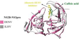
Comparison of allosteric DENV inhibitor (PDB ID: 6MO0) and caffeic acid binding to NS2B-NS3pro from ILHV.
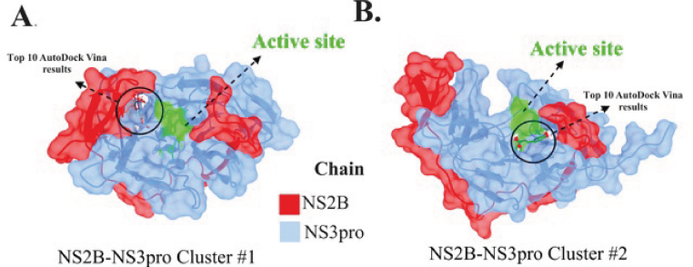
Table I presents docking energy from AutoDock Vina results and PRODIGY binding affinity predictor. As shown in Table I, the docking energy values indicate a higher affinity of caffeic acid for cluster 1 compared to cluster 2. This analysis suggests that cluster 1 structure performed better than cluster 2 about binding affinity. This is an important finding because cluster 1 is the most stable and strongly represented in the MD simulation. Moreover, this may be related to the binding site of CA. While cluster 1 molecules are focused on the hydrophobic allosteric site, cluster 2 ligand binding modes are near active sites that are highly exposed to solvent as can be observed in Figure 6.
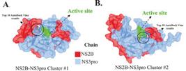
Best ten ligand binding conformations from AutoDock Vina results for (A) NS2B-NS3pro cluster 1 structure on the hydrophobic allosteric site and (B) NS2B-NS3pro cluster 2 structure on ligand modes near active sites highly exposed to solvent.
The PRODIGY scores in Table I of the docking results of NS2B-NS3 cluster #1 are very similar to the experimentally solved structures (PDB IDs: 4N0S and 6YRN) with binding allosteric inhibitors.
This reinforces the hypothesis that CA is a better allosteric inhibitor and that confirmation of cluster #1 is appropriate for its mechanism of action.
Molecular Dynamics Simulations of Complex
To enhance our comprehension of the stability between protein and ligand, we investigated the complexes (NS2B-NS3pro with CA) in solution by molecular dynamics utilizing GROMACS.
The RMSD results indicated that CA enhanced protein stability, with values ranging from 0.1 nm to 0.2 nm for the cluster #1 complex and from 0.3 nm to 0.4 nm for the cluster #2 complex (Figure 7A). The outcome of cluster #2 resembled the conformation of the protein following its stabilization in solution.

MD results of the NS2B-NS3pro protein complex (derived from cluster #1 and cluster #2) with caffeic acid. (A) Root Mean Square Deviation (RMSD) of the protein complexed with CA (B) Cluster analysis employing a limit of 0.25 nm (C) RMSD of the ligand showing its displacement (D) Conformation of cluster #1 obtained from the NS2B-NS3pro complex (Cluster #2).
The outcome of the cluster analysis further corroborates this stability, as the 0.25 nm limit yielded 1 cluster for cluster #1 and 3 clusters for cluster #4, whereas the protein in solution exhibited a total of 10 clusters (Figure 7B). Upon examining the ligand’s retention in the binding site, it is noteworthy that the CA did not stabilize within the allosteric interaction site of cluster #1, whereas in cluster #2, it maintained stability in the pocket throughout most of the simulation, coinciding with a shift in the interaction site (Figure 7C).
The depiction of cluster 1 derived from the NS2B- NS3pro complex (cluster #2) indicates that the location with the highest molecular presence is adjacent to the interaction site of the DENV inhibitor. Nonetheless, the cause of the molecule’s departure from this place and its lack of stability therein warrants investigation.
This outcome indicates that, despite superior affinity in the docking analysis, cluster #2 achieved molecular stability in solution.
In summary, the results indicate that CA primarily binds to allosteric sites of the ILHV NS2B-NS3pro protein without directly interacting with the catalytic site. This confirms the main aim of the study to evaluate the antiviral potential of CA against ILHV in silico and suggests that CA can inhibit enzymatic activity through allosteric mechanisms. This finding not only confirms that CA is a promising antiviral candidate but also indicates that future research should explore allosteric inhibitors and develop derivatives with higher affinity. Further experimental studies will be necessary to confirm this mechanism and evaluate the efficacy of CA in biological situations.
DISCUSSION
The results of this study show that CA preferentially binds to allosteric sites of the NS2B-NS3pro protein of ILHV. This is in line with the main objective of evaluating the antiviral potential of CA against this pathogen in silico, which may offer alternative strategies for the control and treatment of infections caused by this virus. Over the past few decades, the rise of emerging and re-emerging infectious diseases, particularly those associated with viruses, has become a crucial focal point in the realm of public health. Accumulating knowledge across various domains, including antiviral development, is crucial for anticipating and effectively responding to potential future epidemics. RNA viruses like ILHV display frequent mutations and genetic plasticity, potentially leading to increased virulence and transmissibility (Mazeaud, Freppel, Chatel-Chaix, 2018). If this occurs, they are no longer etiological agents of little notoriety restricted to specific endemic regions but rather viruses with wide global distribution, such as the Zika virus, with strains of greater concern (Fulton et al., 2017). Consequently, flavivirus infections pose a substantial burden on public health annually, affecting millions of individuals and imposing significant health, economic, and social challenges on a global scale (Anderson et al., 2007; Pierson, Diamond, 2020).
Here, we have demonstrated through in silico methods possible evidence of the antiviral activity of the natural compound CA against ILHV. Docking results indicated that CA does not interact directly with the catalytic site of the NS2B-NS3pro protein, composed of residues HIS51, ASP75, and SER135. Instead, the observed interactions occur predominantly in allosteric regions, involving residues such as PHE81A, ILE107B, and ARG57B. This finding suggests that CA may exert its inhibitory effect through an allosteric mechanism, modulating the enzyme conformation and, consequently, its catalytic activity indirectly.
The lack of interactions in the active site is particularly significant, as it indicates that CA does not compete directly with the enzyme’s natural substrates. Instead, binding in allosteric regions may induce conformational changes that affect the functionality of the catalytic site and provide an alternative inhibition strategy. This allosteric mechanism could have advantages, such as a lower likelihood of the virus developing resistance, as allosteric targets are usually less conserved and more difficult to mutate without affecting the structural function of the protein (Liu et al., 2020).
Furthermore, a comparison with the structure of DENV NS2B-NS3pro (PDB ID: 6MO0) in a complex with a similar allosteric inhibitor underlines the plausibility of this mechanism of action for CA (Yao et al., 2019). The overlapping binding regions suggest that CA may interact with other known allosteric inhibitors, confirming its potential efficacy as an inhibitor of viral replication.
These findings expand the understanding of possible strategies for the development of inhibitors against the ILHV NS2B-NS3 pro protein, highlighting the relevance of exploring allosteric sites as viable therapeutic targets. Future experimental studies, such as enzymatic activity assays and additional structural analyses, will be essential to confirm the proposed inhibitory mechanism and evaluate the efficacy of CA in more complex biological contexts.
Previous studies have suggested that CA has diverse biological activities, including antibacterial, antiparasitic, anti-inflammatory, and antitumoral activity (Alson et al., 2018; Chao, Hsu, Yin, 2009; Da Cunha et al., 2004; Espíndola et al., 2019). Furthermore, an investigation has provided evidence that this compound has the capability to impede the replication of HCV. The inhibitory effect was found to be facilitated by the induction of heme oxy-genase-1 (HO-1) through the Keap1/Nrf2 pathway. The heightened expression of HO-1 resulted in the activation of the interferon-alpha (IFNα) antiviral response, consequently leading to the suppression of HCV replication (Shen, Wang, Zuo, 2018). This mechanism of action was not explored in the present study; however, a similar mechanism might be involved. Therefore, we encourage further studies from this perspective.
A previous study using in vitro experiments has indicated an antiviral activity of this molecule against ILHV, possibly suggesting the interaction of the molecule with the viral envelope protein (Saivish et al., 2023b). In the present study, through our in silico analyses, we suggest that part of this antiviral activity can perhaps also be attributed to the interaction of CA with the NS2B-NS3pro protein complex. CA, a ubiquitous phenolic compound belonging to the hydroxycinnamates group, is biosynthetically derived from phenylalanine in plants. This compound is naturally present in a variety of agricultural products, including fruits, vegetables, wine, olive oil, and coffee. Additionally, the processing of plant foods gives rise to by-products that serve as abundant reservoirs of phenolic compounds (Moure et al., 2001). A previous study reveals a substantial influence of CA on the infectivity of the assessed virus (Saivish et al., 2023b). These observations align with earlier reports indicating the virucidal activity of additional natural com-pounds, including Curcumin and Luteolin, against ZIKV, CHIKV, and JEV, respectively (Fan et al., 2016; Mounce et al., 2017).
In contrast to previous studies suggesting an interaction of CA with the viral envelope protein (Saivish et al., 2023b), our in silico analysis shows a possible allosteric inhibition of the NS2B-NS3pro protease, consistent with inhibition strategies for other flaviviruses (Lv et al., 2024). These results suggest that CA can act on multiple viral targets, enhancing its antiviral efficacy. Allosteric inhibitors of NS2B-NS3pro have shown promise in combating flavivirus infections, offering benefits such as a reduced likelihood of developing viral resistance. MD simulations revealed that the interaction between CA and the NS2B-NS3pro protein was not entirely stable in solution. Nevertheless, the tests undertaken herein are unable to determine if the duration of complex stability is sufficient to avoid the viral mechanism of action or, at the very least, decrease the efficacy of the protease.
Although docking and molecular dynamics methods provide valuable predictions about the molecular interactions between CA and the ILHV NS2B-NS3pro protein, it is important to recognize some limitations of these in silico approaches. The binding affinity predictions performed by AutoDock Vina and PRODIGY are estimates that, while useful for initial screening, require experimental validation to confirm the inhibitory effect of CA. Molecular dynamics performed on limited time scales may not capture all possible conformations of the protein and ligand, and thus may miss relevant interactions. Additionally, it is essential to conduct multiple MD analyses utilizing replicas to enhance conformational sampling and achieve results with improved statistical reliability. Finally, the lack of direct interactions at the catalytic site observed in the docking results must be interpreted with caution, as indirect interactions and conformational changes may be complex and context-dependent.
Hence, despite the importance of the current data as an initial step, it is known that in vitro experiments should be conducted to elucidate the hypotheses presented here.
CONCLUSION
In conclusion, this study provides valuable insights into the antiviral potential of CA against ILHV through innovative in silico methods. Flaviviruses, exemplified by ILHV, exhibit genetic plasticity and a propensity for increased virulence and transmissibility. This research specifically highlights the interaction between CA and the NS2B-NS3 protease complex, a key component of ILHV’s replication machinery, revealing its potential as a promising antiviral target.
While prior studies have indicated CA’s antiviral potential against ILHV in vitro, our in silico findings elucidate its molecular mechanism, suggesting that CA may inhibit the multifunctional NS2B-NS3 protease. By targeting this critical enzyme, CA demonstrates favorable binding energies, supporting its viability as a potential therapeutic candidate.
The multifunctionality of the NS2B-NS3 complex is crucial for ILHV replication and pathogenesis, making it an attractive target for drug development. This study underscores the importance of computational tools in identifying antiviral candidates, offering a rational approach to evaluating molecules with therapeutic potential. Importantly, our findings lay the groundwork for future experimental validation and the development of effective interventions against neglected tropical diseases like ILHV.
Ultimately, these insights reinforce the utility of in silico approaches in bridging gaps in understanding viral mechanisms and accelerating antiviral discovery, addressing a pressing need in global health.
ACKNOWLEDGEMENTS
This research was funded by grants 2013/21719-3 and 2022/03645-1 from FAPESP (Fundação de Amparo à Pesquisa do Estado de São Paulo). M.L.N. is partially supported by the Centers for Re-search in Emerging Infectious Disease “The Coordinating Research on Emerging Arboviral Threats Encompassing the Neotropics (CREATE- NEO)” grant U01AI151807, awarded by the National Institutes of Health (NIH/USA). M.L.N. is partially supported by INCT Viral Genomic Surveillance and One Health by grant 4057586/2022-0. M.L.N. is a CNPq Research Fellow. M.V.S. was supported by a FAPESP PhD Scholarship, Number 2020/12875-5 and 2023/09590-7. G.D.L.M. was supported by a CAPES PhD Scholarship, process 88887.659079/2021-00. The authors wish to thank LaMCAD (the Multiuser High-Performance Computing Laboratory) at the Universidade Federal de Goiás for allowing them to use their high-performance cluster.
DATA AVAILABILITY STATEMENT
Use of data not disclosed.
REFERENCES
- Abraham MJ, Murtola T, Schulz R, Páll S, Smith JC, Hess B, et al. GROMACS: High performance molecular simulations through multi-level parallelism from laptops to supercomputers. SoftwareX. 2015 Sep;1–2:19–25.
- Alson SG, Jansen O, Cieckiewicz E, Rakotoarimanana H, Rafatro H, Degotte G, et al .In-vitro and invivo antimalarial activity of caffeic acid and some of its derivatives. J Pharm Pharmacol. 2018 Sep 6; 70(10):1349–56.
- Anderson KB, Chunsuttiwat S, Nisalak A, Mammen MP, Libraty DH, Rothman AL, et al. Burden of symptomatic dengue infection in children at primary school in Thailand: a prospective study. Lancet. 2007 Apr;369(9571):1452–9.
- Case DA, Aktulga HM, Belfon K, Cerutti DS, Cisneros GA, Cruzeiro VWD, et al. AmberTools. J Chem Inf Model. 2023 Oct 23;63(20):6183–91.
- Causey OR, Causey CE, Maroja OM, Macedo DG. The isolation of arthropod-borne viruses, including members of two hitherto undescribed serological groups, in the Amazon Region of Brazil. Am J Trop Med Hyg. 1961 Mar;10(2):227–49.
- Chao PC, Hsu CC, Yin MC. Anti-inflammatory and anti-coagulatory activities of caffeic acid and ellagic acid in cardiac tissue of diabetic mice. Nutr Metab (Lond). 2009 Dec;6(1):33.
- Chappell KJ, Nall TA, Stoermer MJ, Fang NX, Tyndall JDA, Fairlie DP, et al. Site-directed Mutagenesis and Kinetic Studies of the West Nile Virus NS3 Protease Identify Key Enzyme-Substrate Interactions. J Biol Chem. 2005 Jan;280(4):2896–903.
- Da Cunha FM, Duma D, Assreuy J, Buzzi FC, Niero R, Campos MM, et al. Caffeic Acid derivatives: In vitro and in vivo anti-inflammatory properties. Free Radic Res. 2004 Jan;38(11):1241–53.
- Espíndola KMM, Ferreira RG, Narvaez LEM, Silva Rosario ACR, Da Silva AHM, Silva AGB, et al. Chemical and pharmacological aspects of caffeic acid and its activity in hepatocarcinoma. Front Oncol. 2019 Jun 21;9:541.
- Fan W, Qian S, Qian P, Li X. Antiviral activity of luteolin against Japanese encephalitis virus. Virus Res. 2016 Jul;220:112–6.
- Fassio AV, Santos LH, Silveira SA, Ferreira RS, de Melo-Minardi RC. nAPOLI: a graph-based strategy to detect and visualize conserved protein-ligand interactions in large-scale. IEEE/ACM Trans Comput Biol Bioinf. 2020 Jul;17(4):1317–28.
- Fulton BO, Sachs D, Schwarz MC, Palese P, Evans MJ. Transposon mutagenesis of the zika virus genome highlights regions essential for RNA replication and restricted for immune evasion. Dermody TS, editor. J Virol. 2017 Aug;91(15):e00698-17.
- Gulcin I. Antioxidant activity of caffeic acid (3,4-dihydroxycinnamic acid). Toxicology. 2006 Jan 16;217(2–3):213–20.
- Huey R, Morris GM, Forli S. Using AutoDock 4 and AutoDock vina with AutoDockTools: a tutorial. The Scripps Research Institute Molecular Graphics Laboratory. 2012;10550(92037):1000.
- Johnson BW, Cruz C, Felices V, Espinoza WR, Manock SR, Guevara C, et al. Ilheus Virus isolate from a human, ecuador. Emerg Infect Dis. 2007 Jun;13(6):956–8.
- Jurrus E, Engel D, Star K, Monson K, Brandi J, Felberg LE, et al. Improvements to the APBS biomolecular solvation software suite. Protein Sci. 2018 Jan;27(1):112–28.
- Laemmert HW, Hughes TP. The Virus of Ilhéus Encephalitis. J Immunol. 1947 Jan 1;55(1):61–7.
- Li J, Lim SP, Beer D, Patel V, Wen D, Tumanut C, et al. Functional profiling of recombinant NS3 proteases from all four serotypes of dengue virus using tetrapeptide and octapeptide substrate libraries. J Biol Chem. 2005 Aug;280(31):28766–74.
- Lindorff-Larsen K, Piana S, Palmo K, Maragakis P, Klepeis JL, Dror RO, et al. Improved side⎕chain torsion potentials for the Amber ff99SB protein force field. Proteins. 2010 Jun;78(8):1950–8.
- Liu X, Lu S, Song K, Shen Q, Ni D, Li Q, et al. Unraveling allosteric landscapes of allosterome with ASD. Nucleic Acids Res. 2020 Jan 8;48(D1):D394–401.
- Lv H, Lu Y, Xin X, Zhou X, Yang S, Jia D, et al. Design, synthesis and biological activity evaluation of novel allosteric inhibitors of the DENV NS2B–NS3 protease. New J Chem. 2024;48(40):17553–63.
- Mazeaud C, Freppel W, Chatel-Chaix L. The Multiples Fates of the Flavivirus RNA Genome During Pathogenesis. Front Genet. 2018 Dec 4;9:595.
- Milhim BHGA, Estofolete CF, Rocha LCD, Liso E, Brienze VMS, Vasilakis N, et al. Fatal outcome of ilheus virus in the cerebrospinal fluid of a patient diagnosed with encephalitis. Viruses. 2020 Aug 29;12(9):957.
- Mounce BC, Cesaro T, Carrau L, Vallet T, Vignuzzi M. Curcumin inhibits Zika and chikungunya virus infection by inhibiting cell binding. Antiviral Res. 2017 Jun;142:148–57.
- Moure A, Cruz JM, Franco D, Domınguez JM, Sineiro J, Domınguez H, et al. Natural antioxidants from residual sources. Food Chem. 2001 Feb;72(2):145–71.
- Musso D, Desprès P. Serological diagnosis of flavivirus-associated human infections. Diagnostics. 2020 May 14;10(5):302.
- O’Boyle NM, Banck M, James CA, Morley C, Vandermeersch T, Hutchison GR. Open Babel: An open chemical toolbox. J Cheminform. 2011 Oct 7;3(1):33.
- Pierson TC, Diamond MS. The continued threat of emerging flaviviruses. Nat Microbiol. 2020 May 4;5(6):796–812.
- Rifaioglu AS, Atas H, Martin MJ, Cetin-Atalay R, Atalay V, Dogan T. Recent applications of deep learning and machine intelligence on in silico drug discovery: methods, tools and databases. Brief Bioinform. 2019 Sep 27;20(5):1878–912.
- Saivish MV, Menezes GDL, Costa VGD, Nebo L, Silva GCDD, Pacca CC, et al. Structural insights into plasticity and discovery of flavonoid allosteric inhibitors of flavivirus NS2B–NS3 protease. Biophysica. 2023a Feb 1;3(1):71–92.
- Saivish MV, Pacca CC, Costa VG da, Menezes G de L, Silva RA da, Nebo L, et al. Caffeic acid has antiviral activity against Ilhéus Virus In Vitro Viruses. 2023b Feb;15(2):494.
- Shen J, Wang G, Zuo J. Caffeic acid inhibits HCV replication via induction of IFNα antiviral response through p62-mediated Keap1/Nrf2 signaling pathway. Antiviral Res. 2018 Jun;154:166–73.
- Smith DR. Waiting in the wings: The potential of mosquito transmitted flaviviruses to emerge. Crit Rev Microbiol. 2017 Jul 4;43(4):405–22.
- Spence L, Anderson CR, Downs WG. Isolation of Ilhéus virus from human beings in Trinidad, West Indies. Trans R Soc Trop Med Hyg. 1962 Nov;56(6):504–9.
- Venegas EA, Aguilar PV, Cruz C, Guevara C, Kochel TJ, Vargas J, et al. Ilheus virus infection in human, Bolivia. Emerg Infect Dis. 2012 Mar;18(3):516–8.
- Williams CJ, Headd JJ, Moriarty NW, Prisant MG, Videau LL, Deis LN, et al. MolProbity: More and better reference data for improved all-atom structure validation. Protein Sci. 2018 Jan;27(1):293–315.
- Wu ZM, Yu ZJ, Cui ZQ, Peng LY, Li HR, Zhang CL, et al. In vitro antiviral efficacy of caffeic acid against canine distemper virus. Microb Pathog. 2017 Sep;110:240–4.
- Yao Y, Huo T, Lin YL, Nie S, Wu F, Hua Y, et al. Discovery, X-ray crystallography and antiviral activity of allosteric inhibitors of flavivirus NS2B-NS3 Protease. J Am Chem Soc. 2019 May 1;141(17):6832–6.