
 An overview of Salmonella biofilms and their inhibition by plant bioactive compounds
Abstract
Microbial biofilms are ubiquitous and highly successful forms of life. Among them, species found in food industries often include human pathogens like Salmonella spp., a significant concern due to its ability to adapt and survive in diverse environmental stresses. This bacterium is a major global public health issue, causing foodborne illnesses with considerable morbidity and economic costs. Combatting bacteria within biofilms necessitates novel strategies, and recent research highlights natural compounds as promising agents due to their antibiofilm, antivirulence, and antimicrobial properties. Specifically, plant-derived compounds have shown potential to modulate biofilm formation either alone or in combination with other substances. This review provides a comprehensive overview of Salmonella biofilms, encompassing their development, composition, resistance mechanisms, and tolerance mechanisms. It focuses particularly on the inhibition of these biofilms by plant-derived compounds.
Keywords:
Antimicrobials; Bioactive molecules; Essential oils; Plant extracts; Quorum sensing
INTRODUCTION
Biofilms are responsible for conducting different biogeochemical cycles of elements in water, soil, sediments, among others, and have been considered as one of the best distributed and successful life forms on the planet (Flemming et al., 2016). They can be found on different abiotic and biotic surfaces and biofilms are affected by several aspects, revealing their dynamic and sometimes unpredictable behaviors. The process of biofilm formation depends on factors such as cellular interactions, the surface where it adheres to, environmental conditions, besides the microbial species within it (Dass, Wang, 2022; Borges et al., 2018; Flemming et al, 2023).
Biofilms also cause the deterioration of the systems in which they are formed, leading to corrosion of pipes and equipment for example, making them a problem in different areas, including the food industry. Biofilms have been found as reservoirs of recurrent bacterial contamination, which can lead to food spoilage and foodborne illnesses (Brooks, Flint, 2008; Carrascosa et al., 2021). Many of the biofilm-forming species found in food factories are human pathogens, which can form biofilms on different surfaces common in food industries such as stainless steel, glass, wood, polypropylene, among others (Galié et al., 2018). These materials are very common in dairy and poultry industries, where various processes and structures such as milk tanks, pasteurizers, pipes, cutters, tables, employee gloves, and packing material act as surfaces for different colonizing species at various temperatures. Food matrices can be contaminated with bacterial cells detached from these biofilms, potentially affecting human health when these contaminants are foodborne pathogens (Carrascosa et al., 2021; Galié et al., 2018).
According to World Health Organization (WHO), every year 1 in 10 people fall ill and 33 million of healthy life years are lost due to foodborne diseases. These ailments can be severe and diarrheal diseases are the most common illnesses resulting from unsafe food, being Salmonella 1 of the 4 key global causes of diarrheal diseases (WHO, 2018). Salmonella is a genus of Gram-negative bacteria that represents one of the major public health problems worldwide, since its diverse group of serotypes mostly cause foodborne illnesses to humans, resulting in significant morbidity and economic impacts (Merino et al., 2019). The Center for Disease Control and Prevention (CDC) estimates that each year in the United States (U.S.), there are about 1.35 million cases of salmonellosis, with 26,500 hospitalizations and 420 deaths, being contaminated food the main source of the cases. Salmonellosis is the second leading cause of foodborne illnesses in the U.S., after norovirus, but is the leading cause of hospitalizations and deaths from food poisoning (FDA, 2023).
In addition to its clinical significance due to causing chronic infections, Salmonella is also of great concern in the food industry, as it can adapt and survive in many stressful environmental conditions. Approximately 50% of the strains isolated from poultry farms have the capacity to produce biofilms in processing areas such as walls, floors, pipes, and drains and in contact surfaces with materials like stainless steel, aluminum, nylon, rubber, plastic, polystyrene, and glass (Marin, Hernandiz, Lainez, 2009; Shatila, Yaça, Yalçin, 2021).
Bacteria living in biofilms have higher tolerance to antibiotics and disinfectants compared to their planktonic state. Thus, novel strategies are needed to combat bacteria in biofilms (Lu et al., 2019; Rossi et al., 2022). This area of research has been extensively explored in recent years, and natural bioactive compounds are commonly reported to hinder biofilm formation alone or in association with other agents due to their antibiofilm, antivirulence, and antimicrobial properties. They comprise plant extracts (Alam et al., 2020), enriched extract fractions (Slobodníková et al., 2016; Zamuz et al., 2021), essential oils (EOs) (Rossi et al., 2022), isolated compounds (Santos et al., 2021), among others.
This review aims to provide an overview of Salmonella biofilms, their development, matrix composition, resistance, and tolerance to biocides and antimicrobials, as well as inhibition by plant-derived compounds.
MATERIAL AND METHODS
In this literature review, studies have been analyzed to elucidate key aspects of Salmonella biofilms, including their development, matrix composition, resistance mechanisms, and inhibition by plant-derived compounds. The goal was to provide an overview of biofilms, with emphasis in their ecological significance and their outstanding ability to persist in diverse environments.
An in-depth literature search was conducted using established databases such as Scopus, PubMed, Web of Science, SciELO, Google Scholar, and ScienceDirect, without imposing data restrictions. However, priority was given to recent studies, particularly those published within the last five years, to ensure that the review reflects the most current advancements in the field. This search culminated in the creation of Table I, which compiles studies that report on the inhibition of Salmonella biofilms by the natural compounds. Key search terms utilized included: “biofilm”, “biofilm inhibition”, “Salmonella”, “quorum sensing inhibition”, “plant-derived compounds”, “phenolic compounds”, and “essential oils”, often combined using the AND operator to refine the results. The selection process involved a critical examination of article abstracts to ensure relevance to the scope of this review.
Overview of studies reporting inhibition of Salmonella biofilms with essential oils (EOs), plant extracts, and plant-derived compounds
The study gathered data regarding the general composition of Salmonella biofilms, identifying the factors that contribute to their antimicrobial tolerance, and exploring the various inhibition mechanisms that have been investigated over the years. Then, the article delved into the specific inhibition of Salmonella biofilms by natural compounds, with a focus on EOs, plant extracts, and isolated compounds. The review also aimed to assess whether natural compounds have been studied in sufficient detail, particularly concerning their potential mechanisms of action and the analytical methods employed to evaluate their efficacy against biofilms.
GENERAL STAGES OF BIOFILM DEVELOPMENT
Bacteria live in association and not simply as autonomous entities. Biofilms are the clearest example of this association as they are organized aggregates of microorganisms attached to an abiotic or biotic surface protected by extracellular polymeric substances (EPS) produced by themselves, forming channels that allow the passage of water, enzymes, nutrients, and residues (Flemming et al., 2023). The EPS is mainly composed of carbohydrates, proteins, lipids, and exogenous DNA (eDNA) (Flemming et al., 2016; Lu et al., 2019). The biofilm architecture has water channels for nutrient transportation in between the EPS matrix. The main role of this matrix is to facilitate the adherence of microbial cells to a surface and to each other, and additionally it provides protection for resident bacteria against a variety of environmental stresses such as adverse temperature, nutritional alterations, osmotic shock, drying, and ultraviolet (UV) radiation (Jamal et al., 2015; Kassinger, van Hoek, 2020).
Due to the properties of the matrix and the physical interactions between the biofilm cells, the lifestyle within a biofilm is completely different from those cells found in the free-living form, called planktonic cells. The EPS matrix enables bacterial cells within the biofilm to stay united and protected, thereby granting them greater resilience against environmental conditions and host defenses compared to planktonic cells (Carrascosa et al., 2021; Dass, Wang, 2022; Flemming et al., 2023).
The biofilm development is a transition process from the planktonic state to the sessile form, and these two life forms are genetically distinct. The genetic transition occurs throughout the biofilm life cycle and involves four distinct stages, as described below, and illustrated in Figure 1.
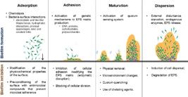
Stages and physicochemical processes involved in biofilm formation and different inhibition strategies in each of the stages.
Stage 1. Adsorption - Reversible attachment
This process begins with the interaction between a few planktonic cells and the substrate and takes only a few seconds to initiate. In this step, a thin layer of organic and inorganic particles is formed. Basically, any substance present in the liquid medium that is transported by Brownian motion, sedimentation, or convection, becomes a conditioning layer that serves as the basis for biofilm growth (Jamal et al., 2015; Mahamuni-Badiger et al., 2020). At this stage, the chemotaxis process is important to direct planktonic cells to the source of nutrients and then promote microbial attachment and growth in the community. When the cells reach the surface, the interaction depends on the sum of attractive and repulsive forces between both surfaces (Muhammad et al., 2020). The planktonic cells adhere to the substrate by nonspecific physical forces, such as electrostatic forces, hydrophobic interactions, and Van Der Waals forces (Crouzet et al., 2014; Khatoon et al., 2018). In this reversible stage, the cells remain in Brownian motion and can be easily detached due to bacterial motility through appendages such as flagella, fimbriae, and pili, or due to the action of opposing forces. This process depends on aspects such as the amount of energy available, temperature, pressure, hydrophobic or hydrophilic characteristics of the contact surface, and whether the repulsive forces on the surface are greater than the attractive ones (Jamal et al., 2015; Khatoon et al., 2018; Muhammad et al., 2020).
Stage 2. Adhesion - Microcolony formation
The attachment of bacteria to the substrate at this stage is irreversible and depends on the bacterial physical appendages such as flagella, fimbriae, and pili, and how these manage to overcome the repulsive physical forces of the conditioning layer. When such bacterial appendages meet the conditioning layer, there is a stimulation of chemical reactions such as oxidation, which consolidates the bonding of the bacteria with the surface. At this stage, more bonding forces appear, such as dipole-dipole, hydrogen bonds, ionic, and covalent bonds, and hydrophobic interactions, all of which are responsible for the so-called irreversible adhesion. The bacteria begin to multiply and release chemical signals that allow them to communicate through a process called quorum sensing (QS), which activates the genetic mechanisms needed to produce EPS and begin the biofilm maturation process (Jamal et al., 2015; Mahamuni-Badiger et al., 2020; Solano, Echeverz, Lasa, 2014).
Stage 3. Biofilm maturation
The bacteria adhered to the surface continue to divide by binary fission and remain aggregated in the form of micro-colonies, decreasing motility. The production of signaling molecules is also continuous at this stage, which keeps the genetic systems for EPS production activated. The growth of the micro-colonies and the EPS matrix allow the development of a three-dimensional structure, normally referred to as a mushroom-like structure, that contains water-filled channels to transport nutrients within the inner parts of the biofilm and remove waste materials, as well as providing greater protection against antimicrobials and host defense mechanisms (Jamal et al., 2015; Muhammad et al., 2020).
Stage 4. Dispersion of bacteria
The dispersal process can occur in response to changes in the microenvironment such as oxygen availability, access to nutrients, and changes in temperature. These factors that were initially important for bacterial survival and optimization of available resources become signals that activate dispersion. The bacteria in the innermost parts of the biofilm face a stressful condition generated by hypoxia, the scarcity of signaling molecules, and the low growth rate (Guzman-Soto et al., 2021). This causes the activation of regulatory mechanisms, remodeling the biofilm structure by the production and release of enzymes that degrade the EPS matrix components, by interruption of non-covalent interactions, and cell death, which produces cavities within the biofilm (Solano, Echeverz, Lasa, 2014). These cavities serve as escape or dispersal routes, releasing cells individually or in groups, leaving them free to spread out and colonize new substrates (Guzman-Soto et al., 2021).
Researchers are now focusing on inhibiting biofilm formation through various strategies that target the different stages of the process of biofilm formation. These approaches involve preventing cell receptors from recognizing surfaces, inhibiting bacterial adhesion, or identifying compounds that can eliminate formed biofilms (Figure 1).
MATRIX COMPONENTS OF Salmonella BIOFILMS
The composition of the bacterial biofilm matrix is complex and can vary greatly between species and even within the same species under different environmental conditions. Its primary component is water, which is responsible for the flow of oxygen and nutrients within the biofilm. The main structural and functional elements of the matrix are carbohydrates, proteins, lipids, lipopolysaccharide (LPS), and eDNA (Flemming et al., 2023; Jamal et al., 2015). Salmonella and other members of the Enterobacteriaceae family produce an EPS matrix composed mainly of curli fimbriae, extracellular cellulose, and biofilm-associated protein (BapA), in response to environmental stress (Sakarikou et al., 2020).
Curli fimbriae is an amyloid-like cell-surface fiber that acts as a promoter of community behavior through the formation of biofilms, being involved in the processes of surface adhesion and cell aggregation (Jonas et al., 2007; Keelara, Thakur, Patel, 2016; Solomon et al., 2005). It is the protein with the highest content in the biofilm matrix produced by many Enterobacteriaceae including Salmonella spp. and Escherichia coli (Smith et al., 2017). In Salmonella spp., the curli fimbriae is encoded by two divergently transcribed operons csgDEFG and csgBAC (agfDEFG and agfBAC depending on the annotation, respectively). The csgBAC operon encodes the structural subunits of curli, CsgB and CsgA, as well as the chaperone CsgC that prevents these subunits from forming an amyloid structure before being transported to the cell envelope. The csgDEFG operon is responsible for encoding the accessory proteins required for curli assembly. CsgE, CsgF, and CsgG participate in translocation and assembly of the structural subunits of curli into the cell envelope. CsgD is known as a master regulator of biofilm development and stimulates the production of curli by transcriptional activation of the csgBAC operon (Evans et al., 2015; Bhoite et al., 2019; Jonas et al., 2007; Keelara, Thakur, Patel, 2016; Solomon et al., 2005).
The expression of a proteinaceous component of the Salmonella EPS matrix, BapA, is synchronized with the expression of curli and extracellular cellulose through the action of the regulatory protein CsgD (Jonas et al., 2007). BapA plays an important role in bacterial aggregation and biofilm formation at the air-liquid interface through homophilic interactions between bacterial cells (Lamas et al., 2021). This protein is secreted through a type I secretion system, BapBCD, all encoded by the same operon, and have been studied specially in Salmonella Enteritidis (Latasa et al., 2005).
Another matrix-component commonly found in Salmonella biofilms is extracellular cellulose, a polysaccharide composed of D-glucose units linked together by β-1-4 glycosidic bonds. Cellulose biosynthesis occurs by expression of the bcsABZD and bcsEFG operons and is also upregulated by the master biofilm regulator CsgD, which stimulates transcription of the diguanylate cyclase AdrA at the post-transcriptional level, important for activating cellulose production (Jonas et al., 2007; Peng, 2016). BcsA and BcsB form the two subunits of the cellulose synthesis complex located in the cell envelope and they are responsible for the conversion of UDP-glucose to cellulose. The other proteins synthesized by the two bcs operons are involved in the regulation of cellulose synthesis and the cellular localization of the synthesis complex (Rõmling, Galperin, 2015). Higher cellulose production has been shown to be associated with increased thermal and chemical resistance of biofilms and it occurs at temperatures ranging from 15 to 25 °C, which are very relevant temperatures in the food industry (Kim, Jyung, Kang, 2022).
The presence or absence of cellulose and/or curli fimbriae determines the four morphotypes that can be observed in Salmonella colonies on Congo red agar plates: strains expressing cellulose and curli appear as red, dry, and rough colonies (RDAR); those expressing cellulose but not curli show the pink, dry, and rough morphotype (PDAR); if only curli is expressed, the colonies show a brown, dry, and rough morphotype (BDAR); when colonies appear smooth and white (SAW) it is possible that the transcriptional regulator CsgD is not active in this strain (Rõmling, 2005; Jain, Chen, 2007; Cwiek, Bugla-Płoskońska, Wieliczko, 2019; Nesse et al., 2021). The detection of these morphotypes can indicate the degree of resistance and survival of Salmonella biofilms to different conditions, since cellulose plays a key role in increasing resistance to heat and chemical compounds (Solano et al., 2002; Villa-Rojas et al., 2017; Kim, Jyung, Kang, 2022).
The presence of different fatty acids and LPS was also noticed in the EPS fraction of RDAR expressing S. Enteritidis strain (Gibson et al., 2006). The LPS also improves surface wettability, required for swarm colony expansion, and LPS mutations may be able to induce alternative pathways leading to EPS matrix production (Steenakers et al., 2012).
INFLUENCE OF SIGNALING MOLECULES ON BIOFILM FORMATION
The cell-cell communication by QS is used by bacteria to collectively adapt by activating or repressing genes involved in collective group behavior (Bassler, 2002; Lima, Winans, Pinto, 2023). This mechanism occurs through signaling molecules, also known as autoinducers (AI), which accumulate in the medium as a function of population density and can regulate the expression of genes (Fuqua, Winans, Greenberg, 1994; Bassler, 2002; Lima, Winans, Pinto, 2023). Many cellular processes are modulated by QS including sporulation, synthesis of antimicrobial peptides, regulation of virulence factors, and biofilm formation (Whitehead et al., 2001; Papenfort, Bassler, 2016; Lima, Winans, Pinto, 2023).
The connection between QS and biofilm formation has been widely described. QS is an important factor in the transition from simple aggregates of microorganisms in the early stages of the biofilm formation to its complex mature structure. Many bacterial species use QS to coordinate some steps in the biofilm formation process like microcolony formation, maturation, and dispersion, and some factors such as biofilm structure, nutrient acquisition, and regulation of antimicrobial resistance (Solano, Echeverz, Lasa, 2014; Guzmán-Soto et al., 2021; Khalid et al., 2022).
In Salmonella spp., different QS systems are known to act in the regulation of virulence factors, including biofilm formation. Communication involving autoinducer type 1 (AI-1) is incomplete for this bacterium, as it does not contain a homologue of the luxI gene that codes for the N-acyl homoserine lactone (AHL) synthase. However, despite not producing its own signaling molecule, Salmonella spp. express a transcriptional regulator homologous to LuxR, known as SdiA, which detects AHLs produced by other bacterial species (Michael et al., 2001; Dyszel et al., 2010; Smith, Ahmer, 2003; Smith et al., 2008; Almeida et al., 2017; Sholpan et al., 2021). Campos-Galvão et al. (2016) showed that the addition of a mixture of AHLs, with carbon chains ranging from six to 12 carbons, did not interfere with growth, but promoted the biofilm formation by S. Enteritidis PT4 578 on polystyrene coupons under anaerobic conditions. Nonetheless, the addition of N-dodecanoyl-homoserine lactone (C12-HSL) induced a denser and more developed biofilm, optimized metabolism, enhanced tolerance to acidic stress, and cationic peptides, while also upregulating the expression of virulence-associated genes hilA, invA, and invF, as well as genes linked to biofilm formation, namely glgC, fliF, lpfA, and fimF (Campos-Galvão et al., 2016; Almeida et al., 2017; Carneiro et al., 2020; Freitas et al., 2020). On the other hand, C12-HSL did not influence biofilm formation by S. Enteritidis PT4 578 at 28 °C under both aerobic and anaerobic conditions. However, at 37 °C, biofilm formation was reduced under aerobic conditions and increased under anaerobic conditions. Furthermore, under anaerobic conditions at 37 °C, the expression of the adrA and luxS genes increased, suggesting an increase in cyclic diguanosine monophosphate (c-di-GMP) levels, a second messenger that controls essential physiological functions in bacteria (Carneiro et al., 2024). Molecular docking analysis confirmed the stronger binding affinity of C12-HSL with the SdiA protein, which may account for the varying levels of regulation by different AHLs (Almeida, Pinto, Vanetti, 2016). Addition of N-butyryl-homoserine lactone (C4-HSL) and N-hexanoyl-homoserine lactone (C6-HSL) also enhanced biofilm formation in S. Typhimurium cultivated aerobically (Bai, Rai, 2016). However, the biofilm formation of S. Typhimurium was not influenced under aerobic conditions by cell-free supernatant (CFS) from Hafnia alvei containing AHLs or by the addition of N-3-oxo-hexanoyl-homoserine lactone (3-oxo-C6-HSL) (Blana, Georgomanou, Giaouris, 2017). It is noteworthy that CFS contained metabolites other than AHLs that may have interfered with cellular responses to these AI (Vanetti et al., 2020). The AHL, C6-HSL and N-octanoyl-homoserine lactone (C8-HSL) increased the invasion of HEp-2 cells by Salmonella enterica serovar Typhi (Nesse et al., 2011) and C8-HSL increased adhesion of this bacterium to HeLa cells (Liu et al., 2014).
Salmonella spp. also uses the AI-2 signaling molecule, synthesized by the LuxS protein, targeting the lsrACDBFG operon, which captures and processes AI-2 itself (Cox, McClelland, Teplitski 2013). It has been shown that the LuxS/AI-2 QS system is required for resistance to bile during biofilm formation by S. Typhimurium (Tsai et al., 2020).
The AI-3 is an aromatic amino compound belonging to the pyrazinone family produced by bacteria from the intestinal microbiota and by some enteric pathogens such as Salmonella and E. coli (Rul, Monnet, 2015). This AI along with epinephrine and norepinephrine secreted by eukaryotic cells are recognized by the same receptor, the sensor kinase QseC. Upon detecting these molecules, QseC phosphorylates the QseB response regulator, which in turn activates its own expression and the expression of target genes. Additionally, QseC also activates the transcription of the two-component QseEF system, which may also play a role in detecting host hormones (Sperandio et al. 2003; Hughes, Sperandio, 2008; Hughes et al., 2009). Hiller et al. (2019) evaluated biofilm formation by S. Enteritidis isolates in the presence of different concentrations of epinephrine and norepinephrine at 12 and 25 °C. They demonstrated a greater number of biofilm-producing isolates at 25 °C than at 12 °C, regardless of treatment. Furthermore, biofilm formation was not influenced by the presence of hormones, except for norepinephrine at 100 μM, which stimulated biofilm formation at 12 °C (Hiller et al., 2019).
Another important regulation system in biofilm formation is c-di-GMP signaling pathway. The c-di-GMP is an important second messenger molecule that exists widely in bacteria and is a central regulator of Salmonella biofilm formation (Tsai et al., 2020; Miller et al., 2022). In brief, CsgD is a key regulator of the csg gene cluster and regulates many genes involved in biofilm formation. CsgD expression is regulated by environmental stimuli and the levels of c-di-GMP. High intracellular levels of c-di-GMP promote CsgD activation, leading to the activation of genes involved in the production of curli and expression of AdrA, which increases cellulose biosynthesis. Thus, c-di-GMP stimulates the production of matrix components such as cellulose and curli, promoting biofilm formation, and inhibits motility (Miller et al., 2022; Ryan et al., 2006). In S. Typhimurium, increased c-di-GMP levels lead to increased cellulose synthesis and biofilm formation (Tsai et al., 2020). c-di-GMP reversely regulates biofilm formation and motility in S. Typhimurium, contributing directly or indirectly to the regulation of the RDAR morphotype, a biofilm phenotype characterized by the expression of cellulose and curli fimbriae (Ahmad et al., 2011).
RESISTANCE AND TOLERANCE OF Salmonella BIOFILMS
Biofilms are renowned for their enhanced ability to withstand various stress conditions compared to planktonic cells (Soto, 2013; Flemming et al., 2023). When faced with starvation, oxygen deprivation, and restricted metabolic flow, bacteria within biofilms can initiate a stringent response that triggers the activation of the SOS system, thereby promoting their survival (Drescher et al., 2019). Additionally, biofilms exhibit a tolerance to antibiotics that is 100 to 1000 times greater than that of free-living cells, which makes treating bacterial infections involving biofilms more challenging. Although some antimicrobial agents may reduce bacterial counts within biofilms, they are rarely effective in completely eradicating the pathogens, which can result in recurrent infections (Dufour, Leung, Lévesque, 2010). Consequently, bacterial resistance to antimicrobial agents, including Salmonella, has emerged as a significant global concern (Ćwiek, Bugla-Płoskońska, Wieliczko, 2019).
Olson et al. (2002) reported that Salmonella spp. in the planktonic form was sensitive to the antibiotics enrofloxacin, gentamicin, ampicillin, oxytetracycline, and trimethoprim, while bacteria in biofilm form were sensitive only to enrofloxacin. Papavasileiou et al. (2010) investigated the susceptibility to antibiotics ampicillin, cefuroxime, cefotaxime, gentamicin, imipenem, cotrimoxazole, ciprofloxacin, and moxifloxacin of 194 Salmonella enterica strains isolated from children with gastroenteritis. Of these, 109 strains formed biofilms, and as expected, they demonstrated that the biofilm form showed increased antimicrobial resistance compared to the planktonic cells.
The resistance of Salmonella biofilms against a wide variety of biocides has also been reported. Tabak et al. (2009) evaluated the susceptibility of S. Typhimurium in both planktonic and biofilm form to triclosan, a biocide included in a wide variety of antiseptic products such as toothpastes, deodorants, soaps, and lotions. The study showed that triclosan at 600 to 2000 ppm was not effective in eradicating this microorganism in biofilm form due to reduced diffusion of the compound caused by the presence of the EPS.
Haubert et al. (2019) showed that 26 isolates of Salmonella were tolerant to benzalkonium chloride disinfectant, in addition to forming biofilms and presenting resistance to streptomycin, sulfonamides, and tetracycline, which are antimicrobials commonly used in animal production.
Antimicrobial tolerance depends on the species, but there are several factors that can contribute to its increase in biofilms, such as the presence of the EPS matrix that limits antimicrobials transport; physiological changes in the bacterial cell due to low multiplication rate, lack of nutrients, and environmental stress; expression of heavy metal resistance genes; QS; the emergence of persister cells that have a broad tolerance to bactericidal agents; and the overexpression of efflux pumps (Mah, O’Toole, 2001; Merritt, Kadouri, O’Toole, 2005; Singh et al., 2017; Ćwiek, Bugla-Płoskońska, Wieliczko, 2019). Some of these factors are detailed below.
Slow penetration of the antimicrobial due to the extracellular polymeric substances (EPS) matrix
Numerous studies have investigated the diffusion of antimicrobial agents through biofilms and have demonstrated that the matrix can serve as a selective barrier that restricts their penetration (Lewis, 2001; Singh et al., 2017; Shree et al., 2023). Some studies have shown that microbial tolerance exists due to a delayed penetration process. When antimicrobial treatment is performed, the biofilm cells at the top of the liquid interface die due a more direct exposure, while bacteria that are in the innermost layers of the biofilm generally survive (Lewis, 2001; Dufour, Leung, Lévesque, 2010).
The matrix can act as an active chemical barrier by binding to and sequestering positively charged antibiotics such as aminoglycosides, toxic heavy metals, and cationic antimicrobial peptides. However, for uncharged antibiotics such as β-lactams, this binding to the matrix is unlikely to occur, which provides little or no barrier to penetration (Dufour, Leung, Lévesque, 2010).
Tabak et al. (2009) demonstrated the effect of triclosan on Salmonella planktonic cells and biofilms. The pathogen’s tolerance to the biocide in the biofilm was attributed to its low diffusion through the EPS matrix and gene expression responses that provided increased tolerance to antimicrobials. Within the biofilm, triclosan positively regulated the bcsA and bcsE genes involved in cellulose synthesis, which caused Salmonella to intensify the exopolysaccharide production (Tabak et al., 2009).
Altered microenvironment and stress response
The environmental conditions within biofilms are not homogeneous throughout the structure. This causes the bacteria in different parts of the biofilm to have distinct physiological and metabolic characteristics (Crabbé et al., 2019). The cells on the surface of the biofilm resemble planktonic cells, while those in the deeper parts, where nutrients and oxygen are limited, have a lower growth rate. This directly influences tolerance to antimicrobials that act on cellular processes involved in bacterial multiplication, such as cell wall synthesis and DNA replication (Dufour, Leung, Lévesque, 2010).
Bacteria can express a coordinated stress response to switch to more tolerant phenotypes when presented with adverse environmental conditions such as starvation, heat or cold shock, cell density, pH, osmolarity, among others (Dufour, Leung, Lévesque, 2010; Banerji et al., 2022). In biofilms, bacteria encounter different microenvironments that can trigger this response. For example, the lack of an important substrate or the accumulation of harmful products can cause some bacteria to enter a non-growth state, which acts as a protective mechanism against death. In addition, changes in the pH or osmotic environment within the biofilm can lead to the induction of a stress response that can result in antibiotic resistance due to reduced porin expression in the cell envelope (Stewart, Costerton, 2001; Rode, Singh, Drescher, 2020).
Paytubi et al. (2017) investigated the impact of culture medium composition on biofilm formation in Salmonella and found that in nutrient-rich growth media like colonization factor antigen (CFA) medium, biofilms were predominantly formed at the air-liquid interface. However, in minimal medium, biofilms formed at the solid-liquid interface. The authors inferred that nutrient deprivation induced transcriptional expression of csgD, resulting in elevated expression of curli and cellulose. This suggests that nutrient availability is a key determinant for the spatial distribution of biofilms.
Persister cells
The formation of persister cells is another mechanism that contributes to antimicrobial tolerance. These specialized cells enter in an inactive state in response to environmental stress that activates toxin-antitoxin systems, allowing them to survive stressful conditions (Jayaraman, 2008; Wang, Wood, 2011). Persister cells constitute a small fraction of the bacterial population in the stationary phase of planktonic cells (up to 1% of the population) and in biofilms of different species and can resume their multiplication when they encounter favorable conditions (Dufour, Leung, Lévesque, 2010; Mah, 2012). These persister cells are phenotypically different from multiplying cells, which make them extremely tolerant to high concentrations of antimicrobials, mainly because most classes of antibiotics in use act on growing cells (Lewis, 2007). Importantly, these cells withstand antimicrobials because they are in an inactive state and not because they possess genetic alterations associated with resistance (Singh et al., 2009).
In Salmonella, this phenomenon was well demonstrated by Drescher et al. (2019), who tested four S. enterica serovars (Schwarzengrund, Agona, Infantis, and Enteritidis) that were able to generate persister cells after exposure to high concentrations of ciprofloxacin and ceftazidime. The persistence levels of these S. enterica serovars varied under different culture conditions, being higher in biofilms when compared to planktonic cells.
Efflux pumps
One mechanism that has also been associated with biofilm resistance to antimicrobials is the overexpression of efflux pumps. Efflux pumps are membrane bound protein structures capable of expelling toxic substances to the external environment, among them antimicrobials. They can be specific for a single antimicrobial agent or be considered multi-drug pumps, eliminating different classes of antimicrobials. Worryingly, their expression can be induced by exposure to sublethal concentrations of antibiotics (Khatoon et al., 2018).
A study conducted by Baugh et al. (2014) aimed to determine if the inhibition of multi-drug efflux pumps could prevent biofilm formation. Using S. Typhimurium mutants lacking different components of the AcrAB-ToIC system, these authors demonstrated that mutants in ToIC and AcrB, but not AcrA, exhibited transcriptional repression of genes involved in biofilm formation, such as those encoding curli, compromising their ability to form biofilms. They concluded that genetic and chemical inactivation of efflux pumps may be a promising antibiofilm strategy.
Salmonella BIOFILMS INHIBITION BY PLANT-DERIVED COMPOUNDS
The biofilm formation process involves different stages that can be targeted by several natural antibiofilm compounds, as illustrated in Figure 1. The initial stage is the most critical for biofilm development due to the need to synthesize structures that allow bacterial adhesion to the surface, such as fimbriae or pilus, which makes this stage a strategic point of inhibition. Another key factor in the biofilm formation process is QS, which has greatly increased academic interest in searching for new products that may interfere with this communication (Ta, Arnason, 2015; Deryabin et al., 2019; Sakarikou et al., 2020; Lima et al., 2023).
Plant extracts and phytochemicals have been widely used as antimicrobial agents due to their cost-effectiveness, great structural diversity, reduced possibility of resistance development because they act at different targets, and their eco-friendly characteristics (Oulahal, Degraeve, 2022; Sakarikou et al., 2020). The plant secondary metabolites exhibit great chemical diversity and important properties such as antimicrobial activity, whether evaluated alone or in combination with other antimicrobials at appropriate concentrations. The groups of phytochemicals with known antimicrobial activity are generally phenolic compounds, terpenoids, EOs, alkaloids, polypeptides, among others (Deryabin et al., 2019; Albuquerque et al., 2021). Here we show the state of the art on the use of EOs, plant extracts, and plant-derived compounds to inhibit Salmonella biofilm formation (Table I).
Essential oils (EOs)
The medicinal properties and antimicrobial activity of many plants are due in part to the presence of a high content of antimicrobial compounds in EOs including thymol, carvacrol, eugenol, menthol, limonene, among others. In many cases, the activity of these substances occurs at sub-minimum inhibitory concentration (sub-MIC) (Luís et al, 2017; Snoussi et al., 2018; Pelarti et al., 2021; Rossi et al., 2022; Sateriale et al., 2023). In addition, they also have the ability to affect cell wall integrity allowing the inactivation of bacterial cells without selecting for resistance, probably as a result of their multiple cellular targets. These compounds can also prevent bacterial adhesion and inhibit cell coaggregation, and can be widely used for their antibacterial, antifungal, antiviral, insecticidal, and antioxidant properties (Maurya et al., 2021; Pelarti et al., 2021; Verešová et al., 2024) thanks to their easy availability, low toxicity, and rapid degradation in the environment, making them safe agents with broad activity (Valeriano et al., 2012; Hakimi Alni, Ghorban, Dadmanesh, 2020; Rossi et al., 2022).
Several studies have demonstrated the effect of EOs on Salmonella biofilm, summarized in Table I. Valeriano et al. (2012) evaluated the effect of a disinfectant solution formulated with the EO of peppermint (Mentha piperita) and lemongrass (Cymbopogon citratus) against biofilm formation by S. Enteritidis S64 on stainless steel surfaces. After 10 min of exposure, a significant reduction in the bacterial populations of the formed biofilm was observed, and a 20 min treatment was sufficient to completely inhibit these bacterial populations. The authors assumed that this result may be due to increased permeability of the cytoplasmic membrane linked to the action of EO that cause a loss of important intracellular contents such as essential ions and molecules that can lead to cell death (Valeriano et al., 2012).
Amaral et al. (2015) evaluated the effect of carvacrol and thymol, the major components of the EO of oregano and thyme on biofilm formation of different Salmonella spp. strains on polypropylene. The microorganisms evaluated were S. Typhimurium ATCC 14028 and three strains isolated from food related to foodborne outbreaks: S. Enteritidis, S. Typhimurium and Salmonella Saint Paul. The cells were quantified during and after biofilm formation in the presence of the evaluated compounds. The results showed that during biofilm formation, the two compounds at sub-MICs were able to reduce the content of planktonic cells between 1-2 logs CFU/mL, while the biofilm formed was reduced between 1-5 logs CFU/cm2. The greatest reduction of biofilms occurred with carvacrol (5 log CFU/cm2) in S. Typhimurium ATCC 14028. Thymol showed the best reduction for S. Enteritidis, approximately 4 log CFU/cm2. These results were confirmed with scanning electron microscopy (SEM) analyses, which showed a break in the structure of the biofilm and a diffuse adherence of the bacterial cells (Amaral et al., 2015). More recently, Sateriale et al. (2023) also observed the inhibition of S. Typhimurium biofilms by thyme EO, suggesting the use of this natural agent as a promising food preservative to counteract biofilm-related contamination in the food industry.
A similar study by Miladi et al. (2017) evaluated the bacterial susceptibility and biofilm eradication by carvacrol, thymol, and eugenol, alone and in combination with nalidixic acid, against 12 strains of S. Typhimurium. The MIC and the minimum biofilm eradication concentration (BEC50) of the evaluated compounds and their combinations were determined. The biofilms were visualized by SEM on stainless steel surfaces after being exposed to the compounds. For most of the evaluated strains, it was observed that the BEC50 were higher than the concentration required to inhibit the multiplication of cells in suspension. All compounds showed synergism with nalidixic acid with a significant reduction in BEC50. SEM analyses showed that with a concentration of 8 μg/mL of nalidixic acid, the bacterial biofilm was reduced. However, when this compound was combined with ½ MIC of each of the compounds, the antibiofilm activity was greater than separately, which could mean that the EOs could facilitate the entry of antibiotics into the biofilms formed by S. Typhimurium.
A recent study showed the effect of Allium sativum and Cuminum cimynum EO against planktonic growth, biofilm formation, and QS of strong biofilm-forming S. Typhimurium strains. The results showed that with a ! MIC of both EO, the expression of QS genes (sdiA and luxS) and cellulose biosynthesis genes (csgD and adr A) were significantly reduced, in addition to a reduction in the amount of biofilm formed. Among the main components found in these EOs are several sulfur compounds and pinene, carene, α- and β-terpineol, which are known to have antimicrobial and antibiofilm activity, interfering with motility as a result of reduced production of EPS (Hakimi Alni, Ghorban, Dadmanesh, 2020).
The EO of clove was tested by Somrani et al. (2022) and Alibi et al. (2022) against S. Enteritidis, achieving inhibitions of 50 and 98% at different concentrations and time of incubation. Alibi et al. (2022) also observed high biofilm inhibitions by EO of cinnamon (99%), thyme (96%), and rosemary (80%), suggesting that sanitizers based on EOs could be a potential strategy to control biofilms in food-related environments.
Guillín et al. (2021) evaluated fifteen EO from medicinal aromatic plants for their anti-biofilm activity against S. Enteritidis ATCC 13076 and S. Typhimurium ATCC 14028. Six EO showed anti-biofilm activity, and EO from Lippia origanoides chemotype thymol-carvacrol II (LTC II) presented the lowest MIC, MBC and the highest percentage of biofilm inhibition (>65%) on both microorganisms, which was confirmed by SEM images. Transcriptional analysis showed that EOs could inhibit the expression of QS-related genes (luxS, qseB, sdiA) and biofilm formation genes (csgA, csgB, csgD, flhD, fliZ, and motB), indicating their potential use as anti-biofilm antimicrobial agents.
Selim et al. (2022) investigated the antimicrobial and antibiofilm effects of EO obtained from Salvia officinalis L. leaves from Saudi Arabia, against many S. enterica isolated from raw milk. The impact of EO on Salmonella biofilm development was qualitatively assessed, and EO at 5% showed an anti-biofilm activity on different isolates. According GC-MS analysis, the most prominent compounds on EO were 1,8-cineole (39.18%), /i-caryophyllene (12.8%), and α-terpineol (10.3%). According to the authors, this was the first report for S. officinalis EO antibiofilm properties against Salmonella, and the EO may be used in the future for the development of antibacterial drugs.
Abdullah et al. (2021) investigated the chemical profile, antimicrobial, and mutagenic activities of the Elletaria cardamomum EO. The major bioactive components found by GC-MS were α-terpinyl acetate (35%), 1,8-cineole (25%), linalool acetate (8%), and sabinene (5%). Green cardamom EO at 0.015, 0.031, 0.062, and 0.125% (v/v) inhibited 6, 45, 50, and 100% of the S. Typhimurium JSG 1748 biofilm. The authors suggest that EO are safe organic antimicrobials and could possibly be used in the food industry as antimicrobials while at the same time imparting a pleasant and appealing aroma for consumers.
Pelarti et al. (2021) evaluated the effect of Artemisia dracunculus EO on S. Typhimurium biofilm formation. The major compound detected by GC-MS was estragole (64.94%). The MBC was 5 μL/mL and an inhibitory and disruption effect on the biofilm was observed at sub-MIC. Besides, significant downregulation of biofilm and QS-related genes (luxS, pfs, and hld) by treatment with ½ MIC was observed. Anti-biofilm, anti-QS, and non-toxicity of A. dracunculus EO was reported for the first time, encouraging their use as antimicrobial in many sectors.
Recently, Somrani et al. (2024) investigated the antibiofilm effects of EOs from cinnamon, garlic, and onion on S. Enteritidis. Their study assessed the impact of these EOs on initial cell adhesion as well as the eradication of preformed biofilms. A dose-dependent effect was observed, with higher EO concentrations exhibiting greater efficacy against preformed biofilms. The EOs effectively inhibited bacterial adhesion and, to a lesser extent, facilitated biofilm removal.
Plant extracts
Plant extracts and plant-derived compounds have been extensively studied against Salmonella biofilm formation, as can be seen in Table I.
Lou et al. (2016) observed a significant inhibitory activity of burdock (Arctium lappa L.) leaves extracts against the growth and biofilm development of E. coli and S. Typhimurium. At a concentration of 2.0 mg/mL, the inhibition reached 78.7 and 69.9%, respectively. The study suggested that phenolic acids contained in burdock leaf fraction, such as chlorogenic acid, rutin, quercitrin, luteolin, p-coumaric acid, caffeic acid, and quercetin, could help control foodborne pathogens and inhibit lipid oxidation, making this product effective against microbial growth and oxidative reactions in meat preservation (Lou et al., 2016).
A study conducted by Wu et al. (2016) determined the effect of Ginkgo biloba extract against biofilm formation of Salmonella spp. and Listeria spp. isolates from poultry. They observed that the antibiofilm activity was concentration dependent; showing that for S. Enteritidis, the G. biloba extract at 100 μg/mL had an inhibitory activity. The extract also reduced swarming motility of S. Enteritidis but induced swimming motility, which suggests that swarming motility influenced the biofilm formation of this bacterium. Flavonoids myricetin and quercetin, and terpenoids ginkgolides and bilobalides were the main functional compounds in the extract (Wu et al., 2016).
Olawuwo et al. (2022) determined the antimicrobial and antibiofilm potential of organic and aqueous extracts of leaves of Alchornea laxiflora, Ficus exasperata, Morinda lucida, Jatropha gossypiifolia, Ocimum gratissimum, and Acalypha wilkesiana against various bacterial pathogens of poultry, such as Salmonella spp. and fungal species, using a crystal violet plate microdilution method. The organic extract of M. lucida showed good antibiofilm activity (> 50% inhibition) against Salmonella Cholerasuis, Salmonella Idikan, Salmonella Kottbus, and S. Enteritidis. Similarly, aqueous extracts of M. lucida also exhibited good antibiofilm activity (> 50% inhibition) against Salmonella Dublin, S. Idikan, S. Kottbus, and S. Typhimurium. The findings of this study provide researchers and chicken breeders with useful information on the use of additives of herbal origin (Olawuwo, Famuyide, McGaw, 2022).
The study by Johnson et al. (2022) examined the antimicrobial and antibiofilm activity of a chitosan-edible coating combined with M. piperita L., Citrus limon, Ocimum sanctum Linn., and Plectranthus amboinicus [Lour.] Spreng to control foodborne pathogens like Salmonella spp. present in chicken. The extracts of M. piperita and P. amboinicus exhibited a higher content of polyphenols, and when these were combined with chitosan, an inhibition of 90% of biofilm formation of Salmonella spp. was seen at ½ concentration of leaf extract with chitosan, while ¼ concentration reduced biofilm formation to 85%. These combinations controlled bacterial growth during a storage period of 15 days, demonstrating that edible coating with phenolic-rich extracts can prolong the shelf life of chicken during refrigerated storage and serve as a substitute for chitosan preservatives (Johnson et al., 2022).
Medicinal plants also have effects in inhibiting Salmonella biofilm formation, as demonstrated by Mulat, Khan and Pandita (2021) with Indian medicinal plants and by Erhabor et al. (2022) by many South African medicinal plants.
Plant derived-compounds
Among plant-derived compounds, the phenolics, especially flavonoids, are extensively researched for their antibacterial, anti-QS, and antibiofilm properties.
The effect of quercetin, a bioactive compound found in a variety of vegetables, on Salmonella biofilms, was investigated by Kim et al. (2022) and Roy, Song and Park. (2022). Kim et al. (2022) observed the inhibitory activities of quercetin against S. Typhimurium and S. Enteritidis on plastic and rubber gloves, and chicken skin during biofilm formation. When quercetin (0–125 μg/mL) was supplemented, the inhibitory effect was 1.50–2.61 log CFU/cm2. The inhibitory impact was observed by microscopies (field-emission scanning electron microscopy and confocal laser scanning microscopy). Quercetin also downregulated the expression levels of virulence (avrA and hilA), stress response (rpoS), and quorum-sensing (luxS) genes. In the study by Roy, Song and Park (2022), quercetin inhibited the S. Typhimurium biofilm by disturbing cell-to-cell connections and inducing cell lysis, resulting in the loss of normal cell morphology, and changes in swarming and swimming motilities at sub-MIC. The authors concluded that quercetin could be used as an antibiofilm agent in the food industry.
Santos et al. (2021) investigated the effect of phenolic compounds on antimicrobial, anti-QS, and anti-biofilm formation against foodborne pathogens. Curcumin (1.5-6 μM) and capsaicin (250-1000 μM) at sub-MIC partially inhibited the biofilm formation by S. Montevideo. According to the authors, at high concentrations (above the MIC), phenolic compounds can exhibit antimicrobial activity, encouraging applications in the food and pharmaceutical industries.
APPLICATION OF PLANT-DERIVED COMPOUNDS
As the text suggests, natural products including plant extracts and plant isolated compounds offer a promising and sustainable way to control microbial growth in the food industry. Plant extracts are rich in several kinds of bioactive compounds such phenolic compounds, EOs and terpenoids which present antimicrobial properties able to inhibit or slow down the growth of spoilage and pathogenic organisms. In practical terms, these compounds can be applied to packing materials, used as natural preservatives or even used directly as edible coatings to the surface of foods (Pinto, Ayala-Zavala, 2024; Oulahal, Degraeve, 2022). These applications may help reduce the use of synthetic preservatives aligning with consumer expectations for clean labels and environmentally friendly products (Pinto et al., 2023).
A few examples can be highlighted on the practical use of natural products in the food sector including the use of phenolic enriched plant extracts as edible coatings to avoid microbial development and increase the shelf life of fresh cut vegetables as well as in meat and meat products (Pinto et al., 2023; Lima et al., 2022; Papuc et al. 2017). Active packaging materials enriched with bioactive compounds may also offer a protective barrier against microorganisms (Alonso, Fernández-Pastor, Guerrero, 2024; Oulahal, Degraeve, 2022). In fact, the use in edible coating and packaging materials seem to be the most promising applications of natural compounds, particularly considering that these compounds tend to lose activity when applied to complex systems such as food matrices. One of the ways to improve their stability and maintain their efficacy is by using micro or nano encapsulated formulations which could expand their applications to be included in cleaning and disinfecting solutions for use at industrial surfaces (Pinto, Ayala-Zavala, 2024):
The mode of action of bioactive compounds is still not fully comprehended. It has been suggested that phenolic compounds act by modifying the permeability of the membrane, inactivating intracellular enzymes, modifying the intracellular pH, interfering with generation of energy (ATP), and inhibiting DNA synthesis (Pinto et al. 2023; Bourab et al. 2019).
Another issue that needs to be further investigated is the potential for sensory interference by bioactive compounds in foods as some plant extracts and/or specific molecules may negatively affect the taste, texture, aroma or the color of the food products in which they may be applied (Pinto, Ayala-Zavala, 2024). A thorough safety evaluation, particularly with a focus on the toxicity of the extracts still need to be further explored. Even though several natural bioactive compounds or plant extracts that contain those have proven antimicrobial properties, their cytotoxic potential have not been evaluated to the same extent. Additionally, the dosage, time of exposure and composition of these extracts may affect their safety. Detailed toxicological studies are needed in order to define safe concentrations and avoid unnecessary risks to consumers’ health (Vilas-Boas, Pintado, Oliveira, 2021; Vettorazzi et al., 2020).
Finally, the development of sustainable production techniques for making these plant extracts is crucial, incorporating production technology that is both environmentally friendly and economically viable for application in large-scale production (Chemat, Vian, Cravotto, 2012).
CONCLUSIONS
This review summarized the important aspects of biofilm formation, with a focus on Salmonella. Additionally, we showed the importance of EOs, plant extracts, and specific compounds in effectively inhibiting biofilm formation by Salmonella spp. Studies involving these sources are becoming increasingly necessary due to the inherent tolerance of bacterial biofilms and the rise in antimicrobial resistant organisms.
Of note, most studies evaluating the use of plant-derived compounds have been carried out in vitro, using only one strain in a monoculture, in addition to using one type of surface, in specific conditions of culture medium, temperature, and atmosphere. In addition, the analytical methods that have been used normally are not able to specifically pinpoint the mechanism of action of these compounds on biofilm inhibition. Another issue that deserves attention is the fact that EOs and plant extracts contain multiple compounds, making it hard to identify a specific molecule responsible for the observed effect. Novel studies should address these issues and involve multispecies or multi-strain biofilms, in addition to combining different antimicrobials in food matrices and industrial relevant conditions.
FUNDING
DATA AVAILABILITY STATEMENT
All data is available within the article or its supplementary materials.
REFERENCES
- Abdullah A, Asghar A, Algburi A, Huang Q, Ahmad T, Zhong H, et al. Anti-biofilm potential of Elettaria cardamomum essential oil against Escherichia coli O157 and Salmonella Typhimurium JSG 1748. Front Microbiol. 2021;12:749.
- Ahmad I, Lamprokostopoulou A, Le Guyon S, Streck E, Barthel M, Peters E, et al. Complex c-di-GMP signaling networks mediate transition between virulence properties and biofilm formation in Salmonella enterica serovar Typhimurium. PLoS One. 2011;6(12).
- Albuquerque BR, Heleno SA, Oliveira MBP, Barros L, Ferreira IC. Phenolic compounds: Current industrial applications, limitations, and future challenges. Food Funct. 2021;12(1):14-29.
- Alam K, Al Farraj DA, Mah-e-Fatima S, Yameen MA, Elshikh MS, Alkufeidy RM, et al. Anti-biofilm activity of plant derived extracts against infectious pathogen-Pseudomonas aeruginosa PAO1. J Infect Public Health. 2020;13(11):1734-1741.
- Alibi S, Selma WB, Mansour HB, Navas J. Activity of essential oils against multidrug-resistant Salmonella Enteritidis. Curr Microbiol. 2022;79(9):1-10.
- Almeida FA, Pinto UM, Vanetti MCD. Novel insights from molecular docking of SdiA from Salmonella Enteritidis and Escherichia coli with quorum sensing and quorum quenching molecules. Microb Pathog. 2016;99:178-190.
- Almeida FA, Pimentel-Filho NJ, Pinto UM, Mantovani HC, Oliveira LL, Vanetti MCD. Acyl homoserine lactone-based quorum sensing stimulates biofilm formation by Salmonella Enteritidis in anaerobic conditions. Arch Microbiol. 2017;199(3):475-486.
- Alonso P, Fernández-Pastor S, Guerrero A. Application of cinnamon essential oil in active food packaging: a review. Appl Sci. 2024;14:6554.
- Amaral VC, Santos PR, da Silva AF, dos Santos AR, Machinski M Jr, Mikcha JMG. Effect of carvacrol and thymol on Salmonella spp. biofilms on polypropylene. Int J Food Sci Technol. 2015;50(12):2639-2643
- Bai AJ, Rai VH. Effect of small chain N-acyl homoserine lactone quorum sensing signals on biofilms of food-borne pathogens. J Food Sci Technol. 2016;53:3609-3614.
- Baugh S, Phillips CR, Ekanayaka AS, Piddock LJV, Webber MA. Inhibition of multidrug efflux as a strategy to prevent biofilm formation. J Antimicrob Chemother. 2014;69:673-681.
- Bassler BL. Small talk: cell-to-cell communication in bacteria. Cell. 2002;109:421-424.
- Banerji R, Karkee A, Kanojiya P, Patil A, Saroj SD. Bacterial communication in the regulation of stress response in Listeria monocytogenes Food Sci Technol - LWT. 2022;154:112703.
- Bhoite S, Van Gerven N, Chapman MR, Remaut H. Curli biogenesis: Bacterial amyloid assembly by the type VIII secretion pathway. EcoSal Plus. 2019;8(2).
- Blana V, Georgomanou A, Giaouris E. Assessing biofilm formation by Salmonella enterica serovar Typhimurium on abiotic substrata in the presence of quorum sensing signals produced by Hafnia alvei Food Control. 2017;80:83-91.
- Borges KA, Furian TQ, Souza SN, Menezes R, Tondo EC, Salle CT, et al. Biofilm formation capacity of Salmonella serotypes at different temperature conditions. Pesq Vet Bras. 2018;38:71-76.
- Campos-Galvão MEM, Ribon AOB, Araújo EF, Vanetti MCD. Changes in the Salmonella enterica Enteritidis phenotypes in presence of acyl homoserine lactone quorum sensing signals. J Basic Microbiol. 2016;56(5):493-501.
- Carneiro DG, Pereira AA, Mantovani HC, Mendes TAO, Vanetti MCD. The quorum sensing molecule C12-HSL promotes biofilm formation and increases adrA expression in Salmonella Enteritidis under anaerobic conditions. Biofouling. 2024;40(1):14-25.
- Carneiro DG, Almeida FA, Aguilar AP, Vieira NM, Pinto UM, Mendes TAO, Vanetti MCD. Salmonella enterica optimizes metabolism after addition of acyl-homoserine lactone under anaerobic conditions. Front Microbiol. 2020;11:1459.
- Carrascosa C, Raheem D, Ramos F, Saraiva A, Raposo A. Microbial biofilms in the food industry - A comprehensive review. Int J Environ Res Public Health. 2021;18(4).
- Chemat F, Vian MA, Cravotto G. Green extraction of natural products: concept and principles. Int J Mol Sci. 2012;13(7):8615-8627.
- Cox CE, McClelland M, Teplitski M. Consequences of disrupting Salmonella AI-2 signaling on interactions within soft rots. Phytopathology. 2013;103(4):352-361.
- CrabbéA, Jensen PØ, Bjarnsholt T, Coenye T.Antimicrobial tolerance and metabolic adaptations in microbial biofilms. Trends Microbiol. 2019;27(10):850-863.
- Crouzet M, Le Senechal C, Brözel VS, Costaglioli P, Barthe C, Bonneu M, et al. Exploring early steps in biofilm formation: Set-up of an experimental system for molecular studies. BMC Microbiol. 2014;14(1):1-12.
- Ćwiek K, Bugla-Ploskońska G, WieliczkoA. Salmonella biofilm development: Structure and significance. Postepy Hig Med Dosw. 2019;73:937-943.
- Dass SC, Wang R. Biofilm through the looking glass: A microbial food safety perspective. Pathogens. 2022;11(3):346.
- Deryabin D, Galadzhieva A, Kosyan D, Duskaev G. Plant-derived inhibitors of AHL-mediated quorum sensing in bacteria: Modes of action. Int J Mol Sci. 2019;20(22):5588.
- Dufour D, Leung V, Lévesque CM. Bacterial biofilm: Structure, function, and antimicrobial resistance. Endod Topics. 2010;22(1):2-16.
- Drescher SPM, Gallo SW, Ferreira PMA, Ferreira CAS, Oliveira SDD. Salmonella enterica persister cells form unstable small colony variants after in vitro exposure to ciprofloxacin. Sci Rep. 2019;9(1):1-11.
- Dyszel JL, Soares JA, Swearingen MC, Lindsay A, Smith JN, Ahmer BMM. E. coli K-12 and EHEC genes regulated by SdiA. PLoS ONE. 2010;5(1).
- Erhabor RC, Erhabor JO, Nkadimeng SM, McGaw LJ. In vitro antimicrobial, antibiofilm and antioxidant activities of six South African plants with efficacy against selected foodborne pathogens. S Afr J Bot. 2022;146:643-652.
- Evans ML, Chorell E, Taylor JD, Åden J, Gõtheson A, Li F, et al. The bacterial curli system possesses a potent and selective inhibitor of amyloid formation. Mol Cell. 2015;57(3):445-455.
-
FDA – U.S. Food and Drug Administration. Get the Facts about Salmonella Content current as of 08/16/2023. Assessed on 12/20/2024. Available at: https://www.fda.gov/animal-veterinary/animal-health-literacy/get-facts-about-salmonella
» https://www.fda.gov/animal-veterinary/animal-health-literacy/get-facts-about-salmonella - Flemming HC, Wingender J, Szewzyk U, Steinberg P, Rice SA, Kjelleberg S. Biofilms: An emergent form of bacterial life. Nat Rev Microbiol. 2016;14(9):563-575.
- Flemming HC, van Hullebusch ED, Neu TR, Nielsen PH, Seviour T, Stoodley P, et al. The biofilm matrix: Multitasking in a shared space. Nat Rev Microbiol. 2023;21(2):70-86.
- Freitas LL, Santos CIA, Carneiro DG, Vanetti MCD. Nisin and acid resistance in Salmonella is enhanced by N-dodecanoyl-homoserine lactone. Microb Pathog. 2020;147:104320.
- Fuqua WC, Winans SC, Greenberg EP. Quorum sensing in bacteria: The LuxR-LuxI family of cell density-responsive transcriptional regulators. J Bacteriol. 1994;176:269-275.
- Galié S, García-Gutiérrez C, Miguélez EM, Villar CJ, Lombó F. Biofilms in the food industry: Health aspects and control methods. Front Microbiol. 2018;9:898.
- Gibson DL, White AP, Snyder SD, Martin S, Heiss C, Azadi P, et al. Salmonella produces an O-antigen capsule regulated by AgfD and important for environmental persistence. J Bacteriol. 2006;188(22):7722-7730.
- Guillín Y, Cáceres M, Torres R, Stashenko E, Ortiz C. Effect of essential oils on the inhibition of biofilm and quorum sensing in Salmonella Enteritidis 13076 and Salmonella Typhimurium 14028. Antibiotics. 2021;10(10):1191.
- Guzmán-Soto I, McTiernan C, Gonzalez-Gomez M, Ross A, Gupta K, Suuronen EJ, et al. Mimicking biofilm formation and development: Recent progress in in vitro and in vivo biofilm models. Iscience. 2021;24(5):102443.
- Hakimi Alni R, Ghorban K, Dadmanesh M. Combined effects of Allium sativum and Cuminum cyminum essential oils on planktonic and biofilm forms of Salmonella Typhimurium isolates. Biotech. 2020;10(7):1-10.
- Haubert L, Zehetmeyr ML, Pereira YMN, Kroning IS, Maia DSV, Sehn CP, et al. Tolerance to benzalkonium chloride and antimicrobial activity of Butia odorata Barb. Rodr. extract in Salmonella spp. isolates from food and food environments. Food Res Int. 2019;116:652-659.
- Hiller CC, Lucca V, Carvalho D, Borsoi A, Borges KA, Furian TQ, et al. Influence of catecholamines on biofilm formation by Salmonella Enteritidis. Microb Pathog. 2019;130:54-58.
- Hughes DT, Sperandio V. Inter-kingdom signalling: Communication between bacteria and their hosts. Nat Rev Microbiol. 2008;6(2):111-120.
- Hughes DT, Clarke MB, Yamamoto K, Rasko DA, Sperandio V. The QseC adrenergic signaling cascade in Enterohemorrhagic E. coli (EHEC). PLoS Pathog. 2009;5(8).
- Jamal M, Tasneem U, Hussain T, Andleeb SA. Bacterial Biofilm: Its Composition, Formation and Role in Human Infections. Res & Rev: J Microbiol Biotechnol. 2015;4(3):1-9.
- Jain S, Chen J. Attachment and biofilm formation by various serotypes of Salmonella as influenced by cellulose production and thin aggregative fimbriae biosynthesis. J Food Prot. 2007;70(11):2473-2479.
- Jayaraman R. Bacterial persistence: Some new insights into an old phenomenon. J Biosci. 2008;33(5):795-805.
- Johnson AM, Thamburaj S, Etikala A, Sarma C, Mummaleti G, Kalakandan SK. Evaluation of antimicrobial and antibiofilm properties of chitosan edible coating with plant extracts against Salmonella and E. coli isolated from chicken. J Food Process Pres. 2022;46(7).
- Jonas K, Tomenius H, Kader A, Normark S, Römling U, Belova LM, Melefors Ö. Roles of curli, cellulose and BapA in Salmonella biofilm morphology studied by atomic force microscopy. BMC Microbiol. 2007;7(70):1-9.
- Kassinger SJ, van Hoek ML. Biofilm architecture: An emerging synthetic biology target. Synth Syst Biotechnol. 2020;5(1):1-10.
- Keelara S, Thakur S, Patel J. Biofilm formation by environmental isolates of Salmonella and their sensitivity to natural antimicrobials. Foodborne Pathog Dis. 2016;13(9):509–516.
- Khatoon Z, McTiernan CD, Suuronen EJ, Mah TF, Alarcon EI. Bacterial biofilm formation on implantable devices and approaches to its treatment and prevention. Heliyon 2018;4(12):e01067.
- Khalid SJ, Ain Q, Khan SJ, Jalil A, Siddiqui MF, Ahmad T, et al. Targeting acyl homoserine lactones (AHLs) by the quorum quenching bacterial strains to control biofilm formation in Pseudomonas aeruginosa Saudi J Biol Sci. 2022;29(3):1673-1682.
- Kim SH, Jyung S, Kang DH. Comparative study of Salmonella Typhimurium biofilms and their resistance depending on cellulose secretion and maturation temperatures. Food Sci Technol – LWT. 2022;154:112700.
- Kim YK, Roy PK, Ashrafudoulla M, Nahar S, Toushik SH, Hossain MI, Mizan MFR, Park SH, Ha SD. Antibiofilm effects of quercetin against Salmonella enterica biofilm formation and virulence, stress response, and quorum-sensing gene expression. Food Control. 2022;137:108964.
- Lamas A, Regal P, Sanjulián L, López-Santamarina A, Franco CM, Cepeda A. An overview of Salmonella biofilms and the use of bacteriocins and bacteriophages as new control alternatives. In: Lamas, A., Regal, P., Franco, C. M. (Eds.), Salmonella spp. - A Global Challenge IntechOpen, 2021.
- Latasa C, Roux A, Toledo-Arana A, Ghigo JM, Gamazo C, Penadés JR, Lasa I. BapA, a large, secreted protein required for biofilm formation and host colonization of Salmonella enterica serovar Enteritidis. Mol Microbiol. 2005;58(5):1322-1339.
- Lewis K. Riddle of biofilm resistance. Antimicrob Agents Chemother. 2001;45(4):999-1007.
- Lewis K. Persister cells, dormancy, and infectious disease. Nat Rev Microbiol. 2007;5(1):48-56.
- Lima EMF, Winans SC, Pinto UM. Quorum sensing interference by phenolic compounds - A matter of bacterial misunderstanding. Heliyon. 2023;9(7):e17657.
- Lima EMF, Almeida FA, Sircili MP, Bueris V, Pinto UM. N-acetylcysteine (NAC) attenuates quorum sensing regulated phenotypes in Pseudomonas aeruginosa PAO1. Heliyon. 2023;9(3):e14152.
- Lima EMF, Matsumura CHS, Da Silva GL, Patrocínio ICS, Santos CA, Pereira PAP, et al. Antimicrobial and Antioxidant Activity of Apricot (Mimusopsis comersonii) phenolic-rich extract and its application as an edible coating for fresh-cut vegetable preservation. biomed res int. 2022;8440304.
- Liu Z, Que F, Liao L, Zhou M, You L, Zhao Q, et al. Study on the promotion of bacterial biofilm formation by a Salmonella conjugative plasmid and the underlying mechanism. PLoS ONE. 2014;9(10):e109808.
- Lou Z, Li C, Kou X, Yu F, Wang H, Smith GM, Zhu S. Antibacterial, antibiofilm effect of burdock (Arctium lappa L.) leaf fraction and its efficiency in meat preservation. J Food Protect. 2016;79(8):1404-1409.
- Lu L, Hu W, Tian Z, Yuan D, Yi G, Zhou Y, et al. Developing natural products as potential anti-biofilm agents. Chin Med. 2019;14(1):1-17.
- Luís Â, Duarte AP, Pereira L, Domingues F. Chemical profiling and evaluation of antioxidant and antimicrobial properties of selected commercial essential oils: A comparative study. Medicines (Basel). 2017;4(2):36.
- Mah TF. Biofilm-specific antibiotic resistance. Future Microbiol. 2012;7(9):1061-1072.
- Mah TFC, O’Toole GA. Mechanisms of biofilm resistance to antimicrobial agents. Trends Microbiol. 2001;9(1):34–39.
- Mahamuni-Badiger PP, Patil PM, Badiger MV, Patel PR, Thorat-Gadgil BS, Pandit A, Bohara RA. Biofilm formation to inhibition: Role of zinc oxide-based nanoparticles. Mater Sci Eng C. 2020;108:110319.
- Marin C, Hernandiz A, Lainez M. Biofilm development capacity of Salmonella strains isolated in poultry risk factors and their resistance against disinfectants. Poult Sci. 2009;88(2):424-431.
- Maurya A, Prasad J, Das S, Dwivedy AK. Essential oils and their application in food safety. Front Sustain Food Syst. 2021;5:653420.
- Merritt JH, Kadouri DE, O’Toole GA. Growing and analyzing static biofilms. Curr Protoc Microbiol. 2005;1–17.
- Merino L, Procura F, Trejo FM, Bueno DJ, Golowczyc MA. Biofilm formation by Salmonella sp. in the poultry industry: Detection, control and eradication strategies. Food Res Int. 2019;119:530-40.
- Michael B, Smith JN, Swift S, Heffron F, Ahmer BMM. SdiA of Salmonella enterica is a LuxR homolog that detects mixed microbial communities. J Bacteriol. 2001;183(19):5733-42.
- Miladi H, Zmantar T, Kouidhi B, Chaabouni Y, Mahdouani K, Bakhrouf A, Chaieb K. Use of carvacrol, thymol, and eugenol for biofilm eradication and resistance modifying susceptibility of Salmonella enterica serovar Typhimurium strains to nalidixic acid. Microb Path. 2017;104:56-63.
- Miller AL, Nicastro LK, Bessho S, Grando K, White AP, Zhang Y, Tükel Ç. Nitrate is an environmental cue in the gut for Salmonella enterica serovar Typhimurium biofilm dispersal through curli repression and flagellum activation via cyclic-di-GMP signaling. ASM Journals / Mbio. 2022;13(1).
- Muhammad MH, Idris AL, Fan X, Guo Y, Yu Y, Jin X, et al. Beyond risk: Bacterial biofilms and their regulating approaches. Front Microbiol. 2020;11:928.
- Mulat M, Khan F, Pandita A. Biochemical composition, antibacterial and anti-biofilm activities of Indian medicinal plants. Anti-Infect Agents. 2021;19(1):91-102.
- Nesse LL, Berg K, Vestby LK, Olsaker I, Djønne B. Salmonella Typhimurium invasion of HEp-2 epithelial cells in vitro is increased by N-acylhomoserine lactone quorum sensing signals. Acta Vet Scand. 2011;53(1):44.
- Nesse LL, Mo SS, Ramstad SN, Witsø IL, Sekse C, Bruvoll AEE, Urdahl AM, Vestby LK. The effect of antimicrobial resistance plasmids carrying bla CMY-2 on biofilm formation by Escherichia coli from the broiler production chain. Microorganisms. 2021;9(1):104.
- Olawuwo OS, Famuyide IM, McGaw LJ. Antibacterial and antibiofilm activity of selected medicinal plant leaf extracts against pathogens implicated in poultry diseases. Front Vet Sci. 2022;9:820304.
- Olson ME, Ceri H, Morck DW, Buret AG, Read RR. Biofilm bacteria: Formation and comparative susceptibility to antibiotics. Can J Vet Res. 2002;66(2):86.
- Oulahal N, Degraeve P. Phenolic-rich plant extracts with antimicrobial activity: An alternative to food preservatives and biocides? Front Microbiol. 2022;12:753518.
- Papenfort K, Bassler BL. Quorum sensing signal–response systems in Gram-negative bacteria. Nat Rev Microbiol. 2016;14(9):576-88.
- Papavasileiou K, Papavasileiou E, Tseleni-Kotsovili A, Bersimis S, Nicolaou C, Ioannidis A, Chatzipanagiotou S. Comparative antimicrobial susceptibility of biofilm versus planktonic forms of Salmonella enterica strains isolated from children with gastroenteritis. Eur J Clin Microbiol Infect Dis. 2010;29(11):1401-5.
- Papuc C, Goran GV, Predescu CN, Nicorescu V, Stefan G. Plant polyphenols as antioxidant and antibacterial agents for shelf-life extension of meat and meat products: Classification, structures, sources, and action mechanisms. Compr Rev Food Sci Food Saf. 2017;16:1243-1268.
- Paytubi S, Cansado C, Madrid C, Balsalobre C. Nutrient composition promotes switching between pellicle and bottom biofilm in Salmonella Front Microbiol. 2017;8:2160.
- Pelarti SM, Zarehshuran LK, Babaeekhou L, Ghane M. Antibacterial, anti-biofilm and anti-quorum sensing activities of Artemisia dracunculus essential oil (EO): A study against Salmonella enterica serovar Typhimurium and Staphylococcus aureus Arch Microbiol. 2021;203(4):1529-37.
- Peng D. Biofilm formation of Salmonella In: Dhanasekaran D, Thajuddin N, editors. Microbial Biofilms - Importance and Applications. IntechOpen; 2016. p. 231-50.
- Pinto L, Ayala-Zavala JF. Application of plant antimicrobials in the food Sector: where do we stand? Foods. 2024;13(14):2222.
- Pinto L, Tapia-Rodríguez MR, Baruzzi F, Ayala-Zavala JF. Plant antimicrobials for food quality and safety: recent views and future challenges. Foods. 2023;12(12):2315.
- Rode DK, Singh PK, Drescher K. Multicellular and unicellular responses of microbial biofilms to stress. Biol Chem. 2020;401(12):1365-74.
- Rõmling U. Characterization of the rdar morphotype, a multicellular behavior in Enterobacteriaceae. Cell Mol Life Sci. 2005;62(11):1234-46.
- Rόmling U, Galperin MY. Bacterial cellulose biosynthesis: Diversity of operons, subunits, products, and functions. Trends Microbiol. 2015;23(9):545-57.
- Rossi C, Chaves-López C, Serio A, Casaccia M, Maggio F, Paparella A. Effectiveness and mechanisms of essential oils for biofilm control on food-contact surfaces: An updated review. Crit Rev Food Sci Nutr. 2022;62(8):2172-91.
- Roy PK, Song MG, Park SY. Impact of quercetin against Salmonella Typhimurium biofilm formation on food-contact surfaces and molecular mechanism pattern. Foods. 2022;11(7):977.
- Rul F, Monnet V. How microbes communicate in food: a review of signaling molecules and their impact on food quality. Curr Opin Food Sci. 2015;2:100-5.
- Ryan RP, Fouhy Y, Lucey JF, Dow JM. Cyclic di-GMP signaling in bacteria: recent advances and new puzzles. J Bacteriol. 2006;188(24):8327-34.
- Sakarikou C, Kostoglou D, Simões M, Giaouris E. Exploitation of plant extracts and phytochemicals against resistant Salmonella spp. in biofilms. Food Res Int. 2020;128:108806.
- Santos CA, Lima EMF, Franco BDGM, Pinto UM. Exploring phenolic compounds as quorum sensing inhibitors in foodborne bacteria. Front Microbiol. 2021;12:735931.
- Sateriale D, Forgione G, De Cristofaro GA, Pagliuca C, Colicchio R, Salvatore P, et al. Antibacterial and antibiofilm efficacy of thyme (Thymus vulgaris L.) essential oil against foodborne illness pathogens, Salmonella enterica subsp. enterica serovar Typhimurium and Bacillus cereus Antibiotics. 2023;12(3):485.
- Selim S, Almuhayawi MS, Alqhtani H, Al Jaouni SK, Saleh FM, Warrad M, Hagagy N. Anti-Salmonella and antibiofilm potency of Salvia officinalis L. essential oil against antibiotic-resistant Salmonella enterica Antibiotics. 2022;11(4):489.
- Shatila F, Yaça İ, Yalçın HT. Biofilm formation by Salmonella enterica strains. Curr Microbiol. 2021;78(4):1150-8.
- Shree P, Singh CK, Sodhi KK, Surya JN, Singh DK. Biofilms: Understanding the structure and contribution towards bacterial resistance in antibiotics. Medicine in Microecology. 2023;16:100084.
- Smith JN, Ahmer BMM. Detection of other microbial species by Salmonella: Expression of the SdiA regulon. J Bacteriol. 2003;185(4):1357-66.
- Smith DR, Price JE, Burby PE, Blanco LP, Chamberlain J, Chapman MR. The production of curli amyloid fibers is deeply integrated into the biology of Escherichia coli Biomolecules. 2017;7(4):75.
- Smith JN, Dyszel JL, Soares JA, Altier EC, Lawhon SD, Adams G, et al. SdiA, an N-acylhomoserine lactone receptor, becomes active during the transit of Salmonella enterica through the gastrointestinal tract of turtles. PLoS ONE. 2008;3(7).
- Singh R, Ray P, Das A, Sharma M. Role of persisters and small-colony variants in antibiotic resistance of planktonic and biofilm-associated Staphylococcus aureus: An in vitro study. J Med Microbiol. 2009;58(8):1067-73.
- Singh S, Singh SK, Chowdhury I, Singh R. Understanding the mechanism of bacterial biofilms resistance to antimicrobial agents. Open Microbiol J. 2017;11(1):53-62.
- Sholpan A, Lamas A, Cepeda A, Franco CM. Salmonella spp. quorum sensing: An overview from environmental persistence to host cell invasion. AIMS Microbiol. 2021;7(2):238.
- Slobodníková L, Fialová S, Rendeková K, Kováč J, Mučaji P. Antibiofilm activity of plant polyphenols. Molecules. 2016;21(12):1717.
- Snoussi M, Noumi E, Punchappady-Devasya R, Trabelsi N, Kanekar S, Nazzaro F, et al. Antioxidant properties and anti-quorum sensing potential of Carum copticum essential oil and phenolics against Chromobacterium violaceum J Food Sci Technol. 2018;55(8):2824-32.
- Solano C, Echeverz M, Lasa I. Biofilm dispersion and quorum sensing. Curr Opin Microbiol. 2014;18(1):96-104.
- Solano C, García B, Valle J, Berasain C, Ghigo JM, Gamazo C, Lasa I. Genetic analysis of Salmonella Enteritidis biofilm formation: critical role of cellulose. Mol Microbiol. 2002;43(3):793-808.
- Solomon EB, Niemira BA, Sapers GM, Annous BA. Biofilm formation, cellulose production, and curli biosynthesis by Salmonella originating from produce, animal, and clinical sources. J Food Protect. 2005;68(5):906-12.
- Somrani M, Debbabi H, Palop A. Antibacterial and antibiofilm activity of essential oil of clove against Listeria monocytogenes and Salmonella Enteritidis. Food Sci Technol Int. 2022;28(4):331-9.
- Somrani M, Huertas JP, Iguaz A, Debbabi H, Palop, A. Biofilm busters: Exploring the antimicrobial and antibiofilm properties ofessential oils against Salmonella Enteritidis. Food Sci Technol Int. 2024;0(0):1-7.
- Soto SM. Role of efflux pumps in the antibiotic resistance of bacteria embedded in a biofilm. Virulence. 2013;4(3):223-9.
- Sperandio V, Torres AG, Jarvis B, Nataro JP, Kaper JB. Bacteria-host communication: The language of hormones. Proc Natl Acad Sci. 2003;100(15):8951-6.
- Stewart PS, Costerton JW. Antibiotic resistance of bacteria in biofilms. Lancet. 2001;358(9276):135-8.
- Ta CAK, Arnason JT. Mini review of phytochemicals and plant taxa with activity as microbial biofilm and quorum sensing inhibitors. Molecules. 2015;21(1):29.
- Tabak M, Scher K, Chikindas ML, Yaron S. The synergistic activity of triclosan and ciprofloxacin on biofilms of Salmonella Typhimurium. FEMS Microbiol Lett. 2009;301(1):69-76.
- Tsai MH, Liang YH, Chen CL, Chiu CH. Characterization of Salmonella resistance to bile during biofilm formation. J Microbiol Immunol Infect. 2020;53(4):518-24.
- Veresová A, Vukic MD, Vukovic NL, Terentjeva M, Ban Z, Li L, et al. Chemical composition, biological activity, and application of rosa damascena essential oil as an antimicrobial agent in minimally processed eggplant inoculated with Salmonella enterica Foods. 2024;13(22):3579.
- Valeriano C, Oliveira TLC, Carvalho SM, Graças Cardoso M, Alves E, Piccoli RH. The sanitizing action of essential oil-based solutions against Salmonella enterica serotype Enteritidis S64 biofilm formation on AISI 304 stainless steel. Food Control. 2012;25(2):673-7.
- Vanetti MCD, Carneiro DG, Almeida FA, Vargas ELG, Lima EMF, Pinto UM. Autoincer-1 quorum sensing communication mechanism in Gram-negative bacteria. In: Rai VR, Bai JA, editors. Trends in Quorum Sensing and Quorum Quenching - New Perspectives and Applications. CRC Press; 2020. p. 9-30.
- Vettorazzi A, López de Cerain A, Sanz-Serrano J, Gil AG, Azqueta A. European Regulatory Framework and Safety Assessment of Food-Related Bioactive Compounds. Nutrients. 2020;12(3):613.
- Vilas-Boas AA, Pintado M, Oliveira ALS. Natural bioactive compounds from food waste: toxicity and safety concerns. Foods. 2021;10(7):1564.
- Villa-Rojas R, Zhu MJ, Paul NC, Gray P, Xu J, Shah DH, Tang J. Biofilm forming Salmonella strains exhibit enhanced thermal resistance in wheat flour. Food Control. 2017;73:689-95.
- Wang X, Wood TK. Toxin-antitoxin systems influence biofilm and persister cell formation and the general stress response. Appl Environ Microbiol. 2011;77(16):5577-83.
- Whitehead NA, Barnard AM, Slater H, Simpson NJ, Salmond GP. Quorum-sensing in Gram-negative bacteria. FEMS Microbiol Rev. 2001;25:365-404.
-
WHO – World Health Organization. Salmonella (non-typhoidal). 20 February 2018. Available at: https://www.who.int/news-room/fact-sheets/detail/salmonella-(non-typhoidal)
» https://www.who.int/news-room/fact-sheets/detail/salmonella-(non-typhoidal) - Wu Y, Park KC, Choi BG, Park JH, Yoon KS. The antibiofilm effect of Ginkgo biloba extract against Salmonella and Listeria isolates from poultry. Foodborne Pathog Dis. 2016;13(5):229-38.
- Zamuz S, Munekata PE, Dzuvor CK, Zhang W, Sant’ Ana AS, Lorenzo JM. The role of phenolic compounds against Listeria monocytogenes in food. A review. Trends Food Sci Technol. 2021;110:385-92.

