ProTaper Ultimate vs WaveOne Gold: instrumentation time, fractures and canal morphology in curved molars
Abstract
Aim This study aimed to compare total working time, active instrumentation time, instrument fracture, and preservation of canal morphology in curved mesiobuccal canals of mandibular molars prepared with ProTaper Ultimate (PTU) or WaveOne Gold (WOG).
Methods Twenty-eight extracted human mandibular molars were randomly assigned to the PTU or WOG groups (n = 14). The canals were prepared by a single calibrated undergraduate operator using an X-Smart Plus motor and the manufacturers’ protocols, with 2.5% NaOCl irrigation. Two independent observers timed total working time (including instrument changes and irrigation) and active instrumentation time using digital stopwatches. In each group, a single instrument kit was reused across all specimens; if an instrument fractured, the corresponding specimen was excluded from the analysis. Pre- and postoperative cone-beam computed tomography enabled qualitative assessment of canal morphology, considering apical transportation, ledges, and canal axis deviation. Shapiro–Wilk, Mann–Whitney test, and Student’s t-test were performed.
Results The WOG group exhibited shorter total working time than PTU (median = 6.310 min; 95% CI: 5.775–7.311 vs median = 10.115 min; 95% CI: 8.241–11.535; p < 0.001), as well as shorter active instrumentation time (2.05 ± 0.576 min vs 3.01 ± 0.765 min; p = 0.001). One fracture occurred in the WOG group (Small 20/.07), and none in PTU. No specimens showed clinically relevant postoperative apical transportation, ledges, or canal deviation; both systems preserved canal morphology, and WOG was more time-efficient despite an isolated instrument fracture.
Conclusion Overall performance was considered safe, and the clinical choice should balance reduced working time against instrument durability.
Keywords
Endodontics; Root canal therapy; Dental instruments
Introduction
The advent of nickel–titanium (NiTi) instruments has transformed endodontic practice by enabling greater predictability and safety in root canal shaping1. The superelasticity and shape memory of these alloys allow instruments to follow complex curvatures, reducing iatrogenic errors commonly associated with stainless-steel files, such as ledges and transportation2. Nevertheless, shaping curved canals remains challenging, as even modern systems can cause apical deviation and loss of original anatomy, particularly in molars3. Studies confirm that, despite advances in instrument design, no system can completely prevent transportation or ensure ideal canal centering in highly curved canals4.
In response to these limitations, various systems have been developed to balance operative efficiency with conservative dentin removal, including instruments designed to preserve pericervical dentin and native root curvature5,6. WaveOne Gold (WOG), a single-file reciprocating system, has gained prominence for its ease of use, reduced chairside time, and favorable apical centering in simulated canals7. ProTaper Ultimate (PTU), a multi-file rotary sequence, was engineered to optimize preservation of structural dentin and to deliver superior mechanical performance relative to its predecessors6,8.
Laboratory comparisons suggest that WOG tends to be more conservative apically, whereas rotary systems such as PTU may better preserve cervical dentin and shape the canal at multiple levels5,6. However, most evidence derives from incisors, premolars, or simulated canals, which do not replicate the anatomical complexity of molars3,6,7.
In mandibular molars, particularly in mesiobuccal (MB) canals, complex anatomy and pronounced curvature pose additional challenges, with higher susceptibility to instrument fracture and trajectory deviations9. Recent studies indicate that even with advanced instruments, substantial portions of the canal remain uninstrumented, underscoring the difficulty of achieving complete canal conformation10. These findings suggest that safety and effectiveness are not yet fully established in this specific clinical scenario.
Despite WOG’s popularity and PTU’s recent introduction, available evidence for PTU remains limited and focuses largely on mechanical analyses or comparisons with earlier generations, highlighting the need for studies in clinically and anatomically demanding contexts11. To date, no studies have directly compared PTU and WOG in shaping MB canals of mandibular molars with pronounced curvature. This gap justifies the present ex vivo investigation, which aimed to compare instrumentation time, instrument fracture, and preservation of canal morphology in curved MB canals of mandibular molars prepared with PTU or WOG.
Materials and Methods
Ethics approval
This study was approved in advance by the Research Ethics Committee of Universidade Federal do Piauí (UFPI) in accordance with Resolution 466/12 of the Conselho Nacional de Saúde (CNS) governing research involving human subjects (CAAE: 26708619.8.0000.5214) and was based on the study by Rocha et al.12 (2013).
Sample selection, specimen preparation, and randomization
Twenty-eight extracted permanent mandibular molars with curved canals were selected and stored in 0.9% sodium chloride solution (normal saline; Farmax, Divinópolis, MG, Brazil) until use. Canal curvature was measured on periapical radiographs using Schneider’s method13. This assessment was performed during specimen screening and standardization, prior to cone-beam computed tomography (CBCT) acquisition. Canals with curvatures between 20° and 40° were included and randomly allocated to two groups (n = 14 each) according to the instrumentation system. After endodontic access, patency was explored and confirmed in all MB canals using a stainless-steel K-file #10 (Dentsply Maillefer, Ballaigues, Switzerland). Working length was determined by subtracting 1 mm from the length at which the file tip was observed exiting the apical foramen under magnification. All working length determinations were performed by a single operator under magnification, using a standardized K-file #10 and a predefined protocol applied uniformly to all specimens.
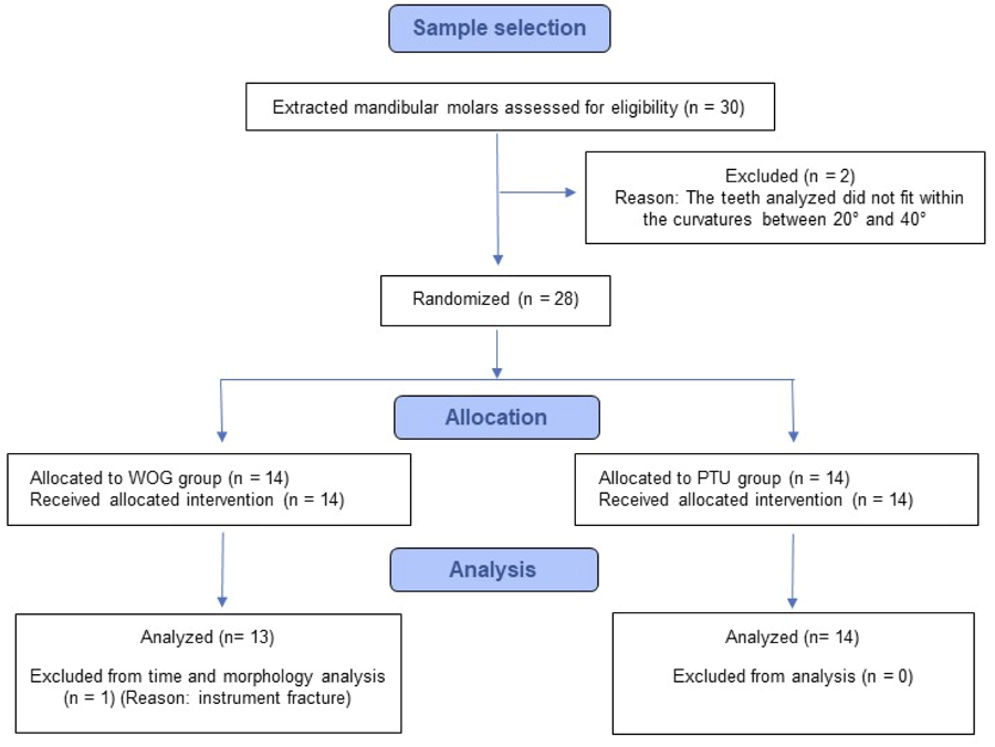
Teeth were pre-numbered (1–28) and assigned by simple randomization (1:1) using a list of random numbers generated in Microsoft Excel 365 (RAND()), sorted in ascending order; the first 14 were assigned to the WOG group and the remaining 14 to the PTU group. The sequence was generated by an independent researcher (G.O.J.) with no involvement in operative procedures14. Thus, the operator was not involved in the allocation process and was blinded to the randomization sequence; however, after allocation, blinding during instrumentation was not feasible because the PTU and WOG protocols differ in file number, kinematics, and sequence and are readily identifiable during the procedure. Sample selection, randomization, group allocation, and exclusions are summarized in a Flow diagram (Figure 1).
The diagram illustrates the number of mandibular molars assessed, excluded, randomized, allocated to each group, and included in the final analyses. Two specimens were excluded before randomization because they did not exhibit curvatures between 20° and 40°. One specimen from the WOG group was excluded from the analyses due to instrument fracture. PTU, ProTaper Ultimate; WOG, WaveOne Gold.
Root canal instrumentation
For preparation of MB canals, a single calibrated operator (undergraduate dental student), after standardized hands-on training with both systems (PTU and WOG) prior to data collection, used NiTi instruments from PTU (Dentsply Maillefer, Ballaigues, Switzerland) and WOG (Dentsply Maillefer, Ballaigues, Switzerland), operated with an X-Smart Plus electric motor (Dentsply Maillefer, Ballaigues, Switzerland). Each system followed the manufacturer’s recommended protocol, speed, and torque. In the PTU group, four instruments were used in sequence: S1 (16/.02), S2 (20/.04), F1 (20/.07), and F2 (25/.08). In the WOG group, three instruments were used: Gold Glider (15/.02), Small (20/.07), and Primary (25/.07). In each group (n = 14), the same instrument kit was used sequentially across all teeth (reuse across specimens) to assess instrument durability until fracture. In both groups, irrigation with 5 mL of 2.5% sodium hypochlorite (Asfer Indústria Química Ltda., São Caetano do Sul, SP, Brazil) was performed after each instrument change. Based on this protocol, the total irrigant volume per canal was 15 mL in the PTU group (three instrument changes) and 10 mL in the WOG group (two instrument changes).
Instrument fracture analysis
If an instrument fractured during preparation, the timers were immediately paused, and the instrument number and size were recorded. Specimens in which instrument fracture occurred before completion of the preparation were excluded from both time and morphological analyses.
Working time analysis
“Total working time” was defined as the interval from the start of canal scouting with the first file of the protocol to removal of the last shaping file, including all irrigations and instrument exchanges. “Active instrumentation time” was defined as the cumulative time during which the instrument was in motion within the canal. Both times were measured using two separate digital stopwatches, Casio HS-3V-1R (Casio Computer Co., Japan).
CBCT image acquisition and comparison
To standardize image acquisition, teeth were placed in horseshoe-shaped templates made of condensation silicone (Yller Biomateriais, Pelotas, Brazil), with four templates holding seven evenly positioned teeth each. Images were obtained by CBCT (Veraviewepocs 3D R100®, J. Morita Corp., Kyoto, Japan) in 360° high-definition scan mode, 2.5 LP/mm resolution, and 80-µm voxel size. Templates were centered in the scanner field of view with adjustments to ensure proper specimen centralization. Preoperative scans were taken with a stainless-steel K-file #10 (Dentsply Maillefer, Ballaigues, Switzerland) inserted into the MB canal to depict the native canal anatomy. Postoperative scans were obtained with a K-file #25 (Dentsply Maillefer, Ballaigues, Switzerland), corresponding to the final apical size in both systems (25/.07–25/.08). Post-instrumentation images were analyzed in isolation, without digital superimposition onto preoperative images.
Assessment of canal morphology preservation
Post-instrumentation canal anatomy was evaluated by visual assessment of CBCT images acquired with a K-file #25 inserted to working length. Images were examined individually according to predefined morphological criteria: apical transportation, ledge formation, and deviation from the original canal axis. A single examiner, an independent endodontist (J.V.V.P.) blinded to group allocation, classified each coded dataset as ‘with deviation’ if one or more alterations were present or ‘without deviation’ when no relevant modifications were observed.
Statistical analysis
Data were organized and tabulated in Microsoft Excel 2010 for Windows, and statistical analyses were performed using JASP 0.19.3 for Windows. Normality was assessed using the Shapiro-Wilk test. The student’s t-test was used for the analysis of the degree of Schneider curvature and active instrumentation time, while the Mann–Whitney test was applied for the analysis of total canal length and total working time. The 95% confidence interval (95% CI) was calculated for each group. Effect sizes were reported using Cohen’s d and rank-biserial correlation (rrb). Data on canal morphology preservation were qualitatively assessed and descriptively reported as the frequency of specimens classified as “with deviation” or “without deviation” in each group. A significance level of 5% was adopted for all analyses.
Results
There was no statistically significant difference between the groups when comparing the degree of Schneider curvature (PTU: mean (± SD) = 30.28 ± 5.63, median = 31.00; WOG: mean (± SD) = 30.14 ± 5.15, median = 31.50; (t(26) = 0.070; p = 0.945) and the total canal length in millimeters (PTU: mean (± SD) = 21.14 ± 0.949, median = 21.00; WOG: mean (± SD) = 21.14 ± 1.406, median = 21.00; (U = 97.000; p = 0.981)).
Total working time and active instrumentation time
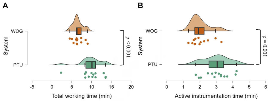
There was a significant difference in total working time between the systems (U = 163.000; p < 0.001; Figure 2A), with a medium effect size (rrb = 0.791), showing a longer time for the PTU group (median = 10.115; 95% CI Upper-lower = 11.535-8.241) compared with the WOG group (median = 6.310; 95% CI Upper-lower = 7.311-5.775).
The data are presented in minutes for both groups. (A) Total working time (PTU: n = 14, WOG: n = 13; Mann-Whitney test); (B) Active instrumentation time (PTU: n = 14, WOG: n = 13; Student’s t-test). One specimen from the WOG group was excluded from time analysis because of instrument fracture. PTU, ProTaper Ultimate; WOG, WaveOne Gold.
Active instrumentation time differed significantly between systems (t(25) = 3.661; p = 0.001; Figure 2B), with a large effect size (Cohen’s d = 1.410). Scores for the PTU group were higher (mean = 3.01; SD = 0.765; 95% CI Upper-lower = 3.452-2.569) than those for the WOG group (mean = 2.05; SD = 0.576; 95% CI Upper-lower = 2.398-1.702).
Instrument fractures and preservation of canal morphology
No instrument fractures occurred in the PTU group. In the WOG group, one fracture occurred during preparation of the 13th canal: a Small 20/.07 file.
No specimens exhibited clinically relevant apical transportation, ledge formation, or deviation from the original canal axis on postoperative CBCT; all canals were classified as “without deviation” in both groups. One specimen was excluded from analysis (WOG, n = 13) due to instrument fracture, per protocol.
Discussion
This study employed canals of MB molars, which are widely recognized as technically complex and are often used for comparisons between endodontic systems12. Both PTU and WOG preserved the original canal anatomy, with no evidence of apical transportation or relevant deviations. However, a difference in preparation time was observed, and a single isolated fracture occurred in the WOG group. Overall, both systems demonstrated safe performance, although their operative profiles differ and warrant critical comparison with the available literature.
Canal preparation time is a relevant parameter to assess the efficiency of endodontic instruments15. Several factors may influence it, including the number, sterilization, and type of instruments, the technique employed, operator experience, and even study methodology16-18. In the present study, WOG exhibited significantly shorter total working and active instrumentation times (Figure 2), which reinforces prior evidence that reciprocating systems tend to be more time-efficient16,19.
The observed divergence may be associated, above all, with the specificities of each system’s clinical protocol. The procedure was meticulously executed in accordance with the technical specifications stipulated by the manufacturers, yielding a range of configurations. In the PTU group, a configuration with four rotary instruments (S1, S2, F1, F2) was utilized, while the WOG group employed a configuration with three reciprocating instruments (Gold Glider, Small, Primary). Despite its classification as a single-file system, anatomical variations may necessitate the utilization of multiple files to adequately enlarge and shape complex canals, as described by Bueno et al.20, (2020). This underscores the importance of clinical flexibility, even in protocols that are considered simplified. Consequently, the observed reduction in preparation time can be attributed to the efficacy of the workflow proposed by each system, rather than to the superiority of a particular instrument in isolation.
Furthermore, the operator’s expertise significantly influences the interpretation of the results. Conceição et al.21 (2020) demonstrated that clinical performance can be influenced by the learning curve. However, despite prior operator calibration, an order-related learning effect due to sequential preparation cannot be excluded, and limited experience may have contributed to the isolated instrument fracture; nevertheless, reinforcing the safe applicability of both systems even in training hands, which is relevant for academic and clinical environments, is important.22
In addition, an isolated fracture was documented in the WOG group, which consequently resulted in the exclusion of the corresponding specimen. The development of reciprocating systems was driven by the necessity of operating continuously below the elastic limit of the instrument. In theoretical terms, this reduces the instrument’s resistance to cyclic fatigue23,24. Nevertheless, failures can occur even in accordance with single-use protocols, although the extant literature indicates a low incidence in these contexts20,25-27. In accordance with this finding, Gürler et al.28 (2024) observed, through a laboratory study with instruments previously used in clinical settings, that WOG exhibits lower resistance to cyclic fatigue when compared to other reciprocating and rotary systems. This observation is noteworthy, despite the absence of clinical fractures documented. Peraça et al.18 (2021) corroborated this trend by demonstrating earlier fracture of WOG compared to Reciproc Blue, even after multiple uses and autoclaving cycles, indicating lower relative durability in simulated canals. As reported by Diaconu et al.8 (2024), PTU demonstrated superior performance in terms of cyclic fatigue resistance when compared to Protaper Gold and M3 systems. This study lends further credence to the notion that variations in instrument design and composition exert a direct influence on their functional longevity.
Regarding the preservation of root canal morphology, none of the specimens showed apical transportation, canal-axis deviation, or ledge formation on post-instrumentation images. Previous studies reinforce PTU’s ability to preserve the original anatomy. Gandhi et al.29 (2025) demonstrated that this system removes less dentin and promotes a smaller volumetric change of the canal compared with ProTaper Next. Additionally, PTU yielded greater preservation of pericervical dentin than WOG, which may have implications for the tooth’s structural longevity6.
In contrast, Medina-Gil et al.30 (2025) observed that WOG presented the highest proportion of uninstrumented areas among the evaluated systems. Although this finding may suggest greater dentin preservation, it may also represent a risk of retaining infected tissue in untouched regions. This limitation tends to intensify under reuse protocols. Peraça et al.18, (2021) observed a reduction in instrumented area after multiple cycles of use and autoclaving with reciprocating instruments, suggesting a progressive loss of cutting efficiency. These findings motivate an analysis of the underlying mechanical determinants, design, kinematics, and heat treatment, that may influence cutting efficiency and the extent of instrumentation. Accordingly, within the qualitative CBCT endpoint adopted, our data corroborate morphological preservation in both systems.
The physicomechanical properties of instruments help contextualize the findings on instrumented area and reuse, providing a basis for interpreting these differences. Martins et al.11 (2025) demonstrated that PTU combines high flexibility with significant torsional resistance, characteristics that favor safe instrumentation of curved canals. WOG, in turn, exhibits increased flexibility due to Gold-Wire heat treatment31. Therefore, although both systems proved effective and safe, WOG may be preferred in situations where clinical speed is prioritized (e.g., emergency pain appointments for symptomatic irreversible pulpitis, or time-restricted visits in anxious/uncooperative patients), whereas PTU may offer a greater mechanical safety margin in severely curved canals.
Among the limitations of the present study, the ex vivo design stands out, as it does not fully simulate clinical complexity. Given the qualitative CBCT assessment of morphology, sensitivity for detecting volumetric changes is limited. In addition, inter- or intra-rater reliability was not formally assessed for morphology classification. Future investigations should include quantitative three-dimensional with microCT analyses and randomized clinical trials to validate these results, as well as evaluate additional parameters such as debris extrusion and cleaning efficacy.
Based on the limitation of this ex vivo study, it can be concluded that both PTU and WOG were equally effective in preserving the anatomy of the MB canals of curved mandibular molars, with no clinically relevant apical transportation or canal-axis deviations. WOG achieved significantly faster preparation but presented one isolated instrument fracture, whereas PTU showed no failures. Overall, both groups demonstrated safe and predictable performance, with differences primarily related to time efficiency and fracture occurrence.
Acknowledgments
The authors gratefully acknowledge the Universidade Federal do Piauí (UFPI), the School of Dentistry, and the Graduate Studies Committee for their support throughout the project. The authors also thank Professor Karla Rovaris, then coordinator of the Undergraduate Program in Dentistry, for her valuable support.
References
-
1 Liang Y, Yue L. Evolution and development: engine-driven endodontic rotary nickel-titanium instruments. Int J Oral Sci. 2022 Feb;14(1):12. doi: 10.1038/s41368-021-00154-0.
» https://doi.org/10.1038/s41368-021-00154-0 -
2 Srivastava S. Root canal instrumentation: current trends and future perspectives. Cureus. 2024 Apr;16(4):e58045. doi: 10.7759/cureus.58045.
» https://doi.org/10.7759/cureus.58045 -
3 Puleio F, Bellezza U, Torre A, Giordano F, Lo Giudice G. Apical transportation of apical foramen by different NiTi alloy systems: a systematic review. Appl Sci. (Switzerland). 2023;13(19):10555. doi: 10.3390/app131910555.
» https://doi.org/10.3390/app131910555 -
4 Sawardeker AV, Tikku AP, Bharti R, Bains R. Evaluation of canal transportation and centering ability in mesiobuccal canals of maxillary molars using two Ni-Ti file systems: An ex-vivo micro-computed tomographic study. Endodontology. 2020;32(4):198-203. doi:10.4103/endo.endo_41_20.
» https://doi.org/10.4103/endo.endo_41_20 -
5 Kim H, Jeon SJ, Seo MS. Comparison of the canal transportation of ProTaper GOLD, WaveOne GOLD, and TruNatomy in simulated double-curved canals. BMC Oral Health. 2021 Oct;21(1):533. doi: 10.1186/s12903-021-01854-z.
» https://doi.org/10.1186/s12903-021-01854-z -
6 Ribeiro G, Martin V, Rodrigues C, Gomes P. Comparative evaluation of the canal shaping ability, pericervical dentin preservation, and smear layer removal of TruNatomy, WaveOne Gold, and ProTaper Ultimate-an ex vivo study in human teeth. J Endod. 2023 Dec;49(12):1733-8. doi: 10.1016/j.joen.2023.09.002.
» https://doi.org/10.1016/j.joen.2023.09.002 -
7 Shi L, Zhou J, Wan J, Yang Y. Shaping ability of ProTaper Gold and WaveOne Gold nickel-titanium rotary instruments in simulated S-shaped root canals. J Dent Sci. 2022 Jan;17(1):430-7. doi: 10.1016/j.jds.2021.08.008. Epub 2021 Aug 30.
» https://doi.org/10.1016/j.jds.2021.08.008 -
8 Diaconu CT, Diaconu AE, Tuculina MJ, Mihai LL, Gheorghi?a M, Gheorghi?a LM, et al. Assessment of the cyclic fatigue performance of the novel protaper ultimate file system used in different kinematics: an in vitro study. J Funct Biomater. 2024 Mar;15(4):85. doi: 10.3390/jfb15040085.
» https://doi.org/10.3390/jfb15040085 -
9 Algarni Y. Fracture incidence of new reciprocating nickel-titanium (NiTi) files: a cross-sectional retrospective study. Cureus. 2024 Aug;16(8):e67762. doi: 10.7759/cureus.67762.
» https://doi.org/10.7759/cureus.67762 -
10 Alsulaiman M, Alsofi L, Alhabib MA. Shaping ability of NiTi reciprocating file systems R-motion and waveOne gold in mesial canals of mandibular molars; micro CT study. Sci Rep. 2025 Jan;15(1):747. doi: 10.1038/s41598-024-84986-w.
» https://doi.org/10.1038/s41598-024-84986-w -
11 Martins JNR, Silva EJNL, Vieira VTL, Costa RP, Baruwa AO, Fernandes FMB, et al. A comparative analysis of protaper ultimate and five multifile systems: design, metallurgy, and mechanical performance. Materials (Basel). 2025 Mar;18(6):1260. doi: 10.3390/ma18061260.
» https://doi.org/10.3390/ma18061260 -
12 Rocha DGP, Alves VO, Martin AS, Fontana CE, Cunha RS, Bueno CES. Comparison of preparation time and ability to maintain canal morphology in curved canals: Pathfile + Protaper Universal versus Twisted Files. Rev Odontol UNESP. 2013;42(2):99-103. doi:10.1590/s1807-25772013000200006.
» https://doi.org/10.1590/s1807-25772013000200006 -
13 Schneider SW. A comparison of canal preparations in straight and curved root canals. Oral Surg Oral Med Oral Pathol. 1971 Aug;32(2):271-5. doi: 10.1016/0030-4220(71)90230-1.
» https://doi.org/10.1016/0030-4220(71)90230-1 -
14 Altman DG, Bland JM. How to randomise. BMJ. 1999 Sep;319(7211):703-4. doi: 10.1136/bmj.319.7211.703.
» https://doi.org/10.1136/bmj.319.7211.703 -
15 Almnea RA, Albeaji SMA, Alelyani AA, AlHarith D, Alshahrani AS, Al Malwi AA, et al. Comparative analysis of three nickel-titanium rotary files in severely curved l-shaped root canals: preparation time, aberrations, and fracture rates. Clin Cosmet Investig Dent. 2024 Feb;16:1-9. doi: 10.2147/CCIDE.S452742.
» https://doi.org/10.2147/CCIDE.S452742 -
16 Bürklein S, Hinschitza K, Dammaschke T, Schäfer E. Shaping ability and cleaning effectiveness of two single-file systems in severely curved root canals of extracted teeth: Reciproc and WaveOne versus Mtwo and ProTaper. Int Endod J. 2012 May;45(5):449-61. doi: 10.1111/j.1365-2591.2011.01996.x. Epub 2011 Dec 22.
» https://doi.org/10.1111/j.1365-2591.2011.01996.x -
17 Lim KC, Webber J. The validity of simulated root canals for the investigation of the prepared root canal shape. Int Endod J. 1985 Oct;18(4):240-6. doi: 10.1111/j.1365-2591.1985.tb00450.x.
» https://doi.org/10.1111/j.1365-2591.1985.tb00450.x -
18 Peraça VO, Xavier SR, Gomes FA, Santos LGP, Souza EM, Pappen FG. Effect of number of uses and sterilization on the instrumented area and resistance of reciprocating instruments. Restor Dent Endod. 2021 Apr;46(2):e28. doi: 10.5395/rde.2021.46.e28.
» https://doi.org/10.5395/rde.2021.46.e28 -
19 You SY, Bae KS, Baek SH, Kum KY, Shon WJ, Lee W. Lifespan of one nickel-titanium rotary file with reciprocating motion in curved root canals. J Endod. 2010 Dec;36(12):1991-4. doi: 10.1016/j.joen.2010.08.040.
» https://doi.org/10.1016/j.joen.2010.08.040 -
20 Bueno CSP, Oliveira DP, Pelegrine RA, Fontana CE, Rocha DGP, Gutmann JL, et al. Fracture incidence of WaveOne Gold files: a prospective clinical study. Int Endod J. 2020 Sep;53(9):1192-8. doi: 10.1111/iej.13349.
» https://doi.org/10.1111/iej.13349 -
21 Conceição I, Ferreira I, Braga AC, Pina-Vaz I. Simulated root canals preparation time, comparing ProTaper Next and WaveOne Gold systems, performed by an undergraduate student. J Clin Exp Dent. 2020 Aug;12(8):e730-e735. doi: 10.4317/jced.56981.
» https://doi.org/10.4317/jced.56981 -
22 Serafini JA, Portugal BN, Scherer MM, Lenzi TL, Luisi SB. Undergraduate dental students'use perception of different endodontic instruments for mechanical-chemical preparation: a systematic review of laboratory studies. Rev ABENO. 2022;22(2):1-13. doi: 10.30979/revabeno.v22i2.1616.
» https://doi.org/10.30979/revabeno.v22i2.1616 -
23 Pedro-Muñoz A, Rico-Romano C, Sánchez-Llobet P, Montiel-Company JM, Mena-Álvarez J. Cyclic fatigue resistance of rotary versus reciprocating endodontic files: a systematic review and meta-analysis. J Clin Med. 2024 Feb;13(3):882. doi: 10.3390/jcm13030882.
» https://doi.org/10.3390/jcm13030882 -
24 Plotino G, Ahmed HM, Grande NM, Cohen S, Bukiet F. Current assessment of reciprocation in endodontic preparation: a comprehensive review--Part II: properties and effectiveness. J Endod. 2015 Dec;41(12):1939-50. doi: 10.1016/j.joen.2015.08.018.
» https://doi.org/10.1016/j.joen.2015.08.018 -
25 Adigüzel M, Capar ID. Comparison of cyclic fatigue resistance of WaveOne and WaveOne Gold small, primary, and large instruments. J Endod. 2017 Apr;43(4):623-7. doi: 10.1016/j.joen.2016.11.021.
» https://doi.org/10.1016/j.joen.2016.11.021 -
26 Oh S, Kum KY, Kim HJ, Moon SY, Kim HC, Chaniotis A, et al. Bending resistance and cyclic fatigue resistance of WaveOne Gold, Reciproc Blue, and HyFlex EDM instruments. J Dent Sci. 2020 Dec;15(4):472-8. doi: 10.1016/j.jds.2019.10.003. Epub 2019 Dec 30.
» https://doi.org/10.1016/j.jds.2019.10.003 -
27 Olcay K, Eyuboglu TF, Erkan E. Cyclic fatigue resistance of waveone gold, protaper next and 2shape nickel titanium rotary instruments using a reliable method for measuring temperature. Niger J Clin Pract. 2019 Oct;22(10):1335-40. doi: 10.4103/njcp.njcp_655_18.
» https://doi.org/10.4103/njcp.njcp_655_18 -
28 Gürler K, Yilmaz S, Dumani A, Yoldas O. Comparison of cyclic fatigue resistance of three different single-file systems after clinical use. BMC Oral Health. 2024 Dec;24(1):1482. doi: 10.1186/s12903-024-05287-2.
» https://doi.org/10.1186/s12903-024-05287-2 -
29 Gandhi PJ, Jobanputra LH, Prajapati MH, Aroori H. Exploration of uninstrumented areas and volumetric precision of ProTaper Ultimate versus ProTaper Next file systems: A micro-computed tomography study. J Conserv Dent Endod. 2025 Jun;28(6):515-21. doi: 10.4103/JCDE.JCDE_202_25.
» https://doi.org/10.4103/JCDE.JCDE_202_25 -
30 Medina-Gil M, Martín-Díaz A, Navarrete N, Aranguren J, Ortolani-Seltenerich PS, Malvicini G, et al. Micro-computed tomography analysis of reciprocating systems in three-dimensional models of mandibular premolars with two canals. Dent J (Basel). 2025 Apr;13(4):175. doi: 10.3390/dj13040175.
» https://doi.org/10.3390/dj13040175 -
31 van der Vyver PJ, Paleker F, Vorster M, Wet FA. Root canal shaping using nickel titanium, M-Wire, and Gold Wire: a micro-computed tomographic comparative study of One Shape, ProTaper Next, and WaveOne Gold instruments in maxillary first molars. J Endod. 2019 Jan;45(1):62-7. doi: 10.1016/j.joen.2018.09.013. Epub 2018 Nov 13.
» https://doi.org/10.1016/j.joen.2018.09.013
-
Data Availability:
Data are available on reasonable request.
-
Funding:
This research received no external funding
Edited by
-
Editor:
Dr. Altair A. Del Bel Cury
Data are available on reasonable request.