Impact of graphene incorporation on the mechanical and optical properties of glass ionomer cements
Abstract
Introduction Failures in orthodontic treatments using bands may be related to deficient properties of GICs. Incorporating graphene into GICs for cementation is an option to improve this material and reduce failures.
Aim The purpose is to evaluate the effect of adding reduced graphene oxide (rGO) on the mechanical and optical properties of glass ionomer cements (GICs) for luting.
Methods Samples of 6 different GICs (Meron, GC Gold Label, GC Fuji Plus, Riva Luting, Vitro Cem, and Vidrion) with and without graphene incorporation at concentrations of 0.25%, 0.5%, and 1% were prepared. Cohesive strength (CoheS), compressive strength (CompS) and Shear Bond Strength (SBS) tests were conducted using a universal testing machine (EMIC). Color change was assessed using the CIELAB system and particle dispersion was observed with Optical Microscopy (OM). Data was tabulated and analyzed with Jamovi software (p<0.05) using descriptive and correlation analyses with One-way ANOVA and Kruskal-Wallis tests, based on the Shapiro-Wilk normality test.
Results The incorporation of rGO significantly increased the compressive strength of Vidrion (0.5%) and the cohesive strength of GC Gold Label and GC Fuji Plus, although it caused perceptible chromatic changes in all tested cements.
Conclusion The addition of rGO to GICs enhanced the mechanical properties of some GICs and altered the color of all GICs.
Keywords
Glass ionomer cements; Graphene oxide; Graphite; Nanostructures
Introduction
Glass ionomer cements (GICs), first introduced by Wilson and Kent1 in 1972, have become widely used in dentistry due to their versatility. Their favorable properties include a compatible coefficient of thermal expansion2, chemical adhesion to enamel and dentin3, fluoride release4, and biocompatibility5. Despite these advantages, GICs exhibit limitations such as low mechanical strength, porosity, and suboptimal aesthetics compared to composite resins6.
To overcome these shortcomings, various modifications have been explored, including the incorporation of silver particles, tartaric acid, titanium, and lead7. More recently, attention has turned to graphene, a material renowned for its exceptional mechanical, thermal, electrical, and optical properties. Preliminary studies suggest that integrating graphene into biomedical and dental materials may enhance both antibacterial activity and mechanical performance8-11.
In the context of orthodontic treatments, where failures in band cementation can negatively impact clinical outcomes12, improving the mechanical resilience of GICs is particularly valuable. Ongoing research has explored the use of graphene in various dental specialties, including periodontology13, implantology14, endodontics15, and polymer-based biomaterials8. Furthermore, Brazil’s abundant graphite reserves—accounting for up to 50% of the global supply—underscore the strategic importance of advancing graphene-based technologies domestically. The incorporation of graphene into glass ionomer cements can offer a promising strategy to enhance their mechanical strength, antibacterial efficacy and overall structural integrity, potentially increasing the material’s durability in clinical applications.
Given this context, this study aims to investigate whether the incorporation of reduced graphene oxide (rGO) into conventional glass ionomer cement for cementation (GIC-C) enhances its mechanical and optical properties.
Materials and Methods
Sample Design
This in vitro study evaluated six commercially available glass ionomer cements for cementation (GIC-C), each tested with and without the incorporation of reduced graphene oxide (rGO) (Table 1). Each material was divided into four subgroups: a control group (without rGO) and three experimental groups containing 0.25%, 0.5%, and 1% (w/w) rGO.
Reduced graphene oxide was synthesized by oxidative treatment of natural graphite. Briefly, 0.5 g of graphite was added to a mixture of concentrated sulfuric acid and nitric acid in a 4:1 (v/v) ratio and refluxed at 80°C under constant stirring for 1 hour. The oxidized material was centrifuged at 4,000 rpm for 10 minutes (GOFLAME GL33 Centrifuge), washed with deionized water, and dried in an oven at 120°C for 1 hour (Marconi MA 030). It was then thermally expanded in a furnace at 1,000°C for 1 minute (QUIMIS Q318M35T Furnace), yielding reduced graphene oxide in powder form.
Incorporation of rGO into GIC-C
Each GIC-C material was weighed and mixed with rGO at concentrations of 0.25%, 0.5%, and 1% (w/w). The mixtures were stored in labeled Falcon tubes and homogenized at 22 rpm for 15 minutes (Phoenix Luferco, Model AP 22/28/32). All samples were coded to ensure blinded testing.
Manipulation of GIC-C
The GIC-C materials were handled using a plastic spatula on disposable waterproof paper pads. The prepared materials were inserted into polymethyl methacrylate (PMMA) and semi-rigid rubber molds, following ISO 9917:2007 and ISO 4049:2019 guidelines.
Preparation of Specimens
Specimens were prepared in various geometries according to the type of mechanical or optical testing:
1. Compressive Strength (CompS) (n = 5 per group)
Samples were placed in PMMA molds (6.0 mm height × 4.0 mm diameter) and covered with a polyester matrix and a 10 mm glass plate during setting.
2. Cohesive Strength (CoheS) (n = 5 per group)
The GIC-C was placed into hourglass-shaped semi-rigid rubber molds, also using a polyester matrix and a 10 mm glass plate.
3. Shear Bond Strength (SBS) (n = 5 per group)
A total of 120 bovine incisors teeth had their roots removed using a metal disk (D5A22), and crowns were embedded in acrylic resin blocks (30 × 20 mm), exposing the buccal surface. These surfaces were standardized using a metallographic polisher (Isomet, Model 11-1280-170, Lake Bluff®, Illinois, USA) with sequential abrasive sandpapers (#100, #220, #400). After polishing, prophylaxis was performed using a rubber cup (KG-Sorensen, São Paulo, Brazil) and a pumice/water slurry (SSWhite®, Juiz de Fora, Brazil) for 10 seconds, followed by rinsing and drying. Brackets for lower incisors (Edgewise Standard 10.30.201, Morelli, Sorocaba, São Paulo) were bonded using the experimental GIC-Cs.
4. Color Evaluation and Optical Microscopy (OM) (n = 1 per group)
Specimens were prepared by compressing the material between two glass slides (26 × 76 mm). After the setting time, samples were stored in Milli-Q water at 37°C for 24 hours prior to testing.
Analysis
Mechanical Testing: Compressive Strength (CompS), Cohesive Strength (CoheS), and Shear Bond Strength (SBS)
All mechanical tests were performed using a universal testing machine (EMIC DL 2000, São José dos Pinhais, PR, Brazil) equipped with a 5000 N load cell and operated at a crosshead speed of 1 mm/min. The following fixtures were used: a compression device (Model OD23, Odeme, Luzerna, SC, Brazil) for CompS, a cohesive fracture clamp (Model OG04, Odeme), and a wedge-shaped micro-shear actuator with a 90° angle (Model OD27, Odeme) for SBS tests. Bovine incisors were used for the SBS evaluation. Results were recorded in Newtons (N) and Megapascals (MPa) for CompS and CoheS, and in kilograms-force (Kgf) and MPa for SBS.
Color Analysis
Colorimetric analysis was performed using the CIELAB color space (L*, a*, b*) to quantify color variation. The L* parameter represents lightness (black-white), a* indicates red-green balance, and b* corresponds to blue-yellow chroma. Measurements were obtained in triplicate using a portable colorimeter (DOHO DR-10, Shenzhen Technology Ltd., China). Total color differences (ΔE00) were calculated using the CIEDE2000 formula according to ISO 28642:201116:
Where: ΔL, ΔC, and ΔH are the differences in lightness, chroma, and hue. RT is a function (known as the rotation function) explaining the interaction between chroma and hue differences in the blue region. The weighting functions SL, SC, and SH are used to adjust the total color difference between the sample and the pure ionomer in L, a, and b coordinates. Parametric factors KL, KC, and KH were set to 1 for calculations.
Optical Microscopy (OM)
Microscopic surface and internal morphology were analyzed using a digital optical microscope (Model YYM-220517-5780, KKMOON, China) at 2000× magnification.
Statistical Analysis
Data were compiled and analyzed using Jamovi® 2.2.5, with significance set at p < 0.05. Depending on normality, determined using the Shapiro–Wilk test, comparisons were made using One-way ANOVA or the Kruskal–Wallis test. Post hoc analyses were conducted for pairwise comparisons when appropriate.
Results
Compressive Strength (CompS) (Table 2)
-
Riva Luting and GC Fuji Plus: No significant differences were observed among control and rGO-modified groups.
-
Vitro Cem: A significant difference was noted between the 0.25% and 1% rGO groups (p = 0.033).
-
Meron: Significant differences occurred between the 0.5% rGO group and both the 0.25% (p = 0.049) and 1% (p < 0.001) groups. The 0.5% group exhibited the lowest average value (63.8 ± 8.69 MPa), while the 1% group showed the highest (85.3 ± 1.71 MPa).
-
GC Gold Label: The control group differed significantly from the 0.5% and 1% rGO groups (p = 0.045).
-
Vidrion C: The 0.5% rGO group differed significantly from both the control and 0.25% rGO groups (p = 0.045). It exhibited a compressive strength seven times higher than the other groups (105 ± 54.7 MPa).
Cohesive Strength (CoheS) (Table 2)
-
GC Gold Label and GC Fuji Plus: Both showed improved strength with rGO. GC Gold Label had the highest value at 0.25% (8.46 ± 1.43 MPa), and Fuji Plus at 0.5% (12.6 ± 0.96 MPa). Control values were the lowest: 4.71 ± 1.66 and 8.04 ± 2.26 MPa, respectively.
-
Meron: A significant difference was observed between the control and 0.25% rGO group (p = 0.021), with the control group showing higher strength.
-
Riva Luting, Vitro Cem, and Vidrion C: No statistically significant differences were found between control and rGO-modified groups.
Shear Bond Strength (SBS) (Table 2)
-
Vitro Cem and Meron: Bracket adhesion was insufficient in the rGO groups, so only control data were recorded.
-
Riva Luting: Measurable results were obtained for the control and 0.25% rGO groups; however, no significant differences were noted.
-
Vidrion C: Significant differences were found between the control and both 0.25% (p = 0.041) and 0.5% (p = 0.044) rGO groups, which exhibited values near zero.
-
GC Gold Label: The control group demonstrated values up to 4.5 times higher (11.6 ± 4.21 MPa) than the 0.25% rGO group (2.59 ± 2.67 MPa), with statistical significance.
-
GC Fuji Plus: Although the 0.5% rGO group had higher values (6.82 ± 1.8 MPa) than the control, the differences were not statistically significant. The lowest result was observed in the 0.25% group (3.61 ± 1.90 MPa).
Color Analysis (Spectrophotometry)
Colorimetric analysis indicated that rGO incorporation significantly altered the color parameters of all GICs. The L* axis showed a decrease (darker appearance), a* values shifted toward green, and b* values shifted toward blue. Overall, ΔE00 values indicated a perceptible and significant color change for all rGO-containing groups (Table 3 and Figure 1).

Optical Microscopy (OM)
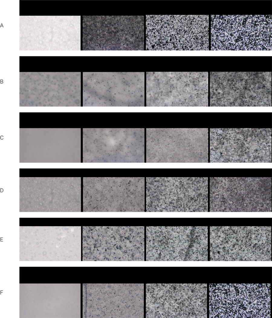
All samples showed visible particle aggregation of rGO, with distribution relatively homogeneous across the GIC matrix. An increase in rGO concentration was associated with a greater number of visible aggregates. Among the tested materials, Vitro Cem, Vidrion C, and Riva Luting exhibited the smallest aggregate areas in the analyzed fields (Figure 2).
Analysis of Optical Microscopy (OM) in GIC-C (A - GC Fuji Plus; B - Vidrion C; C - Vitro Cem; D - Meron; E - Riva Luting; F - GC Gold Label).
Discussion
Glass ionomer cements (GICs) remain widely used in clinical practice due to their chemical adhesion to dental tissues, fluoride release, and biocompatibility, despite known limitations such as fragility, suboptimal mechanical strength, and moisture sensitivity2. Over the years, numerous strategies have been employed to enhance their performance, including the incorporation of various reinforcing agents. This study investigated whether incorporating reduced graphene oxide (rGO) into GICs could improve their mechanical and optical properties, particularly aiming to reduce failures in orthodontic cementation.
The results demonstrate that rGO incorporation, at specific concentrations, can significantly improve mechanical performance in select GIC formulations. Notably, compressive strength increased substantially in Vidrion at 0.5% rGO, and cohesive strength improved in GC Gold Label (0.25% and 1%) and GC Fuji Plus (0.5% and 1%). These enhancements can be attributed to the exceptional tensile and elastic properties of graphene. For example, Lee et al.17 reported a Young’s modulus of approximately 1000 ± 100 GPa in monolayer graphene using atomic force microscopy, closely aligning with the theoretical maximum of 1050 Gpa18. Similarly, Pavithra et al.19 demonstrated that graphene nanocomposites deposited on copper significantly increased both elasticity modulus and hardness. Our findings are consistent with previous research that reported improved mechanical properties following graphene incorporation into GICs9,10,20, though prior studies often used fluorinated graphene, pure graphene, or chemically modified graphene to improve clinical acceptability by altering its inherent dark color. These different forms may yield variable outcomes and should be considered when comparing results across studies.
Among all findings, the most striking was the substantial increase in compressive strength in Vidrion at 0.5% rGO—an approximate 700% increase compared to the control. However, this enhancement was accompanied by an altered setting behavior, with specimens showing a rubbery appearance even 24 hours post-setting. This observation suggests potential interference of rGO with the cement’s acid-base reaction or matrix formation, highlighting the need for further research into the physicochemical interactions between rGO and GIC components.
Color stability was another critical parameter evaluated21,22. All rGO-containing groups showed statistically significant color changes, particularly a reduction in L* (lightness) and shifts toward green (a*) and blue (b*) chromaticity. These results are explained by the inherent dark pigmentation of rGO, which reduces light transmission. According to established perceptibility (ΔE > 1.1) and acceptability (ΔE > 2.8) thresholds23, the color shifts observed (minimum ΔE = 13.33 in GC Fuji Plus 0.25%) would be clearly visible clinically. Although aesthetics are less critical in subgingival or temporary cementation—such as under orthodontic bands—the darkening effect limits the use of rGO-modified GICs in esthetically sensitive areas. That said, colored cements are already used clinically for easy detection and removal, and chemical modifications to render graphene white have also been proposed9, offering a potential pathway for aesthetic adaptation.
Importantly, optical microscopy revealed particle agglomeration in all experimental groups, though distribution was relatively homogeneous in GC Gold Label, GC Fuji Plus, and Meron. Higher rGO concentrations led to more frequent and larger aggregates, potentially contributing to inconsistent mechanical behavior. Poor dispersion may have reduced the effectiveness of rGO reinforcement in some groups, as seen in the decrease of cohesive strength in Meron (0.25%) and shear bond strength in Vidrion (0.25% and 0.5%). These findings support the hypothesis that dispersion quality and particle interaction with the matrix are critical determinants of performance.
Graphene and its derivatives are increasingly studied in dentistry not only for their mechanical benefits but also for their antimicrobial potential8-10. While various synthesis methods exist, including chemical vapor deposition and mechanical exfoliation24,25, this study employed chemical reduction using natural graphite—a cost-effective method well-suited for large-scale applications. Given that Brazil holds approximately 50% of the world’s graphite reserves, this approach could have strategic industrial relevance for the country.
Despite promising results, it is evident that rGO must be used within optimal concentration thresholds. Exceeding these limits may lead to particle agglomeration, void formation, or interference with the cement setting reaction, ultimately compromising performance. Additional studies are needed to identify ideal concentrations, improve dispersion techniques, and explore the long-term effects of rGO-modified GICs under simulated oral conditions. This study also represents one of the first attempts to evaluate the microstructural dispersion of rGO in GICs using optical microscopy. As such, direct comparison with the existing literature remains limited, underscoring the novelty and exploratory nature of the present work.
This study concludes that the incorporation of reduced graphene oxide (rGO) into glass ionomer cements can enhance certain mechanical properties, particularly the compressive strength of Vidrion at 0.5% concentration and the cohesive strength of GC Gold Label (0.25% and 1%) and GC Fuji Plus (0.5% and 1%). Optical testing confirmed that all GICs experienced perceptible color changes, with more homogeneous rGO dispersion observed in GC Gold Label, GC Fuji Plus, and Meron. While promising, the clinical application of rGO-modified GICs requires further optimization, especially in terms of color stability and dispersion control.
References
-
1 Wilson AD, Kent BE. A new translucent cement for dentistry. The glass ionomer cement. Br Dent J. 1972 Feb;132(4):133-5. doi: 10.1038/sj.bdj.4802810.
» https://doi.org/10.1038/sj.bdj.4802810 -
2 Collado-González M, Pecci-Lloret MR, Tomás-Catalá CJ, García-Bernal D, Oñate-Sánchez RE, Llena C, et al. Thermo-setting glass ionomer cements promote variable biological responses of human dental pulp stem cells. Dent Mater. 2018 Jun;34(6):932-43. doi: 10.1016/j.dental.2018.03.015.
» https://doi.org/10.1016/j.dental.2018.03.015 -
3 Nicholson J.W. Adhesion of glass-ionomer cements to teeth: A review. Int. J. Adhes. Adhes. 2016;69:33-8. doi: 10.1016/j.ijadhadh.2016.03.012.
» https://doi.org/10.1016/j.ijadhadh.2016.03.012 -
4 Wiegand A, Buchalla W, Attin T. Review on fluoride-releasing restorative materials--fluoride release and uptake characteristics, antibacterial activity and influence on caries formation. Dent Mater. 2007 Mar;23(3):343-62. doi: 10.1016/j.dental.2006.01.022. Epub 2006 Apr 17.
» https://doi.org/10.1016/j.dental.2006.01.022 - 5 Sidhu SK, Schmalz G. The biocompatibility of glass-ionomer cement materials. A status report for the American Journal of Dentistry. Am J Dent. 2001 Dec;14(6):387-96.
- 6 Khoroushi M, Keshani F. A review of glass-ionomers: from conventional glass-ionomer to bioactive glass-ionomer. Dent Res J (Isfahan). 2013 Jul;10(4):411-20.
-
7 Nicholson JW, Sidhu SK, Czarnecka B. Enhancing the mechanical properties of glass-ionomer dental cements: a review. Materials (Basel). 2020 May;13(11):2510. doi: 10.3390/ma13112510.
» https://doi.org/10.3390/ma13112510 -
8 Xie H, Cao T, Rodríguez-Lozano FJ, Luong-Van EK, Rosa V. Graphene for the development of the next-generation of biocomposites for dental and medical applications. Dent Mater. 2017 Jul;33(7):765-74. doi: 10.1016/j.dental.2017.04.008.
» https://doi.org/10.1016/j.dental.2017.04.008 -
9 Sun L, Yan Z, Duan Y, Zhang J, Liu B. Improvement of the mechanical, tribological and antibacterial properties of glass ionomer cements by fluorinated graphene. Dent Mater. 2018 Jun;34(6):e115-27. doi: 10.1016/j.dental.2018.02.006.
» https://doi.org/10.1016/j.dental.2018.02.006 - 10 Chen J, Zhao Q, Peng J, Yang X, Yu D, Zhao W. Antibacterial and mechanical properties of reduced graphene-silver nanoparticle nanocomposite modified glass ionomer cements. J Dent. 2020 May;96:103332. doi: 10.1016/j.jdent.2020.103332.
-
11 Sari F, Ugurlu M. Reinforcement of resin-modified glass-ionomer cement with glass fiber and graphene oxide. J Mech Behav Biomed Mater. 2023 Jun;142:105850. doi: 10.1016/j.jmbbm.2023.105850.
» https://doi.org/10.1016/j.jmbbm.2023.105850 -
12 Millett DT, Glenny AM, Mattick RC, Hickman J, Mandall NA. Adhesives for fixed orthodontic bands. Cochrane Database Syst Rev. 2016 Oct;10(10):CD004485. doi: 10.1002/14651858.CD004485.pub4.
» https://doi.org/10.1002/14651858.CD004485.pub4 -
13 Kawamoto K, Miyaji H, Nishida E, Miyata S, Kato A, Tateyama A, et al. Characterization and evaluation of graphene oxide scaffold for periodontal wound healing of class II furcation defects in dog. Int J Nanomedicine. 2018 Apr;13:2365-76. doi: 10.2147/IJN.S163206.
» https://doi.org/10.2147/IJN.S163206 -
14 Schünemann FH, Galárraga-Vinueza ME, Magini R, Fredel M, Silva F, Souza JCM, et al. Zirconia surface modifications for implant dentistry. Mater Sci Eng C Mater Biol Appl. 2019 May;98:1294-305. doi: 10.1016/j.msec.2019.01.062.
» https://doi.org/10.1016/j.msec.2019.01.062 -
15 Rosa V, Xie H, Dubey N, Madanagopal TT, Rajan SS, Morin JL, et al. Graphene oxide-based substrate: physical and surface characterization, cytocompatibility and differentiation potential of dental pulp stem cells. Dent Mater. 2016 Aug;32(8):1019-25. doi: 10.1016/j.dental.2016.05.008.
» https://doi.org/10.1016/j.dental.2016.05.008 - 16 ISO/TR 28642:2011. Dentistry: guidance on colour measurement. Geneva: International Organization for Standardization; 2011.
- 17 Lee C, Wei X, Kysar JW, Hone J. Measurement of the elastic properties and intrinsic strength of monolayer graphene. Science. 2008 Jul;321(5887):385-8. doi: 10.1126/science.1157996.
-
18 MacMillan, N.H. The theoretical strength of solids. J Mater Sci 1972 Feb;7, 239-254. doi: https://doi.org/10.1007/BF02403513
» https://doi.org/10.1007/BF02403513 - 19 Pavithra CL, Sarada BV, Rajulapati KV, Rao TN, Sundararajan G. A new electrochemical approach for the synthesis of copper-graphene nanocomposite foils with high hardness. Sci Rep. 2014 Feb;4:4049. doi: 10.1038/srep04049.
-
20 Jordão TRDS, Fernandes LSV, Leite KLF, Alexandria A, Silva EJNL, Maia LC, et al. Physical-mechanical, chemical and biological properties of graphene-reinforced glass ionomer cements. Restor Dent Endod. 2024 Oct;49(4):e37. doi: 10.5395/rde.2024.49.e37.
» https://doi.org/10.5395/rde.2024.49.e37 -
21 Chu SJ, Trushkowsky RD, Paravina RD. Dental color matching instruments and systems. Review of clinical and research aspects. J Dent. 2010;38(Suppl 2):e2-16. doi: 10.1016/j.jdent.2010.07.001.
» https://doi.org/10.1016/j.jdent.2010.07.001 - 22 Commission Internationale de l'Eclairage. Colorimetry. 2nd ed. Vienna: Central Bureau of the CIE; 1986. (CIE Publication; 15.2).
-
23 Paravina RD, Pérez MM, Ghinea R. Acceptability and perceptibility thresholds in dentistry: A comprehensive review of clinical and research applications. J Esthet Restor Dent. 2019 Mar;31(2):103-12. doi: 10.1111/jerd.12465.
» https://doi.org/10.1111/jerd.12465 - 24 Adetayo A, Runsewe D. Synthesis and fabrication of graphene and graphene oxide: a review. J Compos Mater. 2019;9:207-29. doi: 10.4236/ojcm.2019.92012.
- 25 Agudosi ES, Abdullah EC, Numan A, Mubarak NM, Khalid M, Omar N. A review of the graphene synthesis routes and its applications in electrochemical energy storage. Crit. Rev. Solid State Mater. Sci.2020;45: 339-77. doi: 10.1080/10408436.2019.1632793.
-
Data availability:
Datasets related to this article will be available to the corresponding author upon request.
Edited by
-
Editor:
Dr. Altair A. Del Bel Cury
Datasets related to this article will be available to the corresponding author upon request.