Influence of stainless-steel orthodontic brackets on the tomographic planning of dental implants: an ex vivo study
Abstract
Aim This ex vivo study aimed to assess the influence of stainless-steel orthodontic brackets (SSOB) and the acquisition protocol on linear measurements of bone height and width for the planning of dental implants in inter-bracket regions.
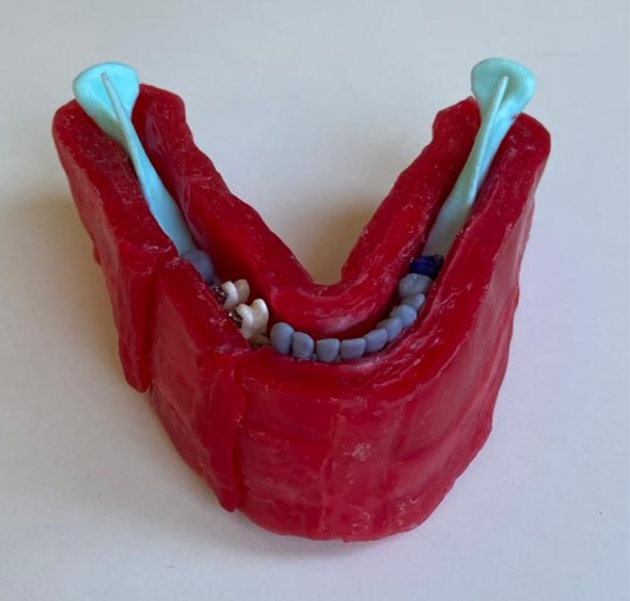
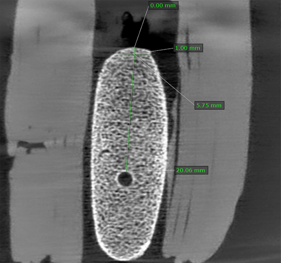
Methods Twenty extracted premolar teeth were adapted in artificial sockets on 10 bovine ribs, then adapted into a 3D printed jaw, coated with wax. Cone beam computed tomography (CBCT) scans with and without SSOB were performed using four acquisition protocols, varying the voxel size (280, 200, 125, and 80 μm). Three trained examiners evaluated all 80 CBCT scans, measuring height and width in the center of the simulated edentulous region between the teeth, in the cross-sectional plane. Intra- and inter-examiner reproducibility were assessed using the Intraclass Correlation Coefficient test. Linear measurements were compared using the Bland-Altman method and ANOVA for repeated measures. A significance level of 5% was used.
Results Intra- and inter-examiner reproducibility presented excellent reliability for height measurements, while inter-examiner presented moderate and intra-examiner presented moderate to good reliability. The measurements showed a statistically significant difference between the examiners (p<0.001), regardless of the voxel size or the presence of SSOB. The Bland-Altman showed discrepancies between -0.18 mm and 1.15 mm in the width measurements.
Conclusion The presence of SSOB and the CBCT voxel size do not affect the planning of dental implants in the inter-bracket region.
Keywords
Dental implants; Cone-beam computed tomography; Artifacts; Orthodontic brackets
 Thumbnail
 Thumbnail