Expression of virulence genes in Enterococcus faecalis is associated with biofilm formation
Abstract
Aim This study compared the expression of virulence genes and the ability to form biofilm in Enterococcus faecalis isolated from patients undergoing different endodontic clinical cases.
Methods For the biofilm-forming capacity, a reference strain (E. faecalis ATCC 29212) and four clinically isolated E. faecalis strains were analyzed by counting the colony-forming units, scanning electron microscopy, and optical density measurement. The gene expression of the collagen-binding protein (ace), aggregation substance (asa), endocarditis antigen (efaA), cytolysin (cylA), protein surface (esp), gelatinase (gelE), and histidine kinase (fsrC) was evaluated by a real-time polymerase chain reaction.
Results All strains were capable of forming biofilms in vitro; however, the reference strain exhibited significantly lower biofilm formation than the clinical isolates. The majority of virulence genes were expressed across all strains, with no significant differences between the clinical groups. Notably, expression of the asa gene was detected only in isolates obtained from root canals associated with apical periodontitis.
Conclusion In conclusion, all E. faecalis strains showed biofilm-forming capacity, but analysis of virulence genes showed the expression of asa only in strains isolated from patients with apical periodontitis.
Keywords
Periapical periodontitis; Virulence; Genes; Biofilms; Real-time polymerase chain reaction; Enterococcus faecalis
Introduction
The presence of microorganisms in the root canal system and the periapical area is the major factor related to endodontic infections1. These microorganisms may colonize the root canal due to procedural errors during the primary clinical intervention, during tooth filling, or coronal sealing2. Even when the endodontic treatment is appropriately realized, an infection can happen, and some microorganisms can persist inside the root canal3. Secondary/persistent endodontic infections are related to microorganisms that survived the chemomechanical preparation (CMP) from the first intervention4. The availability of nutrients, oxygen, and the activity of the local immune system can define the type of microorganisms present in the endodontic site5,6.
The microbiota in secondary/persistent endodontic clinical cases is comprised of Gram-positive bacteria, especially Enterococcus faecalis7,8. E. faecalis has been related to endodontics as well as other kinds of infections9-11. E. faecalis carries a variety of virulence genes transferred horizontally from other organisms, which are related to increased antibiotic tolerance, survival in the latent phase in hostile environments, and the ability to form biofilms10,12-16.
Biofilms are a complex and highly organized aggregation of microorganisms covered by an extracellular polymeric matrix of single or multiple species17. This matrix protects the bacteria from the environmental stress caused by endodontic procedures, including the activity of antimicrobial substances. Biofilm can attach to a biotic (e.g., tooth, host tissue), abiotic surface (e.g., implants, gutta-percha), or form floating aggregates, always immersed in a self-produced matrix18. The biofilm growth is controlled by quorum sensing, chemical signals used as communication between the microorganisms and population control19.
E. faecalis encodes proteins of different virulence factors, among them are collagen-binding protein (ace), aggregation substance (asa), endocarditis antigen (efaA), cytolysin (cylA), protein surface (esp), gelatinase (gelE), and histidine kinase (fsrC)20,21. The virulence genes related to biofilm production by E. faecalis are asa, efa, and esp16,22.
The transcriptional expression analysis of the bacterial genes isolated from patients undergoing different endodontic treatments can help understand the pathogenesis of the infection and allow the development of therapeutic strategies. This study compared the expression of virulence genes and the ability to form biofilm in E. faecalis isolated from four different endodontic cases.
Materials and Methods
Strains
In this study, four E. faecalis strains isolated from root canals were used, two strains were collected from root canals with evident apical periodontitis, and two strains were collected from root canals without evident apical periodontitis, corresponding to #1) E. faecalis ATCC 29212, #2) and #3) E. faecalis isolated from a root canal with apical periodontitis, and #4) and #5) to the E. faecalis isolated from root canal without apical periodontitis. E. faecalis ATCC 29212 was used as a reference strain. This study was approved by the human research ethics committee of Piracicaba Dental School (119/2015). Root canal samples were collected with sterile absorbent paper points (Dentsply Maillefer, Tulsa, USA), which remained in the canal for 60 seconds after removal of the filling material. The paper points were then transferred to VMGA III viability medium23, and 50 µL of the inoculated medium was plated on M-Enterococcus agar (Difco, Sparks, MD, USA) to recover clinical isolates. After incubation and growth of the colonies, the identification of the strains was done using mass spectrometry (Bruker Daltonik MALDI Biotyper System) and confirmed by sequencing the 16S ribosomal RNA24. The gene sequences were compared to the GenBank with a similarity of 97%.
Quantification of biofilm biomass via optical density (OD) measurement
The biofilm-forming capacity of each E. faecalis strain was quantified with the crystal violet assay, in which mature biofilms are stained with crystal violet, unbound dye is removed, the bound dye is eluted with ethanol, and the absorbance of the eluted solution is measured at 575 nm to estimate biomass. Fresh overnight cultures were streaked on M-Enterococcus agar, then transferred to 5 mL Mueller-Hinton broth (MHB, HiMedia Laboratories, Mumbai, India) supplemented with 0.25 % glucose and adjusted to an optical density between 0.20 and 0.25 at 600 nm with a spectrophotometer (Unico, Dayton, NJ, USA). Two hundred microlitres of each standardized suspension were dispensed into a 96-well microtiter plate and incubated for 24 h at 37 °C to allow biofilm formation. After incubation, the medium was removed and each well was gently washed once with 100 µL sterile saline before air-drying at room temperature for 30 min. Biofilms were stained with 1 % crystal violet for 15 min, excess stain was removed with 200 µL saline, and the bound dye was solubilised with 200 µL absolute ethanol for 30 min at room temperature. The ethanol eluate was transferred to a new 96-well plate and its absorbance read at 575 nm in an ELISA reader (VersaMAX, Molecular Devices, San Jose, CA, USA) using ScanPlus software (Loganville, GA, USA). Wells containing sterile MHB served as negative controls.
Quantification of biofilm formation by counting colony-forming units (CFU) and morphologic analysis of the biofilm by scanning electron microscopy (SEM)
The viable population and ultrastructure of Enterococcus faecalis biofilms were assessed after growth on stainless-steel discs (DIN9021, stainless steel A2, size M2, diameter 5.9mm). Overnight cultures grown in Mueller–Hinton broth (MHB, HiMedia Laboratories, Mumbai, India) at 37 °C were adjusted to 10⁵ CFU mL⁻1 in fresh MHB. The metal discs were placed individually in 48-well microplates, 200 µL of the standardized suspension was added to each well, and the plates were incubated for 48 h at 37 °C in a humid chamber25. Wells containing sterile MHB served as negative controls. After incubation, the discs were processed as follows.
For viable-cell enumeration (CFU) assay, the discs were removed from the plates, added to centrifuge tubes containing phosphate-buffered solution (PBS, pH 7.4; Carl Roth GmgH, Karlsruhe, BW, Germany), and sonicated for 3 min at high intensity (Bactosonic, BANDELIN, Berlin, BE, Germany). After sonication, 50 µL of each fluid containing the detached biofilms was plated on Mueller-Hinton agar plates using an automatic spiral plater (WASP 2, Don Whitley Scientific, Shipley, West Yorkshire, UK). The plates were incubated for 24 h at 37°C. Wells containing only Mueller-Hinton Broth were used as negative controls. After the incubation, the colony-forming units (CFU) were counted.
For scanning-electron microscopy, the discs were washed with 400 µL of PBS for the removal of planktonic cells. Following this, the bacteria were fixed in 1 mL of 2.5% glutaraldehyde at 4°C for 24 h. The samples were dehydrated with an ascending ethanol series (50%, 70%, 80%, and 99.9%) and placed on aluminum pins with double-sided carbon tapes (Leit-C, Göcke, Plano GmgH, Wetzlar, HE, Germany). The pins were covered with gold (Agar Sputter Coater, Agar Scientific Ltd, Stansted, Essex, UK) for 45 sec, and analyzed with scanning electron microscopy (SEM, JSM-6010LV, JEOL GmbH, Freising, BY, Germany).
Detection of virulence gene expression by quantitative reverse transcription real-time PCR (RT-qPCR)
Each strain was grown overnight in multi-well plates using MHB (HiMedia Laboratories, Mumbai, India) at 37°C. After incubation, the wells were washed with PBS, and the media was removed. Biofilms were detached from the wells by scratching and washing with PBS. The liquid containing the detached biofilms was transferred to a 1.5 mL tube. Tubes were centrifuged at 12,000 x g (Micro 200R, Hettich, Tuttlingen, BW, Germany) for 10 min at 4°C. Supernatants were discarded, and 1 mL of TRI Reagent (Merck KGaA, Darmstadt, HE, Germany) was added to resuspend the pellet. Samples were added into Lysing Matrix B tubes (MP Biomedicals, Santa Ana, CA, USA) and homogenized in FastPrep 24-5G (MP Biomedicals, Santa Ana, CA, USA) at 10.0 m/s for 35 sec. The homogenization was repeated 3 times. Tubes were placed on ice for 2 min in between the repetitions. The mixtures were centrifuged at 12,000 x g for 5 min at 4°C. The obtained supernatants were placed into a new tube and incubated for 5 min at room temperature. After 5 min, 300 µL of chloroform (Merck, KGaA, Darmstadt, HE, Germany) was added and incubated for an additional 5 min at room temperature. After centrifugation, the supernatants containing the RNA were transferred into new tubes, and the same volumes of Phenol:Chloroform: Isoamyl alcohol solution (25:24:1; Carl Roth GmbH, Karlsruhe, BW, Germany) were added. The solution was homogenized and centrifuged at 12,000 x g, for 5 min at 4°C. This step was repeated twice. The supernatant was placed into new tubes with 500 µL of 100% cold ethanol (Sigma-Aldrich, St. Louis, USA). Samples were frozen for 2 h at -20°C. After 2 h, solutions were centrifuged at 12,000 x g for 15 min at 4°C. The supernatant was again removed and mixed with 500 µL 75% ethanol, centrifuged at 12,000xg for 15 min at 4°C. After the final wash with 75% ethanol and centrifugation, the pellet was briefly air-dried at room temperature to remove residual ethanol.
RNA concentration and purity were assessed spectrophotometrically (NanoVue, GE Healthcare Europe GmbH, Freiburg, BW, Germany), and only samples with a 260/280 ratio between 1.8 and 2.0 were used for cDNA synthesis. Although RNA concentrations varied across samples, all were below 1000 ng/µL. Equal RNA volumes were then reverse transcribed using the iScript Reverse Transcription Supermix (Bio-Rad, Hercules, CA, USA). Gene expression was normalized to 16S rRNA as the reference gene to control for variations in input amounts. The resulting cDNA was quantified and diluted to a final concentration of 50 ng/µL for qPCR analysis.
Quantitative PCR reactions were performed in a final volume of 20 µL, containing 10 µL of iQ SYBR Green Supermix (Bio-Rad), 1 µL of each primer (forward and reverse) at 10 µM working concentration (final concentration 0.5 µM each), 7 µL of nuclease-free water, and 1 µL of diluted cDNA (50 ng/µL). All primers used in this study were purchased from Metabion International (AG, Planegg, Germany), and the sequences are shown in Table 1. All reactions were run on a CFX96 Touch Real-Time PCR Detection System (Bio-Rad), and melting curve analysis was performed to confirm specificity. Relative gene expression levels were analyzed using the 2^–ΔΔCt method with 16S rRNA as the internal control.
Data Analysis
Statistical analysis was performed using one- or two-way ANOVA, followed by Tukey´s post hoc test when data were normally distributed, and the Kruskal-Wallis test followed by Dunn’s post hoc test when data were not normal. Normality was assessed using the Shapiro-Wilk test. All experiments were conducted in duplicates and repeated three times. Analyses were performed using GraphPad Prism 7.0 (GraphPad Software, Inc., San Diego, CA, USA).
Results
Quantification of biofilm biomass by optical density (OD) and by colony-forming units (CFU)
The biofilm biomass was evaluated using crystal violet staining, which binds to the negatively charged bacteria molecule and biofilm matrix based on colorimetric analysis. E. faecalis ATCC 29212 showed a significantly lower capacity to form biofilms in vitro when compared to the clinical isolates (p<0.05). Between the clinically isolated strains, no difference in the biofilm-forming capacity was observed (Figure 1). The biofilm-forming ability was also evaluated by CFU counting. All strains showed an ability to form biofilm on metal discs as substrate with an average of 1.73E+08 CFU/mL (Figure 2). No statistical differences were found between the strains.

Mean and standard deviation of biofilm biomass by optical density (OD) of different strains of E. faecalis. The E. faecalis strains were analyzed by the colorimetric method using crystal violet staining. E. faecalis ATCC 29212 showed a significantly lower capacity to form biofilms in vitro when compared to the clinical isolates (p<0.05). Between the clinically isolated strains, no difference in biofilm-forming capacity was observed. The figure shows data from OD measurements of three independent experiments performed in duplicates. (#1) E. faecalis ATCC 29212; (#2 and #3) two E. faecalis strains isolated from root canals with apical periodontitis; and (#4 and #5) two E. faecalis strains isolated from root canals without apical periodontitis.

Mean and standard deviation of biofilm formation on metal discs of different strains of E. faecalis. The biofilm biomass was analyzed by counting CFU after 48 h at 37°C. No statistical differences were found between the strains. The figure shows data from CFU measurements of three independent experiments performed in duplicates. (#1) E. faecalis ATCC 29212; (#2 and #3) two E. faecalis strains isolated from root canals with apical periodontitis; and (#4 and #5) two E. faecalis strains isolated from root canals without apical periodontitis.
Morphologic analysis of the biofilm by scanning electron microscopy (SEM)
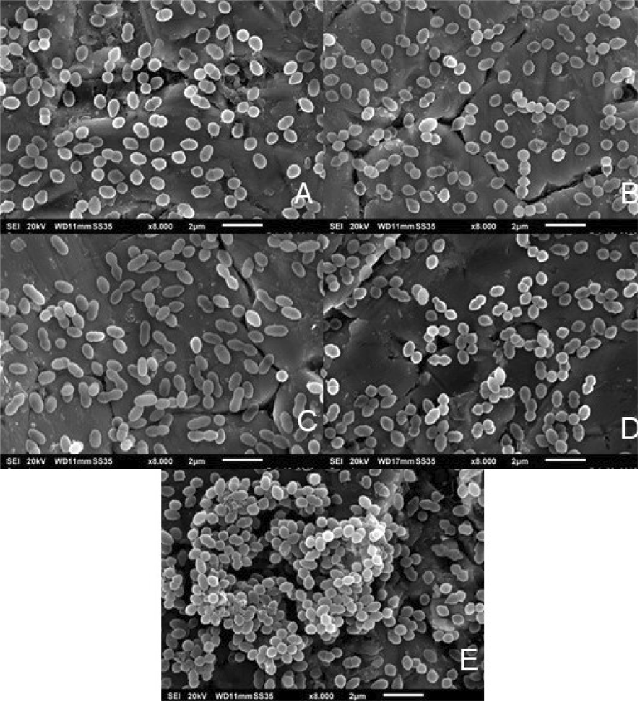
The analysis of the biofilm structure using SEM showed that all strains attached to the metal surface. From all five strains, only one strain isolated from the root canal without apical periodontitis showed a three-dimensional structure after 48 h (Figure 3–E). All the other strains, including the E. faecalis ATCC 29212, formed less massive biofilms with bacterial agglomeration distributed on the surface of the discs (Figure 3 – A to D).

Scanning Electron Microscopy (SEM) analysis of different strains of 48 h biofilms from E. faecalis. (A) E. faecalis ATCC 29212. (B and C) two E. faecalis strains were isolated from root canals with apical periodontitis, and (D and E) two E. faecalis strains were isolated from root canals without apical periodontitis. Magnification: (A, B, C, D, and E) x8.000. The samples were analyzed by SEM (JS-6010V, JEOL GmbH, Freising, Germany).
Expression of virulence genes analyzed by quantitative reverse transcription real-time PCR (RT-qPCR)
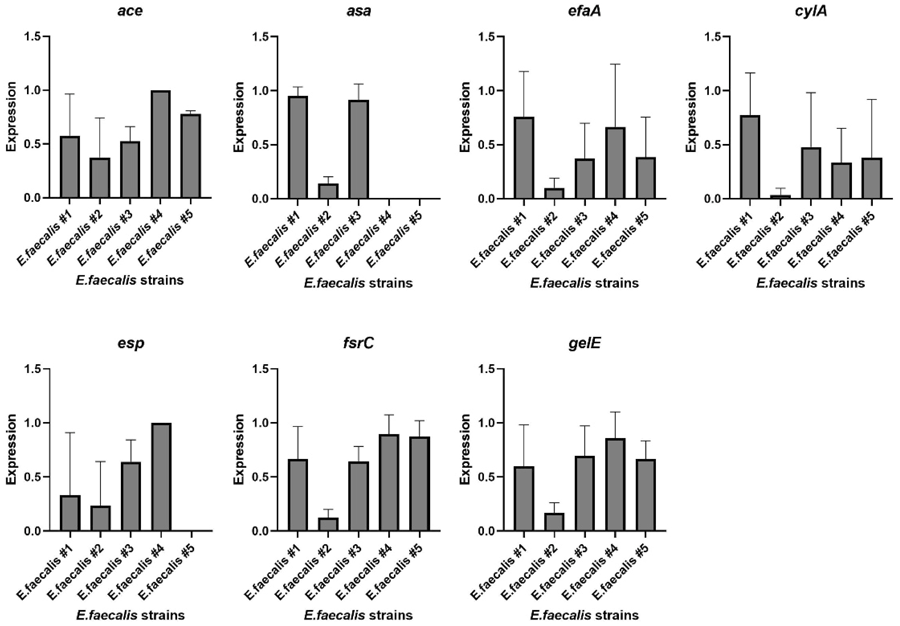
Figure 4 shows that the strains isolated from patients without apical periodontitis, corresponding to E. faecalis #4 and #5, showed no expression of the gene asa. Also, there was no expression of gene esp in E. faecalis #5. Strain #2, isolated from a tooth with evident apical periodontitis, showed low gene expression, especially when compared to the strain isolated from the same clinical case (strain #3).

Mean and standard deviation of virulence gene expression in different strains of 48-hour E. faecalis biofilms. (#1) E. faecalis ATCC 29212; (#2 and #3) two E. faecalis strains isolated from root canals with apical periodontitis; and (#4 and #5) two E. faecalis strains isolated from root canals without apical periodontitis. The experiment was done in duplicates and repeated three times.
No difference in the expression of the virulence genes ace, cylA, esp, fsrC, gelE, and efaA was observed between all the strains tested (p>0.05). Expression of the virulence gene asa revealed differences between E. faecalis ATCC 29212 and strain #4, and between strains #3 and #4 (p<0.05).
Discussion
In the present study, four clinical isolates from endodontically treated teeth with and without radiographic evidence of apical periodontitis were evaluated. The importance of comparing these E. faecalis isolated strains from different clinical situations is to elucidate the pathogenic virulence genes involved in the infection and their relation to the biofilm-forming ability.
All strains tested were able to form biofilms in vitro. However, the reference strain, E. faecalis ATCC 29212 showed less capacity for forming biofilms when compared to the clinical isolates. Similar results were previously described31,32, where the authors observed biofilm formation in different strains. A study33 detected biofilm formation in 73% of E. faecalis isolated from root-filled teeth with apical periodontitis, while another study34 found 47.2% biofilm formation in the clinical isolates.
The biofilm production of E. faecalis is related to the virulence genes for aggregation substances (agg and asa), endocarditis antigen (efa), and enterococcal surface protein (esp)16,20,22 Previous studies have shown that the aggregation substance gene (asa) enhances E. faecalis adherence to and invasion of eukaryotic cells15. In our study, strains carrying this gene were not detected in samples from teeth without apical periodontitis but were consistently present in those obtained from affected patients. The efa gene has been implicated in the adhesion of E. faecalis to cardiac cells during endocarditis15, while esp plays a key role in adherence and colonization of both host cells and abiotic surfaces15. In the present study, however, expression of efa and esp did not differ among the clinical isolates. Another investigation11 reported the presence of agg, gelE, and esp in all vancomycin-resistant E. faecalis strains. The agg gene, in particular, may contribute to biofilm formation, plasmid transfer, and adherence.
Gelatinase (gelE) is an extracellular metalloprotease, able to hydrolyze gelatin, collagen, and hemoglobin, which has also been reported to contribute to bacterial adherence and biofilm formation15,34. Another virulence gene that is associated with collagen-binding protein is ace, which seems to contribute to the adherence to dentin from the root canal35. Our study found no differences between the clinical isolates in these genes, suggesting that it is not directly related to biofilm formation, since all of the tested strains formed biofilm.
Biofilm quorum sensing is a communication between the microorganisms via autoinducers, controlling the behavior and gene expression to the density of the biofilm36,37. The gene histidine kinase (fsr) is related to the quorum sensing system for the upstream of gelE and the expression of fsr genes, which controls biofilm formation through the production of gelatinase. Inactivation of the fsr-controlled gene is related to preventing biofilm formation, which may be an alternative for therapeutic interventions in enterococcal infections38.
Cytolosin (cylA) is a potent bacteriocin that exacerbates enterococcal infections and can cause damage in dentin and periapical tissues via erythrocyte lysis and destruction of host cells15,33. In this study, all strains expressed this gene without difference between clinical isolates. A previous study found that 63.6% of isolates expressed this gene33, whereas another study reported that this gene was expressed in 38% of the samples31. Cytolysin has been shown on highly transmissible plasmids and within the chromosome39.
Stainless-steel discs were chosen because they provide a standardized, reproducible, and cost-effective surface for biofilm formation, and have been previously used in microbiological research for this purpose40,41. Moreover, the rigid and inert properties of stainless steel facilitate the mechanical detachment of biofilms for viable cell counting and enable high-resolution morphological analysis by scanning electron microscopy (SEM) without introducing imaging artifacts.
Our study showed a variation in the expression of virulence genes between all the E. faecalis strains. The strains isolated from root canals without apical periodontitis did not express asa. All strains isolated from patients with apical periodontitis expressed asa. Our data indicate that the expression of asa is associated with the development of apical periodontitis. Although other virulence genes such as efaA, ace, cylA, gelE, and fsrC were expressed across all strains, no significant differences in their expression levels were observed. This uniform expression pattern may reflect their basal roles in biofilm maintenance and general adaptation, rather than a specific correlation with disease severity. In addition, it should be highlighted that all the E. faecalis strains in this study showed biofilm-forming capacity. For a better understanding of the pathogenesis of apical periodontitis, further studies should be carried out, taking into consideration the asa expression and the biofilm formation in vivo.
In conclusion, all E. faecalis strains showed biofilm-forming capacity, but analysis of virulence genes showed the expression of asa only in strains isolated from patients with apical periodontitis.
Acknowledgements
We would like to thank Andrea Windisch and Stephan Steixner for their great technical support. We would like to thank Dr. Hyun (Michel) Koo for his valuable insights that greatly contributed to improving the quality of this study. This study was supported by Coordination for the Improvement of Higher Education Personnel (CAPES, financial code 001), The São Paulo Research Foundation (FAPESP 2015/23479-5; 2021/13871-6; 2017/25090-3) and Brazilian National Council for Scientific and Technological Development (CNPq 303852/2019-4, 421801/2021-2).
References
-
1 Gomes BPFA, Herrera DR. Etiologic role of root canal infection in apical periodontitis and its relationship with clinical symptomatology. Braz Oral Res. 2018 Oct;32(suppl 1):e69. doi: 10.1590/1807-3107bor-2018.vol32.0069.
» https://doi.org/10.1590/1807-3107bor-2018.vol32.0069 -
2 Siqueira JF Jr, Rôças IN. Present status and future directions: Microbiology of endodontic infections. Int Endod J. 2022 May;55 Suppl 3:512-530. doi: 10.1111/iej.13677.
» https://doi.org/10.1111/iej.13677 -
3 Delboni MG, Gomes BP, Francisco PA, Teixeira FB, Drake D. Diversity of Enterococcus faecalis genotypes from multiple oral sites associated with endodontic failure using repetitive sequence-based polymerase chain reaction and arbitrarily primed polymerase chain reaction. J Endod. 2017 Mar;43(3):377-82. doi: 10.1016/j.joen.2016.10.042.
» https://doi.org/10.1016/j.joen.2016.10.042 -
4 Prada I, Micó-Muñoz P, Giner-Lluesma T, Micó-Martínez P, Collado-Castellano N, Manzano-Saiz A. Influence of microbiology on endodontic failure. Literature review. Med Oral Patol Oral Cir Bucal. 2019 May;24(3):e364-72. doi: 10.4317/medoral.22907.
» https://doi.org/10.4317/medoral.22907 -
5 Figdor D, Davies JK, Sundqvist G. Starvation survival, growth and recovery of Enterococcus faecalis in human serum. Oral Microbiol Immunol. 2003 Aug;18(4):234-9. doi: 10.1034/j.1399-302x.2003.00072.x.
» https://doi.org/10.1034/j.1399-302x.2003.00072.x -
6 Francisco PA, Delboni MG, Lima AR, Xiao Y, Siqueira WL, Gomes BPFA. Proteomic profile of root canal contents in teeth with post-treatment endodontic disease. Int Endod J. 2019 Apr;52(4):451-60. doi: 10.1111/iej.13021. Epub 2018 Oct 28.
» https://doi.org/10.1111/iej.13021 -
7 Gomes BPFA, Francisco PA, Godoi EP Jr, Endo MS, Barbosa-Ribeiro M, Delboni MG, et al. Identification of culturable and nonculturable microorganisms, lipopolysaccharides, and lipoteichoic acids from root canals of teeth with endodontic failure. J Endod. 2021 Jul;47(7):1075-86. doi: 10.1016/j.joen.2021.04.011.
» https://doi.org/10.1016/j.joen.2021.04.011 -
8 Deng Z, Lin B, Liu F, Zhao W. Role of Enterococcus faecalis in refractory apical periodontitis: from pathogenicity to host cell response. J Oral Microbiol. 2023 Mar;15(1):2184924. doi: 10.1080/20002297.2023.2184924.
» https://doi.org/10.1080/20002297.2023.2184924 -
9 Alghamdi F, Shakir M. The influence of Enterococcus faecalis as a dental root canal pathogen on endodontic treatment: a systematic review. Cureus. 2020 Mar;12(3):e7257. doi: 10.7759/cureus.7257.
» https://doi.org/10.7759/cureus.7257 -
10 García-Solache M, Rice LB. The Enterococcus: a model of adaptability to its environment. Clin Microbiol Rev. 2019 Jan;32(2):e00058-18. doi: 10.1128/CMR.00058-18.
» https://doi.org/10.1128/CMR.00058-18 -
11 Lins RX, Hirata Junior R, Wilson M, O Lewis MA, Fidel RAS, Williams D. Comparison of genotypes, antimicrobial resistance and virulence profiles of oral and non oral Enterococcus faecalis from Brazil, Japan and the United Kingdom. J Dent. 2019 May;84:49-54. doi: 10.1016/j.jdent.2019.03.002.
» https://doi.org/10.1016/j.jdent.2019.03.002 -
12 Chávez De Paz LE, Dahlén G, Molander A, Möller A, Bergenholtz G. Bacteria recovered from teeth with apical periodontitis after antimicrobial endodontic treatment. Int Endod J. 2003 Jul;36(7):500-8. doi: 10.1046/j.1365-2591.2003.00686.x.
» https://doi.org/10.1046/j.1365-2591.2003.00686.x -
13 Kayaoglu G, Ørstavik D. Virulence factors of Enterococcus faecalis: relationship to endodontic disease. Crit Rev Oral Biol Med. 2004 Sep;15(5):308-20. doi: 10.1177/154411130401500506.
» https://doi.org/10.1177/154411130401500506 -
14 Manson JM, Hancock LE, Gilmore MS. Mechanism of chromosomal transfer of Enterococcus faecalis pathogenicity island, capsule, antimicrobial resistance, and other traits. Proc Natl Acad Sci U S A. 2010 Jul;107(27):12269-74. doi: 10.1073/pnas.1000139107.
» https://doi.org/10.1073/pnas.1000139107 -
15 Anderson AC, Jonas D, Huber I, Karygianni L, Wölber J, Hellwig E, et al. Enterococcus faecalis from Food, clinical specimens, and oral sites: prevalence of virulence factors in association with biofilm formation. Front Microbiol. 2016 Jan;6:1534. doi: 10.3389/fmicb.2015.01534.
» https://doi.org/10.3389/fmicb.2015.01534 -
16 Akbari Aghdam M, Soroush Barhaghi MH, Aghazadeh M, Jafari F, Beomide Hagh M, Haghdoost M, et al. Virulence genes in biofilm producer Enterococcus faecalis isolates from root canal infections. Cell Mol Biol (Noisy-le-grand). 2017 May;63(5):55-9. doi: 10.14715/cmb/2017.63.5.11.
» https://doi.org/10.14715/cmb/2017.63.5.11 -
17 Karygianni L, Ren Z, Koo H, Thurnheer T. Biofilm matrixome: extracellular components in structured microbial communities. Trends Microbiol. 2020 Aug;28(8):668-81. doi: 10.1016/j.tim.2020.03.016.
» https://doi.org/10.1016/j.tim.2020.03.016 -
18 Saeed K, McLaren AC, Schwarz EM, Antoci V, Arnold WV, Chen AF, et al. 2018 international consensus meeting on musculoskeletal infection: summary from the biofilm workgroup and consensus on biofilm related musculoskeletal infections. J Orthop Res. 2019 May;37(5):1007-17. doi: 10.1002/jor.24229.
» https://doi.org/10.1002/jor.24229 -
19 Neelakantan P, Romero M, Vera J, Daood U, Khan AU, Yan A, et al. Biofilms in endodontics-current status and future directions. Int J Mol Sci. 2017 Aug;18(8):1748. doi: 10.3390/ijms18081748.
» https://doi.org/10.3390/ijms18081748 -
20 Georges M, Odoyo E, Matano D, Tiria F, Kyany'a C, Mbwika D, et al. Determination of Enterococcus faecalis and Enterococcus faecium antimicrobial resistance and virulence factors and their association with clinical and demographic factors in Kenya. J Pathog. 2022 Nov;2022:3129439. doi: 10.1155/2022/3129439.
» https://doi.org/10.1155/2022/3129439 -
21 Martinho FC, Chiesa WM, Leite FR, Cirelli JA, Gomes BP. Correlation between clinical/radiographic features and inflammatory cytokine networks produced by macrophages stimulated with endodontic content. J Endod. 2012 Jun;38(6):740-5. doi: 10.1016/j.joen.2012.02.021.
» https://doi.org/10.1016/j.joen.2012.02.021 -
22 Chiniforush N, Pourhajibagher M, Parker S, Benedicenti S, Shahabi S, Bahador A. The effect of sublethal photodynamic therapy on the expression of Enterococcal surface protein (esp) encoding gene in Enterococcus faecalis: Quantitative real-time PCR assessment. Photodiagnosis Photodyn Ther. 2018 Dec;24:311-7. doi: 10.1016/j.pdpdt.2018.10.008.
» https://doi.org/10.1016/j.pdpdt.2018.10.008 -
23 Dahlén G, Pipattanagovit P, Rosling B, Möller AJ. A comparison of two transport media for saliva and subgingival samples. Oral Microbiol Immunol. 1993 Dec;8(6):375-82. doi: 10.1111/j.1399-302x.1993.tb00614.x.
» https://doi.org/10.1111/j.1399-302x.1993.tb00614.x -
24 Ribeiro AC, Matarazzo F, Faveri M, Zezell DM, Mayer MP. Exploring bacterial diversity of endodontic microbiota by cloning and sequencing 16S rRNA. J Endod. 2011 Jul;37(7):922-6. doi: 10.1016/j.joen.2011.04.007.
» https://doi.org/10.1016/j.joen.2011.04.007 -
25 Coraça-Huber DC, Fille M, Hausdorfer J, Pfaller K, Nogler M. Staphylococcus aureus biofilm formation and antibiotic susceptibility tests on polystyrene and metal surfaces. J Appl Microbiol. 2012 Jun;112(6):1235-43. doi: 10.1111/j.1365-2672.2012.05288.x.
» https://doi.org/10.1111/j.1365-2672.2012.05288.x -
26 Fan W, Huang Z, Fan B. Effects of prolonged exposure to moderate static magnetic field and its synergistic effects with alkaline pH on Enterococcus faecalis. Microb Pathog. 2018 Feb;115:117-22. doi: 10.1016/j.micpath.2017.12.028. Epub 2017 Dec 11.
» https://doi.org/10.1016/j.micpath.2017.12.028 -
27 Shepard BD, Gilmore MS. Differential expression of virulence-related genes in Enterococcus faecalis in response to biological cues in serum and urine. Infect Immun. 2002 Aug;70(8):4344-52. doi: 10.1128/IAI.70.8.4344-4352.2002.
» https://doi.org/10.1128/IAI.70.8.4344-4352.2002 -
28 Ran SJ, Jiang W, Zhu CL, Liang JP. Exploration of the mechanisms of biofilm formation by Enterococcus faecalis in glucose starvation environments. Aust Dent J. 2015 Jun;60(2):143-53. doi: 10.1111/adj.12324.
» https://doi.org/10.1111/adj.12324 -
29 Berube BJ, Sampedro GR, Otto M, Bubeck Wardenburg J. The psma locus regulates production of Staphylococcus aureus alpha-toxin during infection. Infect Immun. 2014 Aug;82(8):3350-8. doi: 10.1128/IAI.00089-14.
» https://doi.org/10.1128/IAI.00089-14 - 30 Osafo-Ampadu P, Gaikwad J. Detection of Panton-Valentine Leukocidin (PVL) genes within CA-MRSA carriers of the Oral Roberts University Community. Proc Oklahoma Acad Sci. 2014;94:96-103.
-
31 Sun J, Sundsfjord A, Song X. Enterococcus faecalis from patients with chronic periodontitis: virulence and antimicrobial resistance traits and determinants. Eur J Clin Microbiol Infect Dis. 2012 Mar;31(3):267-72. doi: 10.1007/s10096-011-1305-z. Epub 2011 Jun 10.
» https://doi.org/10.1007/s10096-011-1305-z -
32 Chávez de Paz LE, Davies JR, Bergenholtz G, Svensäter G. Strains of Enterococcus faecalis differ in their ability to coexist in biofilms with other root canal bacteria. Int Endod J. 2015 Oct;48(10):916-25. doi: 10.1111/iej.12501.
» https://doi.org/10.1111/iej.12501 -
33 Saffari F, Sobhanipoor MH, Shahravan A, Ahmadrajabi R. Virulence genes, antibiotic resistance and capsule locus polymorphisms in Enterococcus faecalis isolated from Canals of root-filled teeth with periapical lesions. Infect Chemother. 2018 Dec;50(4):340-5. doi: 10.3947/ic.2018.50.4.340.
» https://doi.org/10.3947/ic.2018.50.4.340 -
34 Zheng JX, Wu Y, Lin ZW, Pu ZY, Yao WM, Chen Z, et al. Characteristics of and Virulence Factors Associated with Biofilm Formation in Clinical Enterococcus faecalis Isolates in China. Front Microbiol. 2017 Nov;8:2338. doi: 10.3389/fmicb.2017.02338.
» https://doi.org/10.3389/fmicb.2017.02338 -
35 Hubble TS, Hatton JF, Nallapareddy SR, Murray BE, Gillespie MJ. Influence of Enterococcus faecalis proteases and the collagen-binding protein, Ace, on adhesion to dentin. Oral Microbiol Immunol. 2003 Apr;18(2):121-6. doi: 10.1034/j.1399-302x.2003.00059.x.
» https://doi.org/10.1034/j.1399-302x.2003.00059.x -
36 Muras A, Mallo N, Otero-Casal P, Pose-Rodríguez JM, Otero A. Quorum sensing systems as a new target to prevent biofilm-related oral diseases. Oral Dis. 2022 Mar;28(2):307-13. doi: 10.1111/odi.13689. Epub 2020 Nov 7.
» https://doi.org/10.1111/odi.13689 -
37 Ali IAA, Lévesque CM, Neelakantan P. Fsr quorum sensing system modulates the temporal development of Enterococcus faecalis biofilm matrix. Mol Oral Microbiol. 2022 Feb;37(1):22-30. doi: 10.1111/omi.12357. Epub 2021 Dec 15.
» https://doi.org/10.1111/omi.12357 -
38 Hancock LE, Perego M. The Enterococcus faecalis fsr two-component system controls biofilm development through production of gelatinase. J Bacteriol. 2004 Sep;186(17):5629-39. doi: 10.1128/JB.186.17.5629-5639.2004.
» https://doi.org/10.1128/JB.186.17.5629-5639.2004 -
39 McBride SM, Fischetti VA, Leblanc DJ, Moellering Jr RC, Gilmore MS. Genetic diversity among Enterococcus faecalis. PLoS One. 2007 Jul 4;2(7):e582. doi: 10.1371/journal.pone.0000582.
» https://doi.org/10.1371/journal.pone.0000582 -
40 Grimus V, Coraça-Huber DC, Steixner SJM, Nagl M. Activity of N-Chlorotaurine against long-term biofilms of bacteria and yeasts. Antibiotics (Basel). 2021 Jul;10(8):891. doi: 10.3390/antibiotics10080891.
» https://doi.org/10.3390/antibiotics10080891 - 41 Ammann CG, Fille M, Hausdorfer J, Nogler M, Nagl M, Coraça-Huber DC. Influence of poly-N-acetylglucosamine in the extracellular matrix on N-chlorotaurine mediated killing of Staphylococcus epidermidis New Microbiol. 2014 Jul;37(3):383-6.
-
Data availability:
Datasets related to this article will be available to the corresponding author upon request.
Edited by
-
Editor:
Dr. Altair A. Del Bel Cury
Datasets related to this article will be available to the corresponding author upon request.