Approach to tongue hyperpigmentation: connecting the dots with associated conditions in general practice
Abstract
Addressing hyperpigmentation in exposed areas of the body can be challenging in general practice and can raise a variety of questions, from possible serious illnesses to aesthetic concerns. Reviewing the case of a 21- year-old female, this study investigates concurrent benign hyperpigmentation of the tongue and conjunctiva while exploring potential links to iron and vitamin D deficiencies. The diagnostic journey involves a comprehensive evaluation, ruling out extrinsic causes and systemic illnesses for tongue hyperpigmentation, and discusses the approach leading to the diagnosis of Pigmented Fungiform Papillae (PFP) on the tongue and Complexion Associated Melanosis (CAM) in the conjunctiva. The article advocates for a cost-effective, minimally invasive approach in managing such cases in general medical practice.
Anemia, iron-deficiency; Melanosis; Glossitis; Conjunctival diseases; Hyperpigmentation
Introduction
Hyperpigmentation is a frequently encountered problem in general practice. Patients may express concerns about hyperpigmentation on prominent areas of their body for cosmetic reasons, or doctors may be concerned because it could indicate a serious illness, such as underlying cancer or systemic sickness. A range of disorders, from common, benign conditions to less common, secondary conditions with potential for malignancy, may be included in the differential diagnosis1. Pigmented Fungiform Papillae (PFP) is a rare idiopathic benign hyperpigmentation condition of the tongue2. Furthermore, Complexion Associated Melanosis (CAM) is another benign hyperpigmented disorder of the ocular conjunctiva3. The simultaneous occurrence of these two conditions is not well documented in the literature.
This report aims to present a case of a young woman, who was determined to have iron deficiency anemia and vitamin D deficiency at the same time that she presented with hyperpigmentation on her tongue and conjunctiva. Moreover, an approach for such patients presenting in general practice is suggested based on recent literature review.
Case Presentation
A 21-year-old woman noticed a hyperpigmented patch on the tongue, along with darkening of the bulbar conjunctiva bilaterally, developed gradually over the past 2-3 years. She denied any associated pain, itching, altered taste sensation, halitosis, or changes in the vision.
However, she experienced shortness of breath for the past 2 to 3 months, particularly with exertion, along with significant hair loss but denied any weight loss. Notably, she did not mention chest pain, palpitations, perspiration, oral ulcers, photosensitivity, joint pains, or alterations in menstrual patterns. Menstrual cycles were described within normal range for both duration and flow. There were no indications of pigmentation or rash on the body, urinary or bowel complaints, or fever.
The patient’s past medical history revealed a previous episode of anemia a few years ago; however, specific details (exact time, hemoglobin levels, and treatment) were not recalled. The family history included Hypertension and Diabetes Mellitus, with no reported instances of hyperpigmentation disorders in the family.
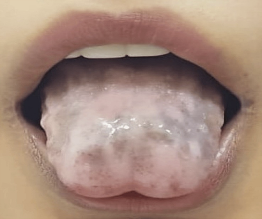
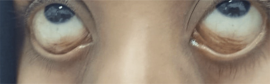
On general examination, she exhibited pallor, with the following vitals: a blood pressure reading of 133/87 mmHg (negative for orthostatic hypotension) and a pulse rate of 98 beats per minute. The oral examination revealed black-pigmented, well-defined, asymmetric patches on the dorsum of the tongue, sparing the rest of the oral mucosa. Ocular examination revealed bilateral brown flat lesions with ill-defined margins on the bulbar conjunctiva (Figure 1, 2). The rest of the systemic review was unremarkable.
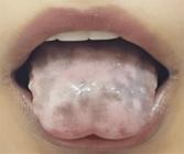
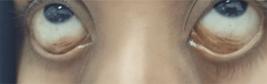
Based on her history and examination findings, following tests were done.
Based on the history, examination, and workup, our assessment included microcytic anemia due to iron deficiency, vitamin D deficiency, with a query regarding the cause of hyperpigmentation of the tongue and bulbar conjunctiva. Treatment with an iron-rich diet and iron supplements was initiated, and vitamin D was replaced. The simultaneous occurrence of unusual hyperpigmentation of the tongue and conjunctiva, along with the concerns expressed by the patient and family, intrigued the authors to explore other possible differentials and causes. The literature was reviewed, and expert opinions were sought. Based on the high likelihood of the benign nature of most hyperpigmentations, a watchful waiting approach was adopted to observe any changes, progression of these lesions, or the onset of new symptoms.
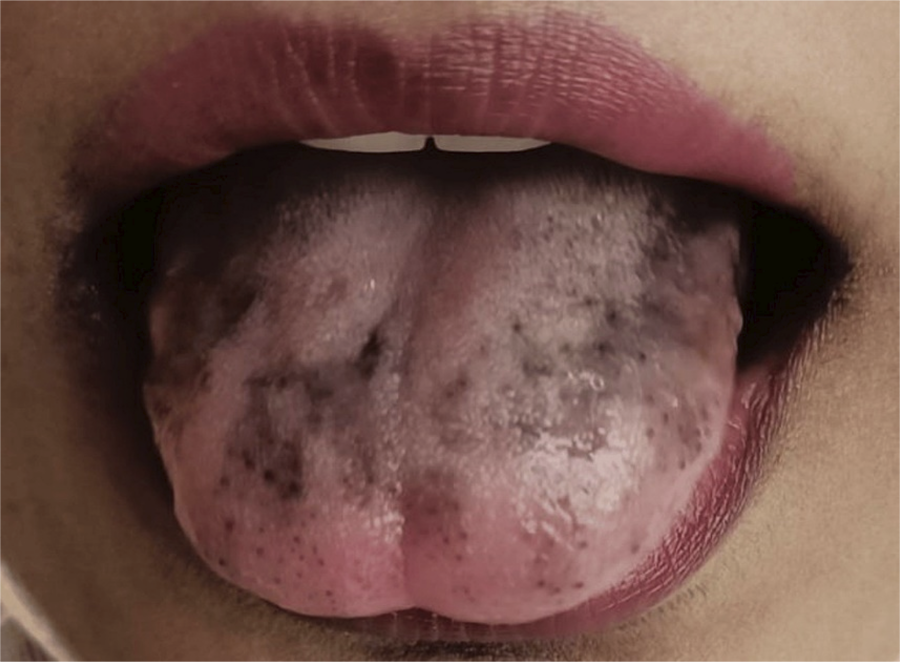
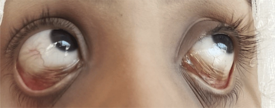
Final follow-up was conducted at 4 months. Her anemia had significantly improved (Hb-11.3 g/dl, MCV-71 fl), along with other symptoms of anemia. However, her tongue and bulbar conjunctival hyperpigmentation did not improve; in fact, the darkening of tongue papillae became mildly prominent, giving the impression.
of Pigmented Fungiform Papillae (PFP) of the tongue. A clinical diagnosis of Complexion Associated Melanosis (CAM) was made for the conjunctival hyperpigmentation (Figure 3,4).
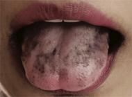
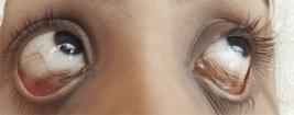
Bilateral Hyperpigmentation of Bulbar Conjunctiva (CAM) on 4 Months Follow-up Complexion Associated Melanosis
Discussion
Many hyperpigmented lesions can be clinically diagnosed based on size, shape, color, and duration, in addition to clinical information. Disorders causing hyperpigmentation can be acquired or inherited. When it comes to acquired hyperpigmentation of tongue, the cause can be either intrinsic/physiologic4,5 or systemic e.g., Addison’s illness, Peutz-Jhegars syndrome, familial Laugier-Hunziker syndrome, etc.1,6.
Increased and reversible production of tongue melanosis has been linked to a number of extrinsic and reversible triggers, such as mechanical injury, medication, radiation, and substance use7-12. A thorough medical history is necessary to ascertain whether acquired tongue hyperpigmentations are the consequence of an external factor. Confirmation of systemic reasons can be accomplished via history, examinations, and investigations8. Once extrinsic reasons and systemic conditions have been ruled out, the option to undergo a biopsy to rule out malignancy can be assessed against the risk of malignancy1,4-5.
In our case, the hyperpigmentation of both tongue and conjunctiva were found to be acquired, irreversible and unrelated to any substance, medication or trauma, ruling out extrinsic causes. Based on the absence of typical symptoms, negative similar family history and lack of characteristic darkening of other parts of the body (such as acanthosis nigricans in Addisons, sparing of lips and remaining oral mucosa in Peutz-Jhegars syndrome) and negative investigations, possible systemic illnesses were ruled out 6,13.
Literature review suggests that the most hyperpigmentation disorders are benign, more common in dark skinned, including Asian population, and can be diagnosed alone on clinical grounds4,5. Given that the anterior and lateral borders of the tongue exhibited hyperpigmented patches along with prominent pointed papules, the appearance more closely resembled with pigmented fungiform papillae (PFP)hence a clinical diagnosis of PFP was established2,14-16. According to the literature, deferring tongue biopsy with 6 months to yearly surveillance is a more cost-effective and preferred approach that can be considered in low risk patients.
Complexion-associated melanosis (CAM), a benign epithelial conjunctival melanosis with a negligibly low risk of malignancy, is the ascertained probable cause for the pigmentation in the eyes due to its characteristic appearance, bilateral distribution and benign course3. PFP and CAM are both melanocytic hyperpigmented lesions, in which the presence of hyperpigmented melanocytes and melanophages has been proposed as a possible histological feature.
The authors searched for a possible explanation or correlation among conjunctival melanosis (CAM) and tongue hyperpigmentation (including PFP), anemia and vitamin D deficiency. It was found that three out of these four conditions (apart from vitamin D deficiency) had been observed and recorded in a pediatric patient in India16. Similar to our case, a recent review article on PFP stated that most cases have been seen in females, primarily under 40 years of age2,14.
Although PFP and CAM are benign physiologic hyperpigmentation conditions that often do not have any special adverse effects on the patient, the precise cause of the physiologic hyperpigmentation on multiple areas at the same time remains unknown. Clinicians should consider and familiarize themselves with these conditions in order to encourage a cost-effective strategy, avoid unnecessary intrusive procedures, and minimize the negative impacts on patients’ perceptions.
In conclusion, a diagnosis of physiological hyperpigmentation, such as PFP and CAM, can be considered on clinical grounds with a watchful waiting approach in cases with hyperpigmentation of the tongue and bulbar conjunctiva, provided that other extrinsic and systemic causes are ruled out based on comprehensive evaluation. It is suggested to carry out additional research for any connections with additional clinical characteristics including anemia and vitamin D deficiency.
Acknowledgements
Acknowledging Esteemed Colleagues for Valuable Consultation on this Case: Dr.Areej Riaz, Senior Clinical Fellow, Russells Hall Hospital, Dudley, UK; Dr.Hamdan Ahmed Pasha, Assistant Professor, Section of Otolaryngology, Head and Neck Surgery, Department of Surgery, Aga Khan University Hospital, Pakistan.
References
-
1 Feller L, Masilana A, Khammissa RA, Altini M, Jadwat Y, Lemmer J. Melanin: the biophysiology of oral melanocytes and physiological oral pigmentation. Head Face Med. 2014 Mar;10:8. doi: 10.1186/1746-160X-10-8.
» https://doi.org/10.1186/1746-160X-10-8 -
2 Surboyo MDC, Samaranayake L, Santosh ABR, Ayuningtyas NF, Wati SM, Rahayu RP, et. al. Pigmented Fungiform Papillae (PFP) of the tongue: a systematic review of current aetiopathogenesis and pathophysiology. Pathophysiology. 2022 Sep;29(3):555-69. doi: 10.3390/pathophysiology29030043.
» https://doi.org/10.3390/pathophysiology29030043 -
3 Alqahtani JM: Conjunctival melanosis: review of the literature. Bahrain Medical Bull. 2013;35(4)1:206-10. doi: 10.12816/0004449.
» https://doi.org/10.12816/0004449 -
4 Chandra S, Keluskar V, Bagewadi A, Sah K. Extensive physiologic melanin pigmentation on the tongue: an unusual clinical presentation. Contemp Clin Dent. 2010 Jul;1(3):204-6. doi: 10.4103/0976-237X.73205.
» https://doi.org/10.4103/0976-237X.73205 -
5 Neiva-Sousa M, Martins M, Bitoque S, Doutel D, Gomes P. Unusual extensive physiologic pigmentation of the tongue: a case report. Cureus. 2022 Jul;14(7):e26767. doi: 10.7759/cureus.26767.
» https://doi.org/10.7759/cureus.26767 - 6 Studdiford J, Seecof OM, Trayes KP, Valko G. Hyperpigmentation of the tongue and systemic symptoms. Am Fam Physician. 2020 Apr;101(8):499-501.
-
7 Sangalli L, Miller CS: Walnut consumption as a cause of black tongue: A case report Journal of Clinical Images and Medical Case Reports. J Clin Images Med Case Rep. 2022 Aug;3(8):1992. doi: 10.52768/2766-7820/1992.
» https://doi.org/10.52768/2766-7820/1992 -
8 Kato M, Kobayashi T, Suzuki H. Case of moxifloxacin-induced black hairy tongue. Am J Case Rep. 2022 Apr;23:e936235. doi: 10.12659/AJCR.936235.
» https://doi.org/10.12659/AJCR.936235 -
9 Gil LM, Marques J, Salgado D. Tongue hyperpigmentation associated with temozolomide as a single agent: case report. Acta Med Port. 2022 Sep;35(9):672-4. doi: 10.20344/amp.15987.
» https://doi.org/10.20344/amp.15987 -
10 Zhao S, Fan L, Feng J, Ma P. Reversible black tongue: a little known side effect of imipenem/cilastatin and evidence for novel mode of action. J Clin Pharm Ther. 2020 Apr;45(2):370-2. doi: 10.1111/jcpt.13066. Epub 2019 Oct 17.
» https://doi.org/10.1111/jcpt.13066 -
11 Sow ML, Moukafih B, Balde S, Elkhoyaali S, Chaibi A, Abahssain H,et al. Vinorelbine and carboplatin-induced black tongue: a case report. Therapie. 2020 May-Jun;75(3):297-301. doi: 10.1016/j.therap.2019.05.006. Epub 2019 Jul 10.
» https://doi.org/10.1016/j.therap.2019.05.006 - 12 Stringer LL, Zitella L. Hyperpigmentation of the tongue. J Adv Pract Oncol. 2014 Jan;5(1):71-2.
-
13 Karwan M, Brzezinski P: Laugier-Hunziker syndrome-a rare cause of acquired hyperpigmentation. Przegl Dermatol. 2022;109:52-7. doi: 10.5114/dr.2022.116733.
» https://doi.org/10.5114/dr.2022.116733 -
14 Lee J, Lee JS, Park SM, Shin K, Ko HC, Kim BS, et al. Pigmented fungiform papillae of the tongue: a single-center experience and review of literature. Ann Dermatol. 2023 Aug;35(4):266-74. doi: 10.5021/ad.22.192.
» https://doi.org/10.5021/ad.22.192 -
15 Surboyo MDC, Ernawati DS, Parmadiati AE, Marsetyo RI. Pigmented fungiform papillae of the tongue and lingual fimbriae as single presentation in adult: a case report and literature review. Eur J Dent. 2020 Oct;14(4):702-6. doi: 10.1055/s-0040-1714041.
» https://doi.org/10.1055/s-0040-1714041 -
16 Sharma S, Sharma S. Pigmentation of the fungiform papillae of the tongue in a child secondary to iron deficiency anaemia: an uncommon occurrence and association. Int J Oral Health Dent. 2016;2:39-42. doi: 10.5958/2395-499X.2016.00005.8.
» https://doi.org/10.5958/2395-499X.2016.00005.8
-
Data availability:
Datasets related to this article will be available to the corresponding author upon request.
-
Disclosures:
Human subjects: I hereby confirm that this case report has received approval from the Head of the Department. Additionally, verbal consent was obtained from the involved patient prior to the submission of the research article for publication. It is important to note that the article does not contain any identifying information, either in the text or images, related to the patients. The research adheres to ethical standards, and all necessary precautions have been taken to protect the privacy and confidentiality of the individuals involved. AI tool (Chat GPT) was used as writing assistance only.
-
Funding sources:
None declared.
Edited by
-
Editor:
Dr. Altair A. Del Bel Cury
Datasets related to this article will be available to the corresponding author upon request.