Morphological and trabecular structure study in oncologic individuals in use of antiresorptive medication
Abstract
Aim Evaluate changes in and the complexity of the bone trabeculae in individuals with cancer using or not using antiresorptive drugs through the panoramic morphometric indices and fractal dimension.
Methods Clinical records and panoramic radiographs of individuals with cancer were analyzed. The risk group (RG) consisted of 22 individuals using zoledronic acid or denosumab for a minimum period of 12 months. The control group (CG) consisted of 26 individuals without any antiresorptive drugs. Linear measurements were performed for the panoramic radiomorphometric indices and the fractal dimension in four different regions of interest (ROI).
Results In RG it was observed that 72.7% of the patients were using zoledronic acid, while 27.3% were using denosumab. In the fractal dimension analysis, the ROIs of the mandibular angle (p = 0.000) and premolar region (p = 0.005) of the RG showed significant changes in bone complexity when compared to the CG. In the analysis of panoramic radiomorphometric indices, the mental index showed a significant increase in linear measurements of the RG compared to the CG (p = 0.008).
Conclusion The fractal dimension analysis of the mandibular angle and premolar regions in the RG indicated greater bone complexity compared to the CG.
Keywords
Fractals; Radiography, panoramic; Mandible; Osteonecrosis
Introduction
Antiresorptive drugs such as zoledronic acid and denosumab are used as supportive treatments for individuals diagnosed with cancer and bone metastases to prevent hypercalcemia and skeletal-related events due to metastatic bone disease1.
The bone remodeling process is impacted by the administration of antiresorptive drugs, with an inhibition of both the cellular differentiation of osteoclast precursor cells and the performance of differentiated osteoclasts, in addition to stimulating their apoptosis2.
Osteoprotective therapies have become an essential component in solid tumor management, where the bone is expected to be a site of metastasis. However, long-term use of zoledronic acid and denosumab increases the risk of compromised bone quality due to excessive bone resorption and reduced angiogenesis3. In this context, medication-related osteonecrosis of the jaw (MRONJ) is the most clinically impacting adverse complication reported from these medications2.
Asymptomatic patients on current or previous treatment with antiresorptive drugs are considered to be at risk for MRONJ3. Therefore, new approaches are needed to detect specific radiographic signs that are incipient in more routinely requested exams, such as panoramic radiography, which can be characterized as markers and predictors of changes in bone architecture.
To date, the panoramic radiomorphometric indices have been suggested as a choice to assess jaw mineral density changes in osteoporotic4, diabetes mellitus5, secondary hyperparathyroidism6 and celiac patients7.
Another method that can be used in the analysis of trabecular bone changes is the fractal dimension8-10. Given that trabecular bone exhibits fractal characteristics, such as self-similarity and a branching structure without a well-defined gap, fractal dimension analysis is appropriate for patients undergoing treatments that affect bone mineral density9.
The determination of early panoramic radiography changes in the bone architecture of cancer patients using antiresorptive drugs can help health professionals in the planning, treatment, and management of these cases to follow the evolutionary and healing process of MRONJ. Therefore, the present study aimed to evaluate the bone architecture of cancer patients using antiresorptive drugs and without a clinical diagnosis of MRONJ through radiomorphometric indices and fractal dimension in panoramic radiographs, which we hypothesized that there are already alterations in the bone architecture of these patients even without the established clinical diagnosis.
Materials and methods
The current study was approved by the Research Ethics Committee of the Federal University of Bahia (n°10477819.0.0000.024). The data were collected using a convenience sampling method composed of panoramic radiographs of cancer patients referred to a private cancer treatment clinic in Salvador, Bahia, Brazil.
Sample selection
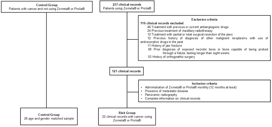
This was a retrospective study that included 237 clinical records of cancer patients initially treated with Zometa® (4 mg zoledronic acid – anhydrous, corresponding to 4.264 mg of zoledronic acid monohydrate; Novartis) or Prolia® (60 mg of denosumab and excipients, Amgen) during the period from 2019 to 2021.
After the initial refinement, based on the exclusion criteria, a second evaluation was performed using the inclusion criteria: monthly administration of Zometa® 4 mg intravenous or subcutaneous Prolia® 120 mg for at least 12 months, presence of metastatic disease, the existence of panoramic radiography of each patient after a minimum period of 12 months of use of the antiresorptive drugs, performed in the same imaging clinic and the presence of relevant information such as age, gender, diagnosis, family cancer history, current use of corticosteroids, type of antiresorptive drugs, dosage and number of cycles, as well as habits related to smoking and alcoholism and possible deaths and their causes. A flowchart illustrating the study participants is presented in Figure 1.

Panoramic radiographs
Panoramic radiography obtained using the digital technique were performed at the same imaging clinic, acquired using the CS K8000 panoramic device (Carestream Dental, Germany), operating at 76 kV and 10 mA, with an exposure time of 12.3 seconds. They had to meet the technical quality criteria to avoid image distortion (the patient’s position was the Frankfort horizontal plane parallel to the horizontal plane and the midsagittal plane perpendicular to the horizontal plane) and to provide full visualization of the mandibular cortical bone. The panoramic radiography used in the present study did not show any lesions or systemic pathologies that interfere with the analyses that justify their exclusion from the sample.
Examiner calibration
The examiner’s calibration for the evaluation of fractal dimension and panoramic radiomorphometric indices in panoramic radiography was performed before the beginning of the analysis of the present study in 20 panoramic radiography not belonging to the sample, selected at random and from the same imaging clinic. After fifteen days, the analyses were repeated by the same examiner. The intraclass correlation coefficient (ICC) was applied to quantitative variables, and the kappa test was used for qualitative variables. The ICC values indicated excellent intraexaminer reproducibility for fractal dimension (1.0) and panoramic radiomorphometric indices (0.968), as well as the values of the weighted kappa test for the qualitative analysis of the Mandibular cortical index (1.0).
Evaluation of panoramic radiomorphometric indices
Linear measurements for panoramic radiomorphometric indices were made in millimeters (mm) on all panoramic radiography using RadioImp® Software (Radio Memory Ltd., Belo Horizonte, MG, Brazil). The evaluated panoramic radiomorphometric indices can be seen in Figure 2.

a) Panoramic radiograph with the measuring method used for the Panoramic mandibular index (PMI), mental index (MI), antegonial index (AI), goniac index (GI), antegonial notch depth (AND); b) Cropped panoramic radiographs showing the C1, C2, and C3 classification of the mandibular cortical index
Mandibular cortical index (MCI)
The inferior mandibular cortical morphology was categorized into three groups: (C1) normal cortex, when the endosteal margin of the mandibular cortex is uniform and tender on both sides; (C2) moderately eroded cortex, when the endosteal margin shows semilunar defects (lacunar resorptions) or appears to form endosteal cortical residues; and (C3), severely eroded or porous cortex, when the mandibular cortical layers include dense endosteal cortical residues and are clearly porous11.
Panoramic Mandibular Index (PMI)
This index was characterized by the ratio of the mandibular cortex thickness to the distance between the inferior portion of the mental foramen and the mandibular cortex12.
Mental Index (MI)
The thickness of the inferior cortex of the mandible was measured in the region of the mental foramen. After identifying the mental foramen, a line perpendicular to the tangent of the lower border of the mandible was drawn and measured13.
Goniac Index (GI)
Measurement of the mandibular cortex in the region of the bisector angle formed by two lines tangent to the base and the mandibular ramus14.
Antegonial Index (AI)
It is characterized by providing a measure of cortical thickness at the location defined by a line extending from the anterior edge of the ascending ramus to the lower border of the mandible15.
Antegonial Notch Depth (AND)
It´s represented by a line tangent to the body and ramus of the mandible with the measurement of the most posterior region of the antegonial notch16.
Fractal Dimension Evaluation
The fractal dimension analysis was performed using box-counting17 and the images were analyzed using ImageJ software, version 1.3 (National Institutes of Health, Bethesda, MD, USA) and saved in JPEG format. Periapical and periodontal sites were not used to avoid misinterpretation caused by inflammatory changes. The regions of interest (ROIs) were selected as follows: region 1: subcortical area in the condyle; region 2: area above the supracortical extent of the angle of the mandible; region 3: area above the mandibular canal on the distal side of the second premolar; and region 4: area anterior to the mental foramen18.
These four ROIs were selected bilaterally, each corresponding to an area of 40 x 40 pixels7. Each ROI was selected from the original image and duplicated; duplicate images were blurred with a Gaussian filter (sigma-35), used to remove brightness variations due to smooth tissue overlap and varying bone thickness; the resulting image was subtracted from the original image of the ROI; a gray value of 128 was added to the subtraction result and transformed into a binary image, where bone marrow and trabeculae spaces were discriminated from each other; the image was then eroded, dilated and finally skeletonized for fractal dimension (Figure 3).

Panoramic radiograph showing a fractal dimension in a selected region of interest. a) Panoramic radiograph. b) Selected ROI. c) Blurred image with Gaussian filter. d) Subtraction of region of interest from the original image. e) Addition of 128 gray value to each pixel location. f) Binarization. g) Erosion. h) Dilatation. i) Skeletonization.
Statistical Analysis
SPSS 10.0 Software (SPSS Inc., Chicago, IL, USA) was used for data analysis. The chi-square test was used to compare demographic variables (age, sex, diagnosis, disease staging, family cancer history, smoking, alcohol, corticosteroid use, number of missing teeth and frequency of death) and evaluation of the Mandibular cortical index. Student’s T-test was used to compare the groups for the analysis of fractal dimension and panoramic radiomorphometric indices, adopting a significance level of 5%.
Results
The final sample consisted of 22 clinical records of individuals diagnosed with cancer, using Zometa® 4 mg or Prolia® 120 mg monthly, by intravenous and subcutaneous administration, respectively, for at least 12 months, considered as a risk group (RG), and 26 clinical records of individuals diagnosed with cancer, but without current or previous antiresorptive drugs, characterized as a control group (CG), totaling 48 patients.
The mean age of the sample was 68 years. In the RG, 17 (77.2%) were women, and 50% were diagnosed with breast cancer. In both groups, smoking was a commonly denied habit, with 20 (77%) and 13 (59%) nonsmoking patients in the CG and RG, respectively. In addition to tobacco consumption, alcohol consumption was also evaluated, with 10 (38.5%) patients from the CG and 8 (36.4%) patients from the RG who reported social consumption.
The average number of missing teeth in the mandible was three dental units, with 14/22 (63.6%) patients in the RG having more than four missing teeth. Regarding the frequency of death, only 1/26 patients in the CG died, and the cause was cancer. In the RG, 8/22 (36.4%) patients died, and all died due the underlying disease (Table 1).
When the variables related to the use of antiresorptive drugs in the RG were analyzed, such as the type of drug used and the number of cycles, 72.7% of the patients used zoledronic acid, while 27.3% used denosumab. The mean number of antiresorptive drugs cycles was 24 ± 8.39, with a mean of months of drug use of 26 ± 10.32 (shortest time of use of 12 months and greater than 52 months).
When comparing the ROIs in the analysis of fractal dimension in CG and RG, the ROIs that showed significant differences were the mandible angle (p = 0.000) and premolar region (p = 0.005) (Table 2).
Regarding the mandibular cortical morphology, evaluated qualitatively through the Mandibular cortical index, it was possible to assess that there was no statistically significant difference between the CG and RG according to the classifications C1, C2 and C3 (p = 0.643). In CG, similarly to the RG, most patients were classified as C2, representing 40 patients (76.9%) and 36 patients (82%), respectively. Four (9%) patients from the RG and 4 (7.7%) from the CG were classified as C1; as C3, 4 patients (9%) were observed in the RG, and 8 patients (15.4%) belonged to the CG.
When analyzing the panoramic radiomorphometric indices, the only statistically significant index between the CG and RG was the Mental index (p = 0.008). However, in the other indices, even though there was no statistically significant difference, the results showed an increase in the mean, minimum and maximum values of RG compared to the CG, except for Antegonial Notch Depth (Table 3).
Discussion
Panoramic radiomorphometric indices and fractal dimension are image analysis tools capable of evaluating the architecture and bone complexity of the maxillary bones. Compared to other bones in the human body, the jaws have a higher concentration of these antiresorptive drugs, justified by the fact that they are deposited in areas with good blood supply and increased cell turnover. Considering that cancer patients using antiresorptive drugs are already classified as being at risk for the development of MRONJ3, it is relevant to study new signs of incipient changes in the trabecular bone to be able to detect, in an increasingly precocious and accurate way, changes in the bone structure of the jaws, since most of the analyses performed include the disease already installed clinically and in more advanced stages19-21.
The present study opted to use panoramic radiography, as it is the most frequently requested imaging test in the initial dental consultation and is routinely used to establish diagnosis and treatment planning22. It is an easy-to-access examination, which makes it possible to perform different measurements on the right and left sides23, in addition to having lower cost and radiation exposure compared to computed tomography21.
To our knowledge, to date, there have been few studies that have simultaneously addressed panoramic radiomorphometric indices and the fractal dimension in the assessment of the bone structure of the jaws and, especially, in cases of osteoporosis24-26 and bone diseases such as osteogenesis imperfecta8.
Considering that the trabecular bone can be characterized as a complex structure8,9, through the selection of ROIs in panoramic radiography, it becomes possible to acquire the box-counting value, the most used method.
In the present study, the ROIs of the mandible angle (p = 0.000) and the premolar region (p = 0.005) corroborate the idea of de Molon et al.10, since in the RG, there was an increase in local bone complexity when compared to the CG. However, in general, for the other ROIs evaluated, there was no significant difference in the mean fractal dimension between the groups. Despite the significant difference in the mandible angle region, we believe that this result may correlate with the insertion of the masseter muscle and the possible excessive masticatory force or stress associated with the density and quality of the bone mineral structure in this region.
Demiralp et al.18 evaluated panoramic radiography of patients diagnosed with metastatic cancer using intravenous bisphosphonates, with a mean administration period of 28.2 ± 16.1 months. Although the authors did not observe any statistically significant difference in the same ROIs of the present study, it can be noted that the study group had a higher mean FD (1.39 ± 0.14) than the control group (1.38 ± 0.07).
When comparing the fractal dimension values of the CG and RG of the present study, without distinction of ROIs, the means obtained and standard deviation (SD) were 1.689570 ± 0.0005524 and 1.689591 ± 0.0005615, respectively (p = 0.700), which suggests an increase in bone complexity in patients using antiresorptive drugs, although without a statistically significant difference. The fractal dimension was also analyzed independently for each ROI to consider variations in bone density at each ROI location. For the different ROIs analyzed, the use of antiresorptive drugs for an average period of 26 ± 10.32 months resulted in changes in the complexity of the mandibular bone structure in the ROIs of the mandible angle and premolar region. Due to the existence of studies that demonstrated the presence of specific radiographic alterations in patients with MRONJ24-26, we believe that the analysis of fractal dimension in patients considered at risk is relevant, especially in the mandible, given the approximately 100 times greater occurrence of MRONJ in the alveolar bone than in other skeletal sitestorres20,27,28.
In the present study, regarding the panoramic radiomorphometric indices, their replicability was observed on both sides, since there was no significant difference between the sides of the RG or CG. The only index that showed a significant difference between the groups was the Mental index (p = 0.008), although all other measures, except for Antegonial Notch Depth, showed increased values in the RG when compared to the CG.
The Mental index is considered the most well-established for evaluating signs of resorption on panoramic radiography to identify osteopenia and osteoporosis, and previous studies have found significant relationships between Mental index and bone mineral density29. In a study carried out by Hastar et al.29, 487 panoramic radiographies of patients diagnosed with osteoporosis were evaluated using the Mental index, Mandibular cortical index and Panoramic Mandibular Index, and the authors observed significant differences between the study group and the control group regarding the Mental index and Panoramic Mandibular Index. Therefore, we believe that the significant difference in the Mental index in the present study is a possible predictor for assessing patients at risk for MRONJ.
In the present study, the Panoramic Mandibular Index values were also higher in the RG, although with no statistically significant difference. Corroborating this finding, Wazzan et al.30 evaluated panoramic radiography changes in the mandibular bone in patients diagnosed with multiple myeloma and did not observe significant differences in Panoramic Mandibular Index. However, the authors observed a direct relationship between MRONJ and the thickness and sharpness of the mandibular cortical border (p = 0.0003) using the Mandibular cortical index. In the present study, this difference was not observed, with 36 patients from the RG (82%) and 40 patients from the CG (76.9%) being considered as a C2 classification. This difference between the studies is probably the result of a homogeneous sample concerning age (p=0.226) in the present study, as well as the fact that these patients are considered only at risk, without the disease clinically installed.
Kubo et al.22 revealed a possible significant association between the definition of Mandibular cortical index categorized as C2 and the use of antiresorptive drugs. In turn, the side affected by osteonecrosis was significantly higher than that in the control group. These results suggest that C2 categorization may not necessarily indicate the presence of a bone defect as in the original definition but that the use of bisphosphonates could cause additional bone formation in the thinner cortex, resulting in the roughness of the endosteal margin, which would be evaluated as C2. The authors concluded that a mandible classified as C2, when it becomes inflamed, has a higher frequency for the development of MRONJ; that is, the Mandibular cortical index classified as C2 should be listed as a possible factor that leads to the development of MRONJ.
In a study carried out by Ozcan et al.19, significant differences were found in the Mandibular cortical index between the left and right sides of the MRONJ group (p = 0.014) and between the side compromised with osteonecrosis and the control group (p<0.0001). Perhaps the fact that the authors chose cone-beam computed tomography to assess the Mandibular cortical index is one of the reasons for the difference observed in comparison with the present study, in addition to the fact that patients already had installed osteonecrosis. Furthermore, the authors evaluated, through panoramic radiography, the condylar angle (CA), gonial angle (GA), antegonial angle (AA) and antegonial depth (AD); however, no statistically significant differences were observed in this analysis of two-dimensional images. These findings corroborate the present study, which evaluated the Goniac Index, Antegonial Index and Antegonial Notch Depth, and it was also not possible to observe significant differences. We believe that these results may suggest that antiresorptive drugs are able to influence internal morphological changes in the mandible. However, these changes were not reflected in values measured in the panoramic radiomorphometric indices under the mandibular external morphology.
In the present study, Antegonial Notch Depth was also evaluated, as it is known that due to remodeling of the antegonial region, while bone resorption is seen along the lower margin of the mandible, deposition is seen on the anterior side of the antegonial area31. Ali et al.32 stated that problems in condylar growth are conducted through the masseter and medial pterygoid muscles to the lower part of the mandible, thus forming the Antegonial Notch Depth. In the present study, the mean Antegonial Notch Depth in the CG was higher than that in the RG, although there was no significant difference. This result is like the work by Busra et al.23, who also evaluated Antegonial Notch Depth in patients using bisphosphonates and found statistically significantly higher values in the control group than in the study group (p < 0.05).
The main limitation of this study was due to the heterogeneity of the population evaluated in terms of the primary site of the disease and the number of different drug cycles. However, the results provide essential trigger points for future research, especially regarding the clinical evaluation of cancer patients concerning harmful oral health conditions and habits, as well as investigation of changes in the trabecular bone of these patients, mainly in the Mental index region, through cone-beam computed tomography, for the possible establishment of panoramic radiomorphometric indices predictor of MRONJ and the use of the fractal dimension as a routine tool in patients who use antiresorptive drugs, with the primary focus on the mandibular angle and premolar regions.
In conclusion, our results showed that in the assessment of fractal dimension, the ROIs of the mandible angle and premolar region of the group of patients at risk showed greater bone complexity when compared to patients in the control group. The measurement of the Mental index through panoramic radiomorphometric indices, together with assessments of fractal dimension, are maybe possible predictors of incipient changes in mandibular trabecular bone in cancer patients using zoledronic acid and denosumab.
References
-
1 Jakob T, Tesfamariam YM, Macherey S, Kuhr K, Adams A, Monsef I, et al. Bisphosphonates or RANK-ligand-inhibitors for men with prostate cancer and bone metastases: a network meta-analysis. Cochrane Database Syst Rev. 2020 Dec;12(12):CD013020. doi: 10.1002/14651858.CD013020.pub2.
» https://doi.org/10.1002/14651858.CD013020.pub2 -
2 Kuroshima S, Sasaki M, Sawase T. Medication-related osteonecrosis of the jaw: A literature review. J Oral Biosci. 2019 Jun;61(2):99-104. doi: 10.1016/j.job.2019.03.005.
» https://doi.org/10.1016/j.job.2019.03.005 -
3 Ruggiero SL, Dodson TB, Aghaloo T, Carlson ER, Ward BB, Kademani D. American Association of Oral and Maxillofacial Surgeons' Position Paper on Medication-Related Osteonecrosis of the Jaws-2022 Update. J Oral Maxillofac Surg. 2022 May;80(5):920-43. doi: 10.1016/j.joms.2022.02.008.
» https://doi.org/10.1016/j.joms.2022.02.008 -
4 Youssif FS, Elshall OA. Reliability of different mandibular radio morphometric indices in prediction of osteoporosis and osteopenia. Al-Azhar Dent J Girls 2020 Apr;7(2):247-53. doi: 10.21608/adjg.2020.9573.1121.
» https://doi.org/10.21608/adjg.2020.9573.1121 -
5 Kursun-Çakmak ES, Bayrak S. Comparison of fractal dimension analysis and panoramic-based radiomorphometric indices in the assessment of mandibular bone changes in patients with type 1 and type 2 diabetes mellitus. Oral Surg Oral Med Oral Pathol Oral Radiol. 2018 Aug;126(2):184-91. doi: 10.1016/j.oooo.2018.04.010.
» https://doi.org/10.1016/j.oooo.2018.04.010 -
6 Queiroz SM, Andrade ALDL, Oliveira PT, Leite Maia PR, Oliveira ÂGRDC, Almeida Freitas R, et al. Correlation of radiomorphometric indices of the mandible and biochemical parameters in patients with secondary hyperparathyroidism due to chronic kidney disease. Eur J Dent. 2019 Jul;13(3):303-9. doi: 10.1055/s-0039-1688738.
» https://doi.org/10.1055/s-0039-1688738 -
7 Neves FS, Barros AS, Cerqueira GA, Cruz GA, Reis AA, Alves LB, et al. Assessment of fractal dimension and panoramic radiomorphometric indices in women with celiac disease. Oral Radiol. 2020 Apr;36(2):141-7. doi: 10.1007/s11282-019-00388-z. Epub 2019 Apr 30.
» https://doi.org/10.1007/s11282-019-00388-z -
8 Apolinário AC, Sindeaux R, de Souza Figueiredo PT, Guimarães AT, Acevedo AC, Castro LC, et al. Dental panoramic indices and fractal dimension measurements in osteogenesis imperfecta children under pamidronate treatment. Dentomaxillofac Radiol. 2016;45(4):20150400. doi: 10.1259/dmfr.20150400.
» https://doi.org/10.1259/dmfr.20150400 -
9 Gumussoy I, Miloglu O, Cankaya E, Bayrakdar IS. Fractal properties of the trabecular pattern of the mandible in chronic renal failure. Dentomaxillofac Radiol. 2016;45(5):20150389. doi: 10.1259/dmfr.20150389.
» https://doi.org/10.1259/dmfr.20150389 -
10 de Molon RS, de Paula WN, Spin-Neto R, Verzola MH, Tosoni GM, Lia RC, et al. Correlation of fractal dimension with histomorphometry in maxillary sinus lifting using autogenous bone graft. Braz Dent J. 2015 Jan-Feb;26(1):11-8. doi: 10.1590/0103-6440201300290.
» https://doi.org/10.1590/0103-6440201300290 -
11 Klemetti E, Kolmakov S, Kröger H. Pantomography in assessment of the osteoporosis risk group. Scand J Dent Res. 1994 Feb;102(1):68-72. doi: 10.1111/j.1600-0722.1994.tb01156.x.
» https://doi.org/10.1111/j.1600-0722.1994.tb01156.x -
12 Benson BW, Prihoda TJ, Glass BJ. Variations in adult cortical bone mass as measured by a panoramic mandibular index. Oral Surg Oral Med Oral Pathol. 1991 Mar;71(3):349-56. doi: 10.1016/0030-4220(91)90314-3.
» https://doi.org/10.1016/0030-4220(91)90314-3 -
13 Ledgerton D, Horner K, Devlin H, Worthington H. Panoramic mandibular index as a radiomorphometric tool: an assessment of precision. Dentomaxillofac Radiol. 1997 Mar;26(2):95-100. doi: 10.1038/sj.dmfr.4600215.
» https://doi.org/10.1038/sj.dmfr.4600215 -
14 Xie Q, Wolf J, Tilvis R, Ainamo A. Resorption of mandibular canal wall in the edentulous aged population. J Prosthet Dent. 1997 Jun;77(6):596-600. doi: 10.1016/s0022-3913(97)70101-x.
» https://doi.org/10.1016/s0022-3913(97)70101-x -
15 Ledgerton D, Horner K, Devlin H, Worthington H. Radiomorphometric indices of the mandible in a British female population. Dentomaxillofac Radiol. 1999 May;28(3):173-81. doi: 10.1038/sj/dmfr/4600435.
» https://doi.org/10.1038/sj/dmfr/4600435 -
16 Isman O. Evaluation of jaw bone density and morphology in bruxers using panoramic radiography. J Dent Sci. 2021 Mar;16(2):676-81. doi: 10.1016/j.jds.2020.09.008. Epub 2020 Oct 3.
» https://doi.org/10.1016/j.jds.2020.09.008 -
17 White SC, Rudolph DJ. Alterations of the trabecular pattern of the jaws in patients with osteoporosis. Oral Surg Oral Med Oral Pathol Oral Radiol Endod. 1999 Nov;88(5):628-35. doi: 10.1016/s1079-2104(99)70097-1.
» https://doi.org/10.1016/s1079-2104(99)70097-1 -
18 Demiralp KÖ, Kursun-Çakmak ES, Bayrak S, Akbulut N, Atakan C, Orhan K. Trabecular structure designation using fractal analysis technique on panoramic radiographs of patients with bisphosphonate intake: a preliminary study. Oral Radiol. 2019 Jan;35(1):23-8. doi: 10.1007/s11282-018-0321-4. Epub 2018 Feb 15.
» https://doi.org/10.1007/s11282-018-0321-4 -
19 Ozcan G, Sekerci AE, Gönen ZB. Are there any differences in mandibular morphology of patients with bisphosphonate-related osteonecrosis of jaws?: a case-control study. Dentomaxillofac Radiol. 2016 Jul;45(6):20160047l. doi: 10.1259/dmfr.20160047.
» https://doi.org/10.1259/dmfr.20160047 -
20 Torres SR, Chen CS, Leroux BG, Lee PP, Hollender LG, Lloid M, et al. Mandibular inferior cortical bone thickness on panoramic radiographs in patients using bisphosphonates. Oral Surg Oral Med Oral Pathol Oral Radiol. 2015 May;119(5):584-92. doi: 10.1016/j.oooo.2015.02.005.
» https://doi.org/10.1016/j.oooo.2015.02.005 -
21 Cardoso CL, Barros CA, Curra C, Fernandes LM, Franzolin SO, Ferreira Júnior JS, et al. Radiographic findings in patients with medication-related osteonecrosis of the jaw. Int J Dent. 2017;2017:3190301. doi: 10.1155/2017/3190301.
» https://doi.org/10.1155/2017/3190301 -
22 Kubo R, Ariji Y, Taniguchi T, Nozawa M, Katsumata A, Ariji E. Panoramic radiographic features that predict the development of bisphosphonate-related osteonecrosis of the jaw. Oral Radiol. 2018 May;34(2):151-60. doi: 10.1007/s11282-017-0293-9. Epub 2017 Jun 12.
» https://doi.org/10.1007/s11282-017-0293-9 -
23 Busra T, Guldane M. Evaluation of mandibular morphometry in the bisphosphonate users. Int J Morphol. 2019;37(2):654-63. doi: 10.4067/s0717-95022019000200654.
» https://doi.org/10.4067/s0717-95022019000200654 -
24 Rocha GC, Jaguar GC, Moreira CR, Neves EG, Fonseca FP, Pedreira EN. Radiographic evaluation of maxillofacial region in oncology patients treated with bisphosphonates. Oral Surg Oral Med Oral Pathol Oral Radiol. 2012 Nov;114(5 Suppl):S19-25. doi: 10.1016/j.tripleo.2011.08.016.
» https://doi.org/10.1016/j.tripleo.2011.08.016 -
25 Bedogni A, Blandamura S, Lokmic Z, Palumbo C, Ragazzo M, Ferrari F, et al. Bisphosphonate-associated jawbone osteonecrosis: a correlation between imaging techniques and histopathology. Oral Surg Oral Med Oral Pathol Oral Radiol Endod. 2008 Mar;105(3):358-64. doi: 10.1016/j.tripleo.2007.08.040.
» https://doi.org/10.1016/j.tripleo.2007.08.040 -
26 Gönen ZB, Yillmaz Asan C, Zararsiz G, Kiliç E, Alkan A. Osseous changes in patients with medication-related osteonecrosis of the jaws. Dentomaxillofac Radiol. 2018 Jan;47(1):20170172. doi: 10.1259/dmfr.20170172. Epub 2017 Nov 1.
» https://doi.org/10.1259/dmfr.20170172 -
27 Allen MR, Burr DB. The pathogenesis of bisphosphonate-related osteonecrosis of the jaw: so many hypotheses, so few data. J Oral Maxillofac Surg. 2009 May;67(5 Suppl):61-70. doi: 10.1016/j.joms.2009.01.007.
» https://doi.org/10.1016/j.joms.2009.01.007 -
28 Bauss F, Pfister T, Papapoulos S. Ibandronate uptake in the jaw is similar to long bones and vertebrae in the rat. J Bone Miner Metab. 2008;26(4):406-8. doi: 10.1007/s00774-007-0837-x.
» https://doi.org/10.1007/s00774-007-0837-x - 29 Hastar E, Yilmaz HH, Orhan H. Evaluation of mental index, mandibular cortical index and panoramic mandibular index on dental panoramic radiographs in the elderly. Eur J Dent. 2011 Jan;5(1):60-7.
-
30 Wazzan T, Kashtwari D, Katz J, Matsumura S. Radiographic bone changes in multiple myeloma patients who developed medication-related osteonecrosis of the jaw: a case control study. Oral Surg Oral Med Oral Pathol Oral Radiol. 2019 Oct;128(4):e170-1. doi: 10.1016/j.oooo.2019.01.052.
» https://doi.org/10.1016/j.oooo.2019.01.052 -
31 Dutra V, Yang J, Devlin H, Susin C. Mandibular bone remodelling in adults: evaluation of panoramic radiographs. Dentomaxillofac Radiol. 2004 Sep;33(5):323-8. doi: 10.1259/dmfr/17685970.
» https://doi.org/10.1259/dmfr/17685970 -
32 Ali IM, Yamada K, Hanada K. Mandibular antegonial and ramus notch depths and condylar bone change. J Oral Rehabil. 2005 Jan;32(1):1-6. doi: 10.1111/j.1365-2842.2004.01381.x.
» https://doi.org/10.1111/j.1365-2842.2004.01381.x