Factors associated with the survival rate of 4,556 dental implants – a practice-based multicenter retrospective cohort study
Abstract
Aim the objective of the study was to evaluate the influence of systemic and local factors as implant and prosthesis characteristics on the survival rate of dental implants through a practice-based multicenter retrospective cohort study.
Methods the sample consisted of 1417 patient records, with 4556 implants placed. Dental records were analyzed considering patients who received dental implants in a period up to 20 years. The complete loss (removal) of the implant was considered a failure. Cox proportional-hazards models were used to evaluate the influence of variables (systemic - age, sex, smoking; local – arch region, previously failed site; implant - shape, length, diameter, thread form, insertion torque; prosthesis - load type, prosthodontics type) on the condition of failed implant. The Backward stepwise selection was then performed based on the probability of the Wald statistic. The measure of effect was the hazard rate (HR).
Results a total of 144 implants were lost by the patients (3.2%). The survival rate of the dental implants was 96.8% in a mean time of 5 years. For both Backward stepwise and Enter method, variables that negatively influenced the survival of dental implants were higher total medications (HR=1.511), male sex (HR=1.733), posterior (HR=1.903) and anterior (HR=1.991) maxillary region, previously failed site (HR=3.012), short length (HR=1.742), immediate load (HR=1.896), and overdenture rehabilitation (HR=15.761). Higher survival rate was observed for anterior mandibular region (HR=0.245) and medium (regular) diameter implants (HR=0.450). Age, total diseases, smoking, implant shape, thread form and torque variables did not influence the models.
Conclusion it was possible to conclude that either systemic and local patient factors or implant and prosthesis characteristics influence the survival of dental implants.
Dental implants; Risk factors; Survival rate
Introduction
The patient’s general health and oral conditions are important factors to be analyzed before the implant-supported prosthetic treatment. In healthy individuals, dental implants might be considered a predictable therapy for replacing lost teeth1-3. It is possible to verify an implant survival rate between 85% and 96% over 10 years of follow-up3-6.
Implant failure is the term used for implants that require removal or have already been lost. The failure or loss of an implant might occur during the osseointegration phase, which may be related to a lack of implant stability, surgical site infection, bone overheating during drilling, or systemic factors of the patient4,7,8. On the other hand, delayed failure occurs after the placement and function of the prosthesis and has been related to systemic factors, occlusal overload, parafunction, oral microbiota, and poor hygiene, among others4,7,8. Successful rehabilitation depends on the correct conduction of the diagnostic and clinical phases, as well as the rigorous evaluation of the patient’s conditions, considering their risk factors7.
Risk factors for implant therapy represent all general and local conditions that negatively influence the short- and long-term implant survival. The factors have been associated with systemic and local conditions and implant and prosthesis characteristics4,7. Examples of some systemic factors that have been considered to significantly reduce the survival rate of dental implants involve habits such as smoking9,10, diabetes mellitus10, and the use of drugs such as bisphosphonates11.
Regarding local factors, there are reports for the negative influence of the previous presence of periodontitis on the survival of dental implants12, as a higher incidence of implant failures for partially dentate patients with recurrent periodontal diseases in comparison with non-recurrent patients13. The location of the implant in the dental arch has shown to influence the survival of implants, with lower survival rates for implants placed in maxilla in comparison with the mandibular sites14,15 and also lower survival rates for implants placed in posterior maxillary or mandibular regions in comparison with the respective anterior region14. Implants placed in a previous lost implant site showed lower survival rates in comparison to implants in pristine sites15,16.
Considering factors related to implants, it has been reported that modification of the surface topography of implants can alter the healing process17. The implant geometry has shown influence on the biomechanical performance, based on implant shape18,19 and thread form18. Short implants have shown different clinical response compared with longer implants (≥ 10 mm)20. Differences in survival rate according to implant diameter was also observed21,22. Differences in survival rate of implants were depicted based on the insertion torque23. For prosthetic factors, the influence of type of prosthesis (single or multiple unit)14, type of retention (cemented or screw-retained)24,25, and type of loading (immediate or delayed)26,27 on the implant survival have been presented.
Although dental implants are applied in systemically compromised patients and in adverse local conditions, it is often unknown whether this therapy is feasible in these patients1, especially considering the great variability of implants and prosthetic techniques available. In a previous study, around 56% of implants were lost in approximately 4.7% of patients, and it was observed that combinations of risk factors tend to decrease the implant survival rate28. Thus, studies with subjects presenting different association of implant risk factors are pertinent, aiming to know groups more propense to implant failure and guiding the clinical practice.
Therefore, the present study aimed to evaluate either the influence of systemic and local factors or implant and prosthesis characteristics on the survival rate of dental implants through a multicenter practice-based retrospective study. The study hypothesis was that systemic, local, or implant and prosthesis factors could negatively influence the implant survival.
Materials and Methods
The present study was approved by the Research Ethics Committee (Number 3.423.981 and CAAE: 13082819.6.0000.5319). The experimental design is a multicenter practice-based retrospective cohort study.
Dental records of private Dental Clinics were analyzed, based on the methods of previous studies29,30 considering patients who received dental implants from 1999 to 2019. The scope of the sample (patients and implants) was obtained by convenience and composed of dental records of six specialists in Implantology with at least 10-years of experience.
Eligibility criteria for inclusion in the study were: complete dental records with data from patients who returned to the Dental office for follow-up no longer than one year from the date of the study collection. Exclusion criteria were incomplete dental records and dental records of patients who did not return for follow-up or did it before than one year of the data collection.
Data collection was carried out by a calibrated researcher who presented an intra-examiner Kappa score = 0.85. The collected data were tabulated in Excel.
Whenever necessary, doubts related to the treatment of the patient or the data of dental records were clarified by the responsible professional. The collected data were later checked by another researcher, and inconsistencies were reviewed and corrected.
The factors evaluated in the study were divided into: (I) Systemic factors, (II) Local factors and (III) Implant and prosthetic factors.
The patient’s systemic factors were collected based on anamnesis and laboratory exams. Data were collected regarding chronic systemic medication (Bisphosphonate, Anti-convulsant, Anti-Hypertensive, Anticoagulant, Antidepressant, hypothyroidism medication, Hormone replacement, Calcium, Gastric Protectant, and cholesterol control, diabetes medication, or Parkinson’s disease medication), sex (female or male), age, smoking (smoker and non-smoker), systemic disease (Osteoporosis, Asthma, Hypertension, Hypercholesterolemia, Hypotension, Gastritis, Hepatitis, Diabetes, Hyperthyroidism, Depression, Arrhythmia, Hypothyroidism, Convulsion, Rhinitis, Sinusitis, Anxiety, Hepatitis A, Asthmatic Bronchitis, Labyrinthitis, Schizophrenia, Hepatic Steatosis, Sjogren Syndrome, Arthrosis, Fibromyalgia, HPV, Lupus Erythematosus). Patients were included irrespectively of systemic condition.
Regarding local factors, information was collected on the location of the implant in the dental arch (mandible or maxilla, anterior or posterior) and implantation in the site of a previous lost implant.
Implant factors were the length (long ≥13mm, regular ≥10mm and <13mm, short <10mm), diameter (wide ≥5mm, regular <5mm and ≥3.75mm, narrow <3.75mm), shape (cylindrical/conical, cylindrical, conical), thread type (trapezoidal and square, triangular, buttress), insertion torque (≥ 32 N and <32 N), and prosthetic connection (morse taper, external hexagon and internal hexagon). Prosthetic factors were the type of loading (immediate/early and late) and type of prosthesis (multi-unit partial, single, full-arch protocol, overdenture).
The follow-up time, based on the date of the last recorded follow-up appointment, as well as information on the cause of eventual implant failures, were also extracted from the dental records.
Either the survival time or the complete loss of the implant (leading to its removal, regardless of its replacement), were considered as primary outcomes. Survival time was based on the date of the last follow-up appointment recorded.
Data were explored using IBM® SPSS® Statistics 25 software (IBM Corporation, Armonk, NY), and all inferences were carried out with two-tailed tests, considering a test power of 80% (type II error, β=1-0.20) and a significance of 95% (type I error, α=0.05). The frequency of patients in each category of variables was determined for well-maintained and failed implant conditions. A chi-square test was conducted to associate the categories of sex, smoking, region, previously failed site, shape, thread form, length, diameter, torque, load, and prosthodontics with implant condition. For region, shape, thread form, length, diameter, and prosthodontics categories, the chi-square test was adjusted for all pairwise comparisons within a row of each innermost sub-table using the Bonferroni correction. Numerical variables (age, follow-up time, total diseases, and total medications) were related to implant condition using the Spearman correlation test (rho). Total diseases or total medications were considered as the sum of all systemic diseases or drugs reported by the patients, respectively. Survival regression analysis through Cox proportional-hazards models was used to evaluate the influence of variables on the condition of failed implant. Initially, the Enter method was applied to analyze all variables in a block entered in a single step. The Backward stepwise selection was then performed based on the probability of the Wald statistic. At each step, the least significant variable was removed from the model until all the remaining variables had a statistically significant contribution to the model. The measure of effect was the hazard rate (HR), which is the risk of failure, given that the implant has survived up to a specific time.
Results
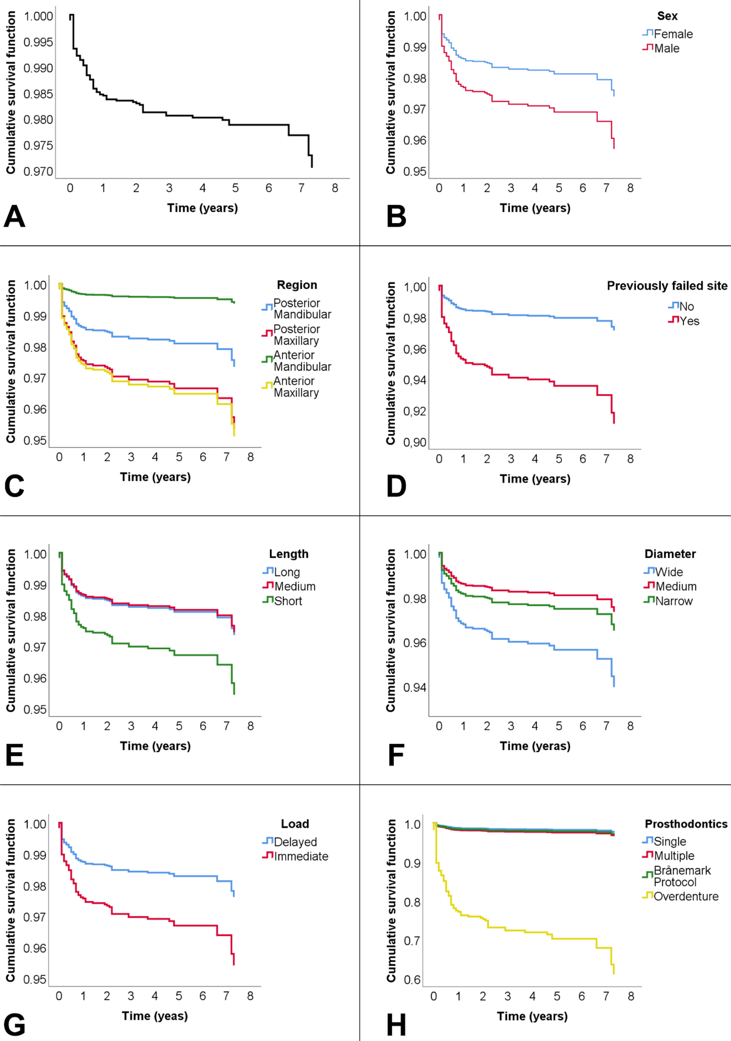
The sample of this study consisted of 1417 patient records, with 4556 implants placed. The median age of patients at the time of implant placement was 62 years (interquartile range 54 – 71 years). Regarding the patient’s sex, 644 (45.5%) were women, and 769 (54.3%) were men. The number of implants placed per patient ranged from a minimum of 1 to a maximum of 20 implants. The mean follow-up time was 5 years. A detailed description of the frequencies and relationships obtained in the study for each covariate can be seen in Table 1. Survival rate of implants at mean of covariates is presented in Figure 1A-H.

Survival rate of implants at mean of covariates (A); Survival Function for Sex (B), Region (C), Previously failed site (D), Length (E), Diameter (F), Load (G), and Prosthodontics (H).
There was a week significant correlation between implant survival and follow-up moment that the implant failed (Rho = -0.214, P = 0.010) (Table 1). In total, 144 (3.2%) implants were lost, accounting for a survival rate of 96.8%. (Figure 1A). By considering the follow-up periods more specifically, as displayed in table 2, it could be observed that most implants (n=40, 28.0%) were lost within 36 days to 6 months (P < 0.0001), and after 9 years, no loss was observed (Table 2).
Association was found between implant condition and sex, previously failed site, thread forms, length, diameter, torque, and prosthodontics variables (P < 0.05) (Table 1). The proportion of failed implants was higher for males (P = 0.032). The percentage of failed implants was also higher when an implant was placed in a previously failed site (P < 0.0001). Ratios of well-maintained and failed implants were similar for square/trapezoidal thread forms. However, the proportion of failed implants was higher for triangular than for buttress forms (P = 0.010). The loss was the same for long and medium length and medium diameter and narrow implants but higher when short or wide implants were installed, respectively (P = 0.001). Higher implant failure was related to torque >32 Ncm (p = 0.004). Moreover, better and worse results were obtained with the Brånemark protocol and overdenture, respectively (P < 0.0001).
For each implant condition, the proportion of patients in each category of smoking, region, shape and load, age, total diseases and total medications were similar (Table 1).
Regression using the Enter method (Table 3) showed that the variables that negatively influenced the survival of dental implants were higher total medications (P = 0.005; HR = 1.511; 95%CI = 1.129 – 2.023), male sex (P = 0.015; HR = 1.733; 95%CI = 1.110 – 2.705), posterior (P = 0.013; HR = 1.903; 95%CI = 1.146 – 3.161) and anterior (P = 0.017; HR = 1.991; 95%CI = 1.130 – 3.510) maxillary region, previously failed site (P = 0.002; HR = 3.012; 95%CI = 1.488 – 6.095), short length (P = 0.048; HR = 1.742; 95%CI = 1.004 – 3.023), immediate load (P = 0.007; HR = 1.896; 95%CI = 1.189 – 3.026), and overdenture rehabilitation (P = 0.000; HR = 15.761; 95%CI = 7.028 – 35.344). Higher survival rate was observed for anterior mandibular region (P = 0.001; HR = 0.245; 95%CI = 0.106 – 0.566) and medium (regular) diameter (P = 0.022; HR = 0.450; 95%CI = 0.228 – 0.891). Age, total diseases, smoking, shape, thread form and torque variables did not influence the model.
After improve model by the Backward stepwise method, the following variables were sequentially excluded in seven steps: age, shape, thread form, total diseases, and torque, respectively (Table 4). It can be seen that the variables that negatively influenced the survival of dental implants were practically the same: higher total medications (P = 0.007; HR = 1.136; 95%CI =1.078 – 1.608), male sex (P = 0.010; HR = 1.770; 95%CI = 1.146 – 2.734), posterior (P = 0.026; HR = 1.765; 95%CI = 1.070 – 2.911) and anterior (P = 0.029; HR = 1.858; 95%CI = 1.065 – 3.242) maxillary region, previously failed site (P = 0.001; HR = 3.178; 95%CI = 1.579 – 6.397), short length (P = 0.044; HR = 1.743; 95%CI = 1.015 – 2.991), immediate load (P = 0.005; HR = 1.943; 95%CI = 1.223 – 3.088), and overdenture rehabilitation (P = 0.000; HR = 20.127; 95%CI = 9.686 – 41.823). Similarly, higher survival rate was observed for anterior mandibular region (P = 0.001; HR = 0.236; 95%CI = 0.103 – 0.542) and medium (regular) diameter (P = 0.015; HR = 0.432; 95%CI = 0.220 – 0.850).
Discussion
The hypothesis tested in the study that systemic and local factors and prosthesis and implant characteristics could affect the survival of dental implants, was accepted. Overall, the survival of dental implants was 96.8% after up to 20 years of follow-up (Figure 1A), corroborating with some studies that show survival of 94.6% after 13 years of follow-up31 and 96.7% after 10 years of follow-up4. Most implants were lost (n=40, 28.0%) within 36 days to 6 months, and after 9 years, no loss was observed. The prevalence of early failures of dental implants has also been presented in the literature3,14,32. This suggests that systemic and local factors of the patient, as well as factors related to the surgical process (such as surgical site infection and bone overheating during drilling), tend to prevail with respect to causes of implant loss3,14,32.
It was observed lower survival rate of dental implants in patients with chronic use of systemic medication. Although, a description of the specific types of medication with the greatest influence was not possible due to the low percentage of each type of medication in this analysis. Previous studies corroborate the findings regarding the influence of systemic conditions on implant failure. Studies have reported that patients who use antidepressants showed an association with implant failure due to serotonin being present in the bone and regulating the activation and differentiation of the osteoclasts, which may negatively influence the osseointegration process33,34. Chrcanovic et al.28 showed an association between the intake of proton pump inhibitors and an increased probability of dental implant failure. Reduced acidity in the stomach impairs intestinal absorption of dietary calcium. Thus, there may be a decrease in calcium absorption, as calcium balance is essential for the maintenance of bone health, it seems reasonable to believe that the imbalance may, to some degree, affect osseointegration35. On the other hand, a cohort study showed that the implant survival rate was significantly higher when patients were on antihypertensive medication. The failure rate was almost seven times lower in antihypertensive drug users (0.6%) than in non-users (4.1%). These drugs exert their effect on blood pressure by inhibiting the β-adrenergic receptors responsible for bone resorption, resulting in increased bone accumulation36.
The present analysis has shown that the male sex presented a significant factor for implant failure compared to women (Figure 1B). Another study also observed the same outcomes37. This might be associated to factors such as bite force38, oral hygiene, and alcohol consumption39.
Implants placed in the maxilla (anterior and posterior regions) have shown significantly lower implant survival than those in the mandibular arch (Figure 1C). Moreover, the highest survival rate in this study was observed for the anterior mandibular region. Two previous studies also agreed with such outcomes14,15. The improved survival rate of implants placed in the anterior mandible in relation to the maxilla may be related to the usually improved bone quality, and greater bone volume found in the anterior mandible, even years after teeth extraction in this region14.
Dental implants replacing failed implants had lower survival rates than the rates reported for the previous attempts of implant placement (Figure 1D). This agrees with two previous retrospective studies15,16, which suggested that a site-specific negative effect may be associated with this phenomenon. One study also observed that other factors might potentially influence the failure rate, such as the intake of antidepressants and antithrombotic agents16. It might be worth to mention that another study has shown that replaced implants, after osseointegrated, showed the same pattern of marginal bone loss than implants placed in pristine sites40.
Short implants showed a significant lower survival rate in this study (Figure 1E). Another retrospective study has also shown that short implants have lower survival rates20. Shorter implants seem to fail more often than longer ones because of decreased initial stability, lower resistance to bending moment forces, and an increased risk of movement at the interface28. Moreover, obviously, any eventual initial bone loss would be more deleterious to short implants than to longer ones. However, a meta-analysis showed similar survival rates for extra-short (≤6 mm) and longer (≥10 mm) dental implants at 1 and 3 years of follow up. The authors concluded that the long-term effectiveness of extra-short dental implants should be further explored41.
Immediately loaded implants showed significant lower survival rates in this study than those subjected to delayed load (Figure 1G). The literature shows specific data about immediate loading on different types of prosthetic treatment. Immediate loading in the fully edentulous jaw by means of a fixed prosthesis is a well-documented treatment concept. In the mandible, the use of four implants leads to a failure rate of 0 - 3.3%, being a predictable treatment26. However, immediately loaded single implants have lower survival rates than the delayed approach, ranging from 85.7 - 100% in studies included in a critical review26. In fact, a meta-analysis demonstrated a five times higher risk of failure for immediately loaded single implants compared with delayed loading27. The implants immediately loaded by practitioners in this study were those that, in general, achieved an initial toque > 32 N.cm. However, despite the adequate initial implant stability, other factors play a significant role in the success of osseointegration for immediate implants, such as prosthetic design, occlusal adjustement, biofilm control and presence of parafunctional habits.
Overdentures have been associated with significantly lower implant survival than single- or multi-unit fixed partial prostheses in this study (Figure 1H). The failure rate of implants associated with overdentures was 17.6% (22 failures out of 147 implants). This high failure rate agrees with a previous retrospective study that evaluated implants with follow-ups of at least 20 years, in which the failure rate for implants retaining overdentures was 27.2% (9 failures out of 33 implants)14. It is important to note that the proper use of overdentures is more patient-dependent than the other types of (fixed) prostheses. This might have influenced the outcomes of this treatment.
Regular diameter implants were at a greater survival rate in the present study (Figure 1F). It might be considered that they tend to preserve more surrounding bone in comparison to wide implants and have greater bone-to-implant contact for stress dissipation in comparison to narrower implants. A previous meta-analysis has shown that narrower implants (<3.3 mm) had significantly lower survival rates compared with wider implants (≥3.3 mm)21. A review showed that narrow implants <3.0 mm performed clinically inferiorly than regular ones, which was not the case for those for other categories (3 - 3.25 mm or 3.3 - 3.5 mm)22. Another point to consider is that, in the present study, the wider implants are frequently those of shorter length, which might help to explain the significant lower survival in comparison to the implants of regular diameter.
As limitations of this study, it can be pointed out that only medical records were evaluated, and not the patients clinically, so only the survival of dental implants was evaluated and not the success rate. Specific systemic alterations or medications might alone influence implant survival, but in the present analysis, they could not be evaluated separately due to the limited number of individuals in each category. The study included patients treated from 1999 and significant evolution occurred at implant systems since them, which might also affect the survival rate.
Through the analysis of the data of this study, it was possible to conclude that: (I) Systemic and local factors and implant and prosthetic characteristics influence the survival of dental implants; (II) Variables that negatively influenced the survival of dental implants were higher total medications, male sex, posterior and anterior maxillary region, previously failed site, short length implants, immediate load, and overdenture rehabilitation; (III) A higher survival rate was observed for the anterior mandibular region and regular-diameter implants; (IV) Age, total diseases, smoking, implant shape, thread form, and torque variables did not influence the models.
Acknowledgements
Coordenação de aperfeiçoamento de pessoal de nível superior (CAPES) – finance code 001
References
-
1 Vissink A, Spijkervet F, Raghoebar GM. The medically compromised patient: are dental implants a feasible option? Oral Dis. 2018 Mar;24(1-2):253-60. doi: 10.1111/odi.12762.
» https://doi.org/10.1111/odi.12762 -
2 Manor Y, Simon R, Haim D, Garfunkel A, Moses O. Dental implants in medically complex patients-a retrospective study. Clin Oral Investig. 2017 Mar;21(2):701-8. doi: 10.1007/s00784-016-1937-6. Epub 2016 Sep 8.
» https://doi.org/10.1007/s00784-016-1937-6 -
3 Jemt T, Nilsson M, Olsson M, Stenport VF. Associations between early implant failure, patient age, and patient mortality: a 15-year follow-up study on 2,566 patients treated with implant-supported prostheses in the edentulous jaw. Int J Prosthodont. 2017 Mar-Apr;30(2):189-97. doi: 10.11607/ijp.4933.
» https://doi.org/10.11607/ijp.4933 -
4 Hasegawa T, Kawabata S, Takeda D, Iwata E, Saito I, Arimoto S, et al. Survival of Brånemark System Mk III implants and analysis of risk factors associated with implant failure. Int J Oral Maxillofac Surg. 2017 Feb;46(2):267-73. doi: 10.1016/j.ijom.2016.10.014. Epub 2016 Nov 14.
» https://doi.org/10.1016/j.ijom.2016.10.014 -
5 Chrcanovic BR, Kisch J, Larsson C. Retrospective evaluation of implant-supported full-arch fixed dental prostheses after a mean follow-up of 10 years. Clin Oral Implants Res. 2020 Jul;31(7):634-45. doi: 10.1111/clr.13600.
» https://doi.org/10.1111/clr.13600 -
6 Crespi R, Toti P, Covani U, Crespi G, Brevi B, Menchini-Fabris GB. Bone assessment in grafted and ungrafted sockets after dental implant placement: a 10-year follow-up study. Int J Oral Maxillofac Implants. 2020 May-Jun;35(3):576-84. doi: 10.11607/jomi.7969.
» https://doi.org/10.11607/jomi.7969 -
7 Donos N, Calciolari E. Dental implants in patients affected by systemic diseases. Br Dent J. 2014 Oct;217(8):425-30. doi: 10.1038/sj.bdj.2014.911.
» https://doi.org/10.1038/sj.bdj.2014.911 -
8 Chrcanovic BR, Kisch J, Albrektsson T, Wennerberg A. Factors influencing early dental implant failures. J Dent Res. 2016 Aug;95(9):995-1002. doi: 10.1177/0022034516646098.
» https://doi.org/10.1177/0022034516646098 -
9 Strietzel FP, Reichart PA, Kale A, Kulkarni M, Wegner B, Küchler I. Smoking interferes with the prognosis of dental implant treatment: a systematic review and meta-analysis. J Clin Periodontol. 2007 Jun;34(6):523-44. doi: 10.1111/j.1600-051X.2007.01083.x.
» https://doi.org/10.1111/j.1600-051X.2007.01083.x -
10 French D, Ofec R, Levin L. Long term clinical performance of 10 871 dental implants with up to 22 years of follow-up: a cohort study in 4247 patients. Clin Implant Dent Relat Res. 2021 Jun;23(3):289-97. doi: 10.1111/cid.12994.
» https://doi.org/10.1111/cid.12994 -
11 Ruggiero SL, Woo SB. Biophosphonate-related osteonecrosis of the jaws. Dent Clin North Am. 2008 Jan;52(1):111-28, ix. doi: 10.1016/j.cden.2007.09.002.
» https://doi.org/10.1016/j.cden.2007.09.002 -
12 Sgolastra F, Petrucci A, Severino M, Gatto R, Monaco A. Periodontitis, implant loss and peri-implantitis. A meta-analysis. Clin Oral Implants Res. 2015 Apr;26(4):e8-e16. doi: 10.1111/clr.12319. Epub 2013 Dec 31.
» https://doi.org/10.1111/clr.12319 -
13 Guarnieri R, Di Nardo D, Di Giorgio G, Miccoli G, Testarelli L. Longevity of teeth and dental implants in patients treated for chronic periodontitis following periodontal maintenance therapy in a private specialist practice: a retrospective study with a 10-year follow-up. Int J Periodontics Restorative Dent. 2021 Jan-Feb;41(1):89-98. doi: 10.11607/prd.4674.
» https://doi.org/10.11607/prd.4674 -
14 Chrcanovic BR, Kisch J, Albrektsson T, Wennerberg A. A retrospective study on clinical and radiological outcomes of oral implants in patients followed up for a minimum of 20 years. Clin Implant Dent Relat Res. 2018 Apr;20(2):199-207. doi: 10.1111/cid.12571. Epub 2017 Dec 6.
» https://doi.org/10.1111/cid.12571 -
15 Leisner L, Kronsteiner D, Rammelsberg P. Effect of previous implant failure on the prognosis of subsequent implants: a retrospective study. Clin Oral Implants Res. 2021 Jul;32(7):863-70. doi: 10.1111/clr.13762.
» https://doi.org/10.1111/clr.13762 -
16 Chrcanovic BR, Kisch J, Albrektsson T, Wennerberg A. Survival of dental implants placed in sites of previously failed implants. Clin Oral Implants Res. 2017 Nov;28(11):1348-53. doi: 10.1111/clr.12992. Epub 2016 Oct 14.
» https://doi.org/10.1111/clr.12992 - 17 Wennerberg A, Albrektsson T, Chrcanovic B. Long-term clinical outcome of implants with different surface modifications. Eur J Oral Implantol. 2018;11 Suppl 1:S123-36.
-
18 Tretto PHW, Dos Santos MBF, Spazzin AO, Pereira GKR, Bacchi A. Assessment of stress/strain in dental implants and abutments of alternative materials compared to conventional titanium alloy-3D non-linear finite element analysis. Comput Methods Biomech Biomed Engin. 2020 Jun;23(8):372-83. doi: 10.1080/10255842.2020.1731481.
» https://doi.org/10.1080/10255842.2020.1731481 -
19 Dos Santos MBF, Meloto GO, Bacchi A, Correr-Sobrinho L. Stress distribution in cylindrical and conical implants under rotational micromovement with different boundary conditions and bone properties: 3-D FEA. Comput Methods Biomech Biomed Engin. 2017 Jun;20(8):893-900. doi: 10.1080/10255842.2017.1309394.
» https://doi.org/10.1080/10255842.2017.1309394 -
20 Yang Y, Hu H, Zeng M, Chu H, Gan Z, Duan J, et al. The survival rates and risk factors of implants in the early stage: a retrospective study. BMC Oral Health. 2021 Jun;21(1):293. doi: 10.1186/s12903-021-01651-8.
» https://doi.org/10.1186/s12903-021-01651-8 -
21 Ortega-Oller I, Suárez F, Galindo-Moreno P, Torrecillas-Martínez L, Monje A, Catena A, et al. The influence of implant diameter on its survival: a meta-analysis based on prospective clinical trials. J Periodontol. 2014 Apr;85(4):569-80. doi: 10.1902/jop.2013.130043. Epub 2013 Aug 1.
» https://doi.org/10.1902/jop.2013.130043 -
22 Schiegnitz E, Al-Nawas B. Narrow-diameter implants: a systematic review and meta-analysis. Clin Oral Implants Res. 2018 Oct;29 Suppl 16:21-40. doi: 10.1111/clr.13272.
» https://doi.org/10.1111/clr.13272 -
23 Darriba I, Seidel A, Moreno F, Botelho J, Machado V, Mendes JJ, et al. Influence of low insertion torque values on survival rate of immediately loaded dental implants: a systematic review and meta-analysis. J Clin Periodontol. 2023 Feb;50(2):158-69. doi: 10.1111/jcpe.13733. Epub 2022 Oct 22.
» https://doi.org/10.1111/jcpe.13733 -
24 Wittneben JG, Joda T, Weber HP, Brägger U. Screw retained vs. cement retained implant-supported fixed dental prosthesis. Periodontol 2000. 2017 Feb;73(1):141-51. doi: 10.1111/prd.12168.
» https://doi.org/10.1111/prd.12168 -
25 Kraus RD, Espuelas C, Hämmerle CHF, Jung RE, Sailer I, Thoma DS. Five-year randomized controlled clinical study comparing cemented and screw-retained zirconia-based implant-supported single crowns. Clin Oral Implants Res. 2022 May;33(5):537-47. doi: 10.1111/clr.13913.
» https://doi.org/10.1111/clr.13913 -
26 De Bruyn H, Raes S, Ostman PO, Cosyn J. Immediate loading in partially and completely edentulous jaws: a review of the literature with clinical guidelines. Periodontol 2000. 2014 Oct;66(1):153-87. doi: 10.1111/prd.12040.
» https://doi.org/10.1111/prd.12040 - 27 Atieh MA, Atieh AH, Payne AG, Duncan WJ. Immediate loading with single implant crowns: a systematic review and meta-analysis. Int J Prosthodont. 2009 Jul-Aug;22(4):378-87.
-
28 Chrcanovic BR, Kisch J, Albrektsson T, Wennerberg A. Analysis of risk factors for cluster behavior of dental implant failures. Clin Implant Dent Relat Res. 2017 Aug;19(4):632-42. doi: 10.1111/cid.12485.
» https://doi.org/10.1111/cid.12485 -
29 Schoenbaum TR, Moy PK, Aghaloo T, Elashoff D. Risk factors for dental implant failure in private practice: a multicenter survival analysis. Int J Oral Maxillofac Implants. 2021 Mar-Apr;36(2):388-94. doi: 10.11607/jomi.8983.
» https://doi.org/10.11607/jomi.8983 -
30 Boboeva O, Kwon TG, Kim JW, Lee ST, Choi SY. Comparing factors affecting dental-implant loss between age groups: a retrospective cohort study. Clin Implant Dent Relat Res. 2021 Apr;23(2):208-15. doi: 10.1111/cid.12967. Epub 2020 Dec 13.
» https://doi.org/10.1111/cid.12967 -
31 Moraschini V, Poubel LA, Ferreira VF, Barboza ES. Evaluation of survival and success rates of dental implants reported in longitudinal studies with a follow-up period of at least 10 years: a systematic review. Int J Oral Maxillofac Surg. 2015 Mar;44(3):377-88. doi: 10.1016/j.ijom.2014.10.023. Epub 2014 Nov 20.
» https://doi.org/10.1016/j.ijom.2014.10.023 -
32 Carr AB, Arwani N, Lohse CM, Gonzalez RLV, Muller OM, Salinas TJ. Early implant failure associated with patient factors, surgical manipulations, and systemic conditions. J Prosthodont. 2019 Jul;28(6):623-33. doi: 10.1111/jopr.12978. Epub 2018 Oct 22.
» https://doi.org/10.1111/jopr.12978 -
33 Wu X, Al-Abedalla K, Rastikerdar E, Abi Nader S, Daniel NG, Nicolau B, et al. Selective serotonin reuptake inhibitors and the risk of osseointegrated implant failure: a cohort study. J Dent Res. 2014 Nov;93(11):1054-61. doi: 10.1177/0022034514549378.
» https://doi.org/10.1177/0022034514549378 -
34 Chappuis V, Avila-Ortiz G, Araújo MG, Monje A. Medication-related dental implant failure: Systematic review and meta-analysis. Clin Oral Implants Res. 2018 Oct;29 Suppl 16:55-68. doi: 10.1111/clr.13137.
» https://doi.org/10.1111/clr.13137 -
35 Schinke T, Schilling AF, Baranowsky A, Seitz S, Marshall RP, Linn T, et al. Impaired gastric acidification negatively affects calcium homeostasis and bone mass. Nat Med. 2009 Jun;15(6):674-81. doi: 10.1038/nm.1963.
» https://doi.org/10.1038/nm.1963 -
36 Wu X, Al-Abedalla K, Eimar H, Arekunnath Madathil S, Abi-Nader S, Daniel NG, et al. Antihypertensive medications and the survival rate of osseointegrated dental implants: a cohort study. Clin Implant Dent Relat Res. 2016 Dec;18(6):1171-82. doi: 10.1111/cid.12414.
» https://doi.org/10.1111/cid.12414 -
37 Grisar K, Sinha D, Schoenaers J, Dormaar T, Politis C. Retrospective analysis of dental implants placed between 2012 and 2014: indications, risk factors, and early survival. Int J Oral Maxillofac Implants. 2017 May-Jun;32(3):649-54. doi: 10.11607/jomi.5332.
» https://doi.org/10.11607/jomi.5332 -
38 Takaki P, Vieira M, Bommarito S. Maximum bite force analysis in different age groups. Int Arch Otorhinolaryngol. 2014 Jul;18(3):272-6. doi: 10.1055/s-0034-1374647.
» https://doi.org/10.1055/s-0034-1374647 -
39 Lipsky MS, Su S, Crespo CJ, Hung M. Men and oral health: a review of sex and gender differences. Am J Mens Health. 2021 May-Jun;15(3):15579883211016361. doi: 10.1177/15579883211016361.
» https://doi.org/10.1177/15579883211016361 -
40 Oh SL, Shiau HJ, Ashour I, Chen H, Cruz C. Early crestal bone loss around implants placed at previously failed sites compared with initially integrated implants: a retrospective cohort study. Clin Implant Dent Relat Res. 2022 Apr;24(2):233-41. doi: 10.1111/cid.13076.
» https://doi.org/10.1111/cid.13076 -
41 Ravidà A, Wang IC, Barootchi S, Askar H, Tavelli L, Gargallo-Albiol J, et al. Meta-analysis of randomized clinical trials comparing clinical and patient-reported outcomes between extra-short (=6 mm) and longer (=10 mm) implants. J Clin Periodontol. 2019 Jan;46(1):118-42. doi: 10.1111/jcpe.13026. Epub 2018 Nov 29.
» https://doi.org/10.1111/jcpe.13026
Edited by
-
Editor:
Dr. Altair A. Del Bel Cury