Morphometric evaluation of cerebral and cerebellar structures in long-term unilateral sensorineural hearing loss
Objective Structural changes in the brain have been identified in patients with hearing loss. However, the effect of long-term Unilateral Sensorineural Hearing Loss (USNHL) on cerebral and cerebellar morphology is not fully understood.
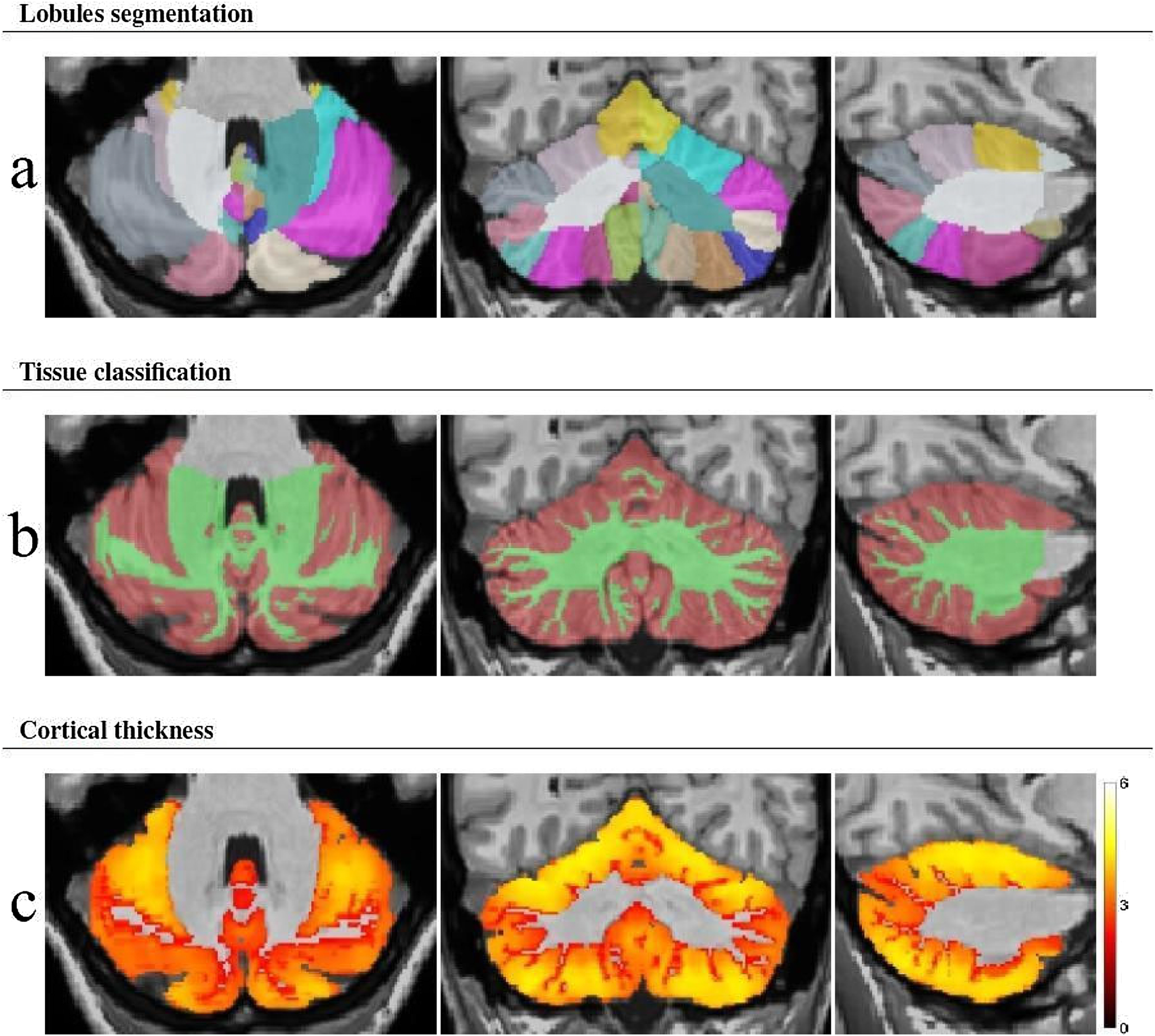
Methods In this study, Magnetic Resonance Imaging (MRI) of 12 long-term USNHL adults was compared with their Normal-Hearing (NH) side using Voxel-Based Morphometry (VBM). The USNHL group was also compared to a control group of 12 hearing-healthy individuals. Volume and cortical thickness changes in cerebral and cerebellar structures, possibly related to hearing loss severity and duration, were assessed using VBM.
Results The study found that, compared to the control group, USNHL patients had significantly higher measurements in the Frontal Operculum (FO) Gray Matter (GM) volume (cm3) (p = 0.024), Superior Temporal Gyrus (STG) thickness (p = 0.041), and cerebellum IX GM volume (cm3) (p = 0.025). No significant differences were observed between the intact and NH sides in the USNHL group (p > 0.05).
Conclusion Cerebral and cerebellar structures, which are essential for perceiving and interpreting sound, integrating information, and coordinating movement, may undergo morphological changes due to neuroadaptive mechanisms in long-term USNHL patients. Larger case series are needed to explore the clinical implications of these changes for diagnosis, treatment, and prognosis.
Levels of evidence Level 3.
Keywords
Hearing loss; Unilateral; Gray matter; Brain morphometry; Auditory cortex
 Thumbnail
 Thumbnail
 Thumbnail