P16INK4a protein expression associated with Head and Neck Squamous Cell Carcinoma: A perspective of a public health reference service in São Paulo
Abstract
Objectives Human Papillomavirus (HPV) may be a predictive biomarker predictor for clinical outcome and influence treatment decisions in patients with Head and Neck Squamous Cell Carcinoma (HNSCC).
Methods We evaluated 253 patients with HNSCC from state of São Paulo, Brazil. The influence of p16INK4a expression was analyzed with epidemiological and clinical variables.
Results In total, 32.4% of tumors studied had positive (+) p16INK4a protein expression, and 67.6% had negative. The variables were similar in both groups being the mostly with age under 64-years, male, white race, functional illiterate, smokers and alcoholics. The most affected primary site was oral cavity with T3/T4 tumoral stage, N1/N2/N3 nodals, M0 metastasis and III/IV clinical stage. Patients with oropharyngeal primary site and (+) p16INK4a, clinical staging III and chemotherapy treatment had worse survival. The median time of distant metastasis-free was 38.3-months in oropharyngeal (+) p16INK4a and 15.1-months in negative (−) p16INK4a.
Conclusion In the present study, the epidemiological variables are similar in both groups (Positive and negative p16INK4a expression): age under 64-years, male, white, functionally illiterate, smokers and alcoholics. There is no association of p16INK4a expression with primary site, however, the (−) p16INK4a shows better overall survival, higher frequencies in distant metastasis and less free time of the disease. Although the literature shows a greater survival in oropharynx (+) p16INK4a, our results are contradictory. It is suggested that future studies in different regions and with a larger sample size should be carried out to confirm these findings, because the patients who participated in the present study are only from a specific region of Brazil.
Level of evidence 2B.
Keywords
Epidemiology; Head and Neck Neoplasms; Human Papillomavirus Viruses
Introduction
According to World Health Organization, Head and Neck Cancer is one of the ten most common cancers worldwide, with 700 thousand new diagnoses annually.1 The most frequent pathological type is squamous cell carcinoma (HNSCC) with increase of incidence every year.1-3
HNSCC includes oral cavity, larynx, pharynx, paranasal sinuses, nasal cavity, and salivary glands.2 These regions have functional impact in swallowing, breathing and speech.4 The main risk factors for HNSCC are alcohol, tobacco and HPV virus.5 The synergistic effect of alcohol and tobacco with HPV infection6 leads to a worse prognosis,7 possibly due to recurrent DNA breaks induced by tobacco during the process of integration of HPV genome.8
HPV is transmitted through skin-to-skin contact, exposure to contaminated objects10 and sexual contact.9 The overexpression of p16INK4a protein is used as an indicator for the presence of high-risk HPV in immunohistochemical detection method, however, this method can induces to misclassify if another technique cannot be used.11-13 Despite, this option makes sense considering the high demand for testing, as it is more cost-effective and less technically unwieldy than HPV-specific testing.14
HPV status is an important prognostic factor that influences treatment decisions in HNSCC in which HPV-positive tumors have better survival outcomes.15-18
There are different methodologies for identify HPV, as Polymerase Chain Reaction (PCR) testing and Immunohistochemical (IHC) staining for p16INK4a, and newer techniques that are currently under investigation.15-18
The comparison of Immunohistochemistry (IHC) and real-time PCR methods relies on the observation that p16INK4a protein is not a determinant for the presence of HPV in immunohistochemistry, since there are uncertainty regarding the sensitivity and specificity of the method when used alone, without detection of viral DNA. However, IHC has been used because it is more accessible in terms of costs.17
Based on the above data, the objectives of the research were to perform an epidemiological analysis related to the presence of p16INK4a protein, in patients with HNSCC, to establish epidemiological, sociodemographic and clinical profile of these patients, and verify the association of the p16INK4a protein with survival time and recurrence.
Methods
Medical records of patients at the Otorhinolaryngology and Head and Neck Surgery service of a university hospital in the northwest of the state of São Paulo from January 2016 to May 2021 were used in the study. This research was approved by the Ethics and Research Committee of the São José do Rio Preto Medical School (FAMERP).
The inclusion criteria were patients diagnosed with squamous cell carcinoma that had p16INK4a protein expression analysis. The exclusion criteria were patients with neoplasias originating in other anatomical sites.
A database was created with epidemiological variables: age, gender, race, literacy level, and smoking and alcohol status. The variable race was classified based on the self-declaration of each patient, following the National Classification Commission of the Brazilian Institute of Geography and Statistics.19 The guidelines provided by the National Indicator of Functional Literacy20 were used to classify individuals into functionally literacy (complete primary education) and functionally illiterate (incomplete primary education history) groups. Six different definitions pertain to what it means to be a smoker.21
In the present study, smokers were considered patients who had smoked for a certain period. For defining alcoholism, the same rationale was used. The clinical variables evaluated included primary anatomical site, classified as the oral cavity, oropharynx, hypopharynx, nasopharynx, nasal cavity, larynx, and occult primary site. Tumor staging was evaluated through the classification of Malignant Tumors (TNM) according to the Union for International Cancer Control and American Joint Committee on Cancer (AJCC) 7th, to standardize the observed data. This system comprises three categories: T, characteristics of the tumor at the primary site (may be based on location, size, or both), with TX and T0 indicating the non-accessibility and absence of evidence of the primary tumor, respectively; N is the degree of lymph node involvement, indicated by N1, N2, N3, and N0 to indicate absence; and M is the absence (M0) or presence (M1) of distant metastases. A numerical status (I, II, III, or IV) of clinical staging is tabulated specifically for each patient.22
The p16INK4a protein expression was evaluated by IHC technique performed at the Hospital Pathology Service. The patients included in the study were classified in two groups: Group 1: Patients with HNSCC and positive p16INK4a protein expression; Group 2: Patients with HNSCC and negative p16INK4a protein expression.
Patients considered alive were those whose medical records did not present a declaration or notification of death.
Statistical analysis
Descriptive statistical was performed by calculating the measures of central tendency, dispersion, and frequency counts with Microsoft Excel (2016).
Quantitative variables were tested for normality using the Kolmorov-Smirnov test. Frequency comparisons were performed using Chi-Square test with with Statistical Package for Social Sciences (SPSS, IBM, version 24.0); p-value ≤0.05 was considered significant. The construction of the spreadsheet was carried out as follows: p16INK4a+ (reference: p16INK4a−), mean age of patients ≥64-years (reference: <64-years), male (reference: female), white (reference: non-white), illiterate (reference: literate), smokers (reference: non-smokers), and alcoholics (reference: non-alcoholics), oral cavity (reference: oropharynx, larynx and other sites), oropharynx (reference: oral cavity, larynx and other sites) larynx (reference: oral cavity, oropharynx and other sites), other sites: nasopharynx, nasal cavity, hypopharynx and hidden primary site (reference: oral cavity, oropharynx and larynx), T3, T4 (reference: Tis, T0, T1, and T2), N1, N2, N3 (reference: N0), M1 (reference: M0), and III/ IV (reference: I/II) using the Statistical Package for Social Sciences program (SPSS, IBM, version 24.0). We evaluated the association between primary sites, epidemiological variables and treatment performed with p16INK4a protein expression.
To evaluate the influence of the HNSCC on survival, curves were constructed using Prisma 6.07 (2015) program, through the Kaplan-Meier method. The outcome to assess survival rates included the period from the diagnosis of the disease until the date of 05/25/2021 or death, for 253 patients.
The GraphPad Instat 3.10 (2009) was used to prepare the time of distant metastasis-free and recurrence-free. The period analyzed was from diagnosis to the date of metastasis/recurrence diagnosis or 05/25/2021, for 253 patients. All data are from the last medical appointment performed, thus demonstrating their evolution, until 05/25/2021, when the data were accessed for the last time.
We evaluated overall survival according to anatomical sites and p16INK4a protein expression; overall survival according to clinical staging regardless of p16INK4a protein expression; overall survival according to treatment performed regardless of p16INK4a protein expression; time of distant metastasis-free and reccurence-free according to anatomical sites and p16INK4a protein expression.
Results
A total of 253 patients with HNSCC diagnosis were evaluated in the present study. Three patients showed two different tumors with opposite expressions and were included in both groups, totalizing 256 tumors. Thus 83 (32.4%) had a positive expression and 173 (67.6%) had negative expression for p16INK4a protein.
The primary sites more frequent were oral cavity (34.9%) and oropharynx (30.1%) in patients with positive p16INK4a protein expression as well as in patients with negative p16INK4a protein expression (Oral cavity: 35.8% and Oropharynx: 30.1%) (Table 1).
The results about the epidemiological and clinical variables of patients according to their anatomical sites with positive and negative expression of the p16INK4a protein are described in Table 2. Regarding Group 1, the majority was patients with age <64-years, male gender, white, illiterate with smoking and alcohol habits, with T3/T4 tumor stage, with N1/N2/N3 nodal stage and absence of metastasis. For Group 2, the majority was patients with age <64-years, male gender, white, illiterate with smoking and alcohol habits, with T3/T4 tumor stage, with N1/N2/N3 nodal stage and absence of metastasis.
The treatment performed in patients with HNSCC associated with p16INK4a protein expression are shown in Table 3 in which concomitant chemotherapy with radiotherapy was the most common treatment in both groups.
A total of 60 patients evaded the treatment and did not show an explanation or notification of death and, therefore, were considered alive in this study. Nineteen of this present positive expression of p16INK4a protein (Oropharynx = 5; All other sites = 14), and forty-one present negative expression of p16INK4a protein (Oropharynx = 10; All other sites = 31).
A total of 36 (14.1%) patients received palliative treatment, 9 (10.8%) with positive expression of p16INK4a protein (Oropharynx = 3; All other sites = 6) and 27 (15.6%) with negative expression of p16INK4a protein (Oropharynx = 8; All other sites = 19).
A total of 46 (17.9%) patients had distant metastasis, 14 (16.9%) with positive expression of p16INK4a protein (Oropharynx = 8; All other sites = 6) and 32 (26.4%) with negative expression of p16INK4a protein (Oropharynx = 11; All other sites = 21), while a total of 23 patients had recurrence, 5 (6.0%) with positive expression of p16INK4a protein (Oropharynx = 1; All other sites = 4) and 18 (10.5%) with negative expression of p16INK4a protein (Oropharynx = 3; All other sites = 15). Patients with distant metastasis and recurrences also had brief evolution, as detailed in Table 4, and the anatomical sites involved in distant metastasis are shown in Table 5.
Overall survival according to anatomical sites, clinical staging and received treatment and association with p16INK4a expression, respectively are illustrated in Fig. 1.

(A) Overall survival according to anatomical sites and p16INK4a expression: The survival curve compares according to the anatomical site of the oropharynx and the other anatomical sites (oral cavity, hypopharynx, nasopharynx, nasal cavity, larynx and hidden primary site) in relation to the expression of the p16INK4a protein. (B) Overall survival according to clinical stage: The survival curve compares the different tumor stages (I, II, III and IV), regardless of p16INK4a protein expression; and (C) overall survival according to treatment: The survival curve compares the different treatments performed (surgery, chemotherapy, radiotherapy and concomitant chemotherapy and radiotherapy), regardless of p16INK4a protein expression. ns, not significant (p > 0.05). Note: In the Fig. 1.C, the analysis of the untreated group is not verified as there are no data on patients who did not adhere to the treatment on their own.
No significance was observed for overall survival according to anatomical sites and p16INK4a protein expression (Fig. 1A). The median survival to the group positive expression of p16INK4a protein was 20.0-months in oropharynx and 44.3-months in other anatomical sites, while to the group negative expression of p16INK4a protein was 15.5-months in oropharynx and 29.0-months in other anatomical sites. The frequency of patients with HNSCC in all primary sites who died after the diagnosis of the disease with positive expression of the p16 INK4a protein were 30.1% of cases in total, with 44.0% cases of tumors in oropharynx and 24.1% cases of tumors in other anatomical sites, while 24.9% of patients was negative in total, with 23.1% cases of tumors in oropharynx and 25.6% cases of tumors in other anatomical sites, regardless of survival time.
On overall survival, according to the clinical stage (Fig. 1B) no significance was assessed either. The median survival in I, II, III and IV clinical stages were respectively, 38.6-, 27.5-months, 31.6-, and 28.1-months, unrelated to p16INK4a protein expression. The frequence of patients with HNSCC, independent of the primary site, who died after the diagnosis of the disease with positive expression of the p16INK4a protein with clinical stage I, II, III, and IV were, respectively, 31.6, 40.0, 30.0 and 36.7%, while with negative expression of the p16INK4a protein were, respectively, 12.0, 16.7, 27.6 and 29.8%, regardless of survival time and primary anatomic site.
The overall survival according received treatment (Fig. 1C), also did not show significance. The median survival in surgery, chemotherapy, radiotherapy and concomitant chemotherapy and radiotherapy were, respectively, 34.0-, 30.9-, 31.7- and 32.9-months, unrelated to p16INK4a protein expression. The frequency of patients with HNSCC, independent of the primary site, who died after surgery, chemotherapy, radiotherapy and concomitant chemotherapy and radiotherapy with positive expression of the p16INK4a protein were, respectively, 23.5, 47.8, 25.0 and 21.2%, while with negative expression of the p16INK4a protein were, respectively, 18.2, 30.9, 68.3 and 20.8%, regardless of survival time and primary anatomic site.
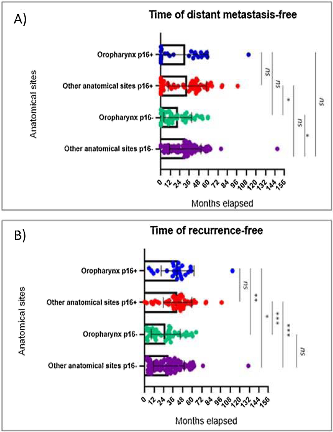
The median time of distant metastasis-free and recurrence-free according to anatomical sites and p16INK4a expression were described in Fig. 2.

The times of distant metastasis-free (2A) and recurrence-free (2B) were represented as scatter plot with bar (Mean + Standard Deviation (SD). Differences among groups were assessed by Kruskal-Wallis plus Dunn’s post hoc test. ns, not significant (p > 0.05); *, p < 0.05; **, p < 0.01; ***, p < 0.001. Note: Time 0 was considered for those patients who came from other institutions with failure in the primary treatment (cases of recurrence), since no old information is available in the system, or who received the HNSCC primary diagnosis with the distant metastasis, at the same time.
The median time of distant metastasis-free (2.A) to the positive expression of p16INK4a protein group was 38.3-months in oropharynx and 36.7-months in other anatomical sites, while to the group negative expression of p16INK4a protein was 15.1-months in oropharynx and 30.4-months in other anatomical sites.
About to the time of recurrence-free (2.C), the median time to the group positive expression of p16INK4a protein was 41.7-months in oropharynx and 42.6-months in other anatomical sites, while to the group negative expression of p16INK4a protein was 27.7-months in oropharynx and 30.4-months in other anatomical sites.
Discussion
Understanding
Our results corroborate with international studies regarding p16INK4a expression, in which p16INK4a protein expression by IHC is highly variable and cannot be recommended to predict HPV-presence.23,24
HPV-associated with HNSCC has an attribution rate that depends on the world region, which is higher in high-income countries.9 In USA, the rate of HPV-associated with HNSCC is 59.3%, in Europe is 31.1% and in Brazil is 4.1%, that confirm the geographic disparities26 and the association in economic sphere.
Regarding to primary site showed we did not found significant association of cancer and p16INK4a expression. Oral cavity, oropharynx, and larynx were the most affected sites in both groups (p16INK4a positive and negative), as study of Rygalski et al. (2021).27
The average age of patients was 64-years. In positive p16INK4a expression group, the percentage of patients under the age of 64-years was predominant in oropharynx cases (64.0%). Historically, HPV-positive in HNSCC is associated with younger individuals; however, recently, incidences in older adults have also been observed,26 as also shown in our results. According to the results of Baijens et al. (2021),28p16INK4a negative expression is predominant in older patients (≥65-years) HNSCC induced by alcohol and tobacco, while younger patients (25-64 years) with this type of cancer were associated with HPV virus.
Males were the most affected in group with positive expression of p16INK4a protein, as well as white individuals that were illiterate, smokers, and alcoholics. Men are three to five times more likely to develop HNSCC than women.27 White individuals make up most of the population affected by HPV-positive oropharyngeal carcinoma.29 The clinical and epidemiological characteristics observed reflect the profile of patients assisted by the public health service in Brazil.
Primary prevention measures for HNSCC include smoking cessation and passive exposure to smoke, in addition to environmental exposure to carcinogens and limited alcohol consumption. The role of HPV vaccines has also been advocated in preventing cervical cancer in girls, however, given the increase in HPV-positive HNSCC in men, vaccination in boys may also play a role in the future prevention and reduction of this cancers.28
Our results showed that p16INK4a expression had no significant association with overall survival or clinical stage. The literature shows that HPV-positive cases tend to involve extensive and early nodal metastasis,30 with approximately 85% of patients presenting them at some point.29
Treatment of HNSCC, including chemotherapy, radiotherapy and surgery, remains the standard, regardless of p16INK4a expression.16 The treatment of patients is complex, because they comprise diverse groups of cancers with different behavior and prognosis, necessitating different treatment approaches.31
The analysis of overall survival according to anatomical sites and p16INK4a expression showed no significant association, though it showed a lower survival in oropharyngeal when compared to other anatomical sites, in positive cases of p16INK4a expression. Our results are contradictory to scientific literature, this fact is probably due to the sample's low-income background and unified health system in Brazil. Furthermore, although widely used in diagnostic practice, the IHC technique is subject to limitations that may affect the accuracy of results, including the occurrence of both false-positive and false-negative findings.25
It is important to emphasize that all patients that participate of this study received the follow up according to Brazilian Unified Health System (SUS). As a result, the exclusion of individuals who did not undergo IHC analysis may have led to a selection bias, as well as the number of patients that evaded the treatment that was high.
Additionally, based on database of our study, most of the patients died as a result of the disease, due to reports of cachexia.
Palliative care was used in 10.8% of patients with positive p16INK4a expression and 15.6% in negative patients, reflecting better data than that found in the literature.18
Despite the multidisciplinary approach, the clinical evolution of patients with HNSCC is unsatisfactory clinical outcome, with approximately 50% local recurrence at 3-years.36 Some other pathological risk factors, which also have been associated with an increased risk of recurrence after surgery. The following risk factors, especially when two or more occur together, are indicators for recommending postoperative radiotherapy: nodules >3 cm, having two or more lymph nodes involved, perineural invasion, closed mucosal margins, and stage T3‒T4 disease classification.37
We found association of distant metastasis and p16INK4a expression (Fig. 2a) when we compare other anatomical sites positive p16INK4a expression and oropharynx negative p16INK4a expression, and other anatomical sites and oropharynx, both with negative p16INK4a expression. Also, was found association of recurrence and p16INK4a expression (Fig. 2b), except in other anatomical sites with oropharynx, in positive and negative p16INK4a expression. Wittekindt et al. (2018),10 declares the failure of primary treatment in patients with HPV-negative carcinomas often leads to the development of locoregional recurrence, while primary treatment failure in patients with HPV-induced carcinomas often leads to the development of distant metastasis, however, our results showed the negative p16INK4a expression has the most frequency and lower time free of distant metastasis and recurrence.
According to Duprez et al. (2017),38 the most diagnosed visceral organs in HNSCC metastasis are the lungs, bones, and liver, while in our study, including not just visceral organs, were lymph nodes, cervical region and lungs, respectively. In cases of unknown primary HNSCC, positive p16INK4a expression should be interpreted with caution, as approximately 20% of metastatic squamous cell skin and lung cancers, without associated high-risk HPV, show more than 70% nuclear staining and cytoplasmic positive in the IHC of p16INK4a.39
Comparison to literature
According to Khanna et al. (2020),32 patients with HPV-positive oropharyngeal cancer demonstrate a better prognosis, better survival, and greater response to treatment, regardless of treatment modality, when compared to HPV-negative patients. However, Paver et al. (202029 found that patients with HPV-positive carcinomas in other regions of the head and neck do not have the same favorable prognosis. Additionally, Maléřová et al. (202024 concluded that p16INK4a positive may be a poor prognostic factor for oral cancers.
In relation to overall survival and clinical stage (Fig. 1B), our results show no association with p16INK4a expression, however, shows the clinical stage III, with the worst survival, differing from the literature in terms of more aggressive tumors having better prognosis and survival in p16INK4a positive cases.15-33
The overall survival and treatment also did not show significance with p16INK4a expression, however, it corroborates with the literature when concomitant chemoradiotherapy remains the treatment of choice for most patients with advanced HNSCC, while surgery remains the preferred modality for surgically respectable cancers of the oral cavity.34 Another issue raised for Mittal et al. 202234 is the role of chemotherapy treatment in early or locally advanced HNSCC in non-organ preservation, as well as toxicity after radiotherapy, which are defined as adverse effects,35 which may be one of the explanations why patients who received chemotherapy and radiotherapy singly treatment had a lower survival rate.
Limitations and strengths
The study's limitations include the lack of IHC in 502 samples and absence of HPV type confirmation in 256 cases. However, the study allowed observing that the rate of HNSCC associated with HPV is increased in the casuistic evaluated.
Conclusion
We found that patients with oropharyngeal cancer and positive for p16INK4a protein expression are typically male, smokers, alcoholics, under 64-years old, white, and illiterate. There is no association between p16INK4a expression and primary site, however patients with negative p16INK4a expression have better overall survival and higher rates of distant metastasis with and less free time of the disease in the casuistic evaluated. Despite literature suggesting better survival in p16INK4a positive cases, our results are contradictory. It is important to emphasize that all patients that participate of this study received the follow up according to Brazilian Unified Health System (SUS). As a result, the exclusion of individuals who did not undergo IHC analysis may have led to a selection bias, as well as the number of patients that evaded the treatment that was high. Moreover, factors such as late diagnosis, limited access to specialized care and lack of adequate treatment also may be interfered in our results. It is suggested that future studies in different regions and with a larger sample size should be carried out to confirm these findings, because the patients who participated in the present study are only from a specific region of Brazil.
-
Funding
This work was supported by the Coordination for the Improvement of Higher Education Personnel ‒ CAPES (Financial Code 001); National Council for Scientific and Technological Development - CNPq (Grant number: 310987/2018-0), and São Paulo Research Foundation - FAPESP (Grant number: 2018/26166-6). We thank the Faculty of Medicine of São José do Rio Preto (FAMERP) and the Hospital de Base of São José do Rio Preto, Foundation Faculty of Medicine of São José do Rio Preto (HB-FUNFARME), Brazil for their institutional support and infrastructure in conducting this study.
Data availability statement
The authors declare that all data are available in repository.
References
-
1 GLOBOCAN - Cancer Today: Estimated number of new cases in 2020, lip, oral cavity, oropharynx, nasopharynx, hypopharynx, both sexes, all ages. International Agency for Research on Cancer (IARC). 2020. https://gco.iarc.fr/today/online-analysis-table?v=2020&mode=population&mode_population=income&population=900&populations=900&key=asr&sex=0&cancer=1_3_4_5&type=0&statistic=5&prevalence=0&population_group=0&ages_group%5B%5D=0&ages_group%5B%5D=17&group_cancer=1&include_nmsc=0&include_nmsc_other=1. Accessed 11/13/2025
» https://gco.iarc.fr/today/online-analysis-table?v=2020&mode=population&mode_population=income&population=900&populations=900&key=asr&sex=0&cancer=1_3_4_5&type=0&statistic=5&prevalence=0&population_group=0&ages_group%5B%5D=0&ages_group%5B%5D=17&group_cancer=1&include_nmsc=0&include_nmsc_other=1. Accessed 11/13/2025 - 2 Qiao XW, Jiang J, Pang X, Huang M-C, Tang Y-J, Liang X-H, et al. The Evolving Landscape of PD-1/PD-L1 Pathway in Head and Neck Cancer. Front Immunol. 2020;11:1721.
- 3 Vasicek SM, Pondorfer P, Holzmeister C, Graupp M, Weiland T, Wolf A, et al. Head and neck cancer in Styria: An epidemiologic and clinical audit. Wien Klin Wochenschr. 2020;132:444-51.
- 4 Shibata H, Zhou L, Xu N, Egloff AM, Uppaluri R. Personalized cancer vaccination in head and neck cancer. Cancer Sci. 2021;112:978-988.
- 5 Tumban E. A Current Update on Human Papillomavirus-Associated Head and Neck Cancers. Viruses. 2019;11:922.
- 6 Kumar R, Rai AK, Das D, Das R, Suresh Kumar R, Sarma A, et al. Alcohol and Tobacco Increases Risk of High Risk HPV Infection in Head and Neck Cancer Patients: Study from North-East Region of India. PLoS One. 2015;10:e0140700.
- 7 Descamps G, Karaca Y, Lechien JR, Kindt N, Decaestecker C, Remmelink M, et al. Classical risk factors, but not HPV status, predict survival after chemoradiotherapy in advanced head and neck cancer patients. J Cancer Res Clin Oncol. 2016;142(10):2185-96.
- 8 Aguayo F, Muñoz JP, Perez-Dominguez F, Carrillo-Beltrán D, Oliva C, Calaf GM, et al. High-Risk Human Papillomavirus and Tobacco Smoke Interactions in Epithelial Carcinogenesis. Cancers (Basel). 2020;12:2201
- 9 Aupérin A. Epidemiology of head and neck cancers: an update. Curr Opin Oncol. 2020;32:178-86.
- 10 Wittekindt C, Wagner S, Sharma SJ, Würdemann N, Knuth J, Reder H, et al. HPV - A different view on Head and Neck Cancer. Laryngorhinootologie. 2018;97(S 01):S48-S113.
- 11 Augustin JG, Lepine C, Morini A, Brunet A, Veyer D, Brochard C, et al. HPV Detection in Head and Neck Squamous Cell Carcinomas: What Is the Issue? Front Oncol. 2020;10:1751.
- 12 Wai KC, Strohl MP, van Zante A, Ha PK. Molecular Diagnostics in Human Papillomavirus-Related Head and Neck Squamous Cell Carcinoma. Cells. 2020;9:500.
- 13 Stephen JK, Divine G, Chen KM, Chitale D, Havard S, Worsham MJ. Significance of p16 in Site-specific HPV Positive and HPV Negative Head and Neck Squamous Cell Carcinoma. Cancer Clin Oncol. 2013;2:51-61.
- 14 Shelton J, Purgina BM, Cipriani NA, Dupont WD, Plummer D, Lewis JS. p16 immunohistochemistry in oropharyngeal squamous cell carcinoma: a comparison of antibody clones using patient outcomes and high-risk human papillomavirus RNA status. Mod Pathol. Sep 2017;30:1194-203.
- 15 Hoffmann M, Quabius ES. Relevance of Human Papillomaviruses in Head and Neck Cancer-What Remains in 2021 from a Clinician's Point of View? Viruses. 2021;13:1173.
- 16 Rosenberg AJ, Vokes EE. Optimizing Treatment De-Escalation in Head and Neck Cancer: Current and Future Perspectives. Oncologist. 2021;26:40-8.
- 17 Simoens C, Gheit T, Ridder R, Gorbaslieva I, Holzinger D, Lucas E, et al.; HPV-AHEAD study group. Accuracy of high-risk HPV DNA PCR, p16(INK4a) immunohistochemistry or the combination of both to diagnose HPV-driven oropharyngeal cancer. BMC Infect Dis. 2022;22:676.
- 18 Mayland CR, Ho QM, Doughty HC, Rogers SN, Peddinti P, Chada P, et al. The palliative care needs and experiences of people with advanced head and neck cancer: A scoping review. Palliat Med. 2021;35:27-44.
- 19 IBGE. Instituto Brasileiro de Geografia e Estatística.
- 20 INAF. Indicador Nacional de Alfabetismo Funcional.
- 21 Barbosa A.S BLS, Rodrigues L, Oliveira K.L, Argimon I.I.L. Múltiplas definições de ser fumante e diagnóstico de tabagismo: uma revisão sistemática. Aletheia.
- 22 Commitee for Head and Neck Surgery and Oncology ea. TNM staging of head and neck cancer and neck dissection classification.
- 23 Mooren JJ, Gültekin SE, Straetmans JMJAA, Haesevoets A, Peutz-Kootstra CJ, Huebbers CU, et al. P16(INK4A) immunostaining is a strong indicator for high-risk-HPV-associated oropharyngeal carcinomas and dysplasias, but is unreliable to predict low-risk-HPV-infection in head and neck papillomas and laryngeal dysplasias. Int J Cancer. 2014;134:2108-17.
- 24 Maléřová S, Kalfeřt D, Grega M, Tachezy R, Klozar J. The significance of p16 protein expression in oral squamous cell carcinoma. Epidemiol Mikrobiol Imunol. 2020;69:64-72.
- 25 Gown AM. Diagnostic Immunohistochemistry: What Can Go Wrong and How to Prevent It. Arch Pathol Lab Med. 2016;140:893-898.
- 26 Menezes FDS, Fernandes GA, Antunes JLF, Villa LL, Toporcov TN. Global incidence trends in head and neck cancer for HPV-related and -unrelated subsites: A systematic review of population-based studies. Oral Oncol. 2021;115:105177.
- 27 Rygalski CJ, Zhao S, Eskander A, Zhan KY, Mroz EA, Brock G, et al. Time to Surgery and Survival in Head and Neck Cancer. Ann Surg Oncol. 2021;28:877-85.
- 28 Baijens LWJ, Walshe M, Aaltonen L-M, Arens C, Cordier R, Cras P, et al. European white paper: oropharyngeal dysphagia in head and neck cancer. Eur Arch Otorhinolaryngol. 2021;278:577-616.
- 29 Paver EC, Currie AM, Gupta R, Dahlstrom JE. Human papilloma virus related squamous cell carcinomas of the head and neck: diagnosis, clinical implications and detection of HPV. Pathology. 2020;52:179-91.
- 30 Glastonbury CM, Hodler J, Kubik-Huch RA, von Schulthess GK, editors. Head and Neck Squamous Cell Cancer: Approach to Staging and Surveillance. In: Diseases of the Brain, Head and Neck, Spine 2020-2023: Diagnostic Imaging [Internet]. Cham (CH): Springer; 2020. Chapter 17. IDKD Springer Series.
- 31 Kaidar-Person O, Gil Z, Billan S. Precision medicine in head and neck cancer. Drug Resist Updat. 2018;40:13-16.
- 32 Khanna S, Palackdharry S, Roof L, Wicker CA, Mark J, Zhu Z, et al. Determining the molecular landscape and impact on prognosis in HPV-associated head and neck cancer. Cancers Head Neck. 2020;5:11.
- 33 Taberna M, Mena M, Pavón MA, Alemany L, Gillison ML, Mesía R. Human papillomavirus-related oropharyngeal cancer. Ann Oncol. 2017;28:2386-98.
- 34 Mittal A, Sharma A. Current status of systemic therapy in head and neck cancer. J Chemother. 2022;34:9-24.
- 35 Alfouzan AF. Radiation therapy in head and neck cancer. Saudi Med J. 2021;42:247-54.
- 36 Alterio D, Marvaso G, Ferrari A, Volpe S, Orecchia R, Jereczek-Fossa BA. Modern radiotherapy for head and neck cancer. Semin Oncol. 2019;46:233-45.
- 37 Evans M, Beasley M. Target delineation for postoperative treatment of head and neck cancer. Oral Oncol. 2018;86:288-95.
- 38 Duprez F, Berwouts D, De Neve W, Bonte K, Boterberg T, Deron P, et al. Distant metastases in head and neck cancer. Head Neck. 2017;39:1733-43.
- 39 Rahimi S. HPV-related squamous cell carcinoma of oropharynx: a review. J Clin Pathol. 2020;73:624-9.
Edited by
-
Edited by
Dr C Chone.