 Across-surface distances after one- and two-stage palatoplasty in children with oral cleft
Abstract
The goal of this study was to digitally evaluate the development of maxillary dental arches of children with unilateral cleft lip and palate treated with one- and two-stage palatal closure. One hundred and sixty-eight digitized dental models of cheiloplasty and one-stage palatoplasty (G1) and cheiloplasty and two-stage palatoplasty (G2) were evaluated at preoperative time 1 (T1), preoperative time 2 (T2), and postoperative (T3). The following surface distances were evaluated: across surface distance; cleft widths anterior (P-P′) and posterior (U-U′) cleft widths, intercanine width (C-C′), and intertuberosity width (T-T′); smallest (P′-T′) and largest (P-T) segment lengths; and smallest (C′-D′) and largest (C-D) segment cleft depths. In G1, P-P′, U-U′, and C-C′ reduced at T2, unlike P′-T′ (P<0.05). P-T and C′-D′ distances increased at T3 (P<0.05), while C-D increased at all stages (P<0.001). In G2, U-U′ and C-C′ reduced at T2 (P<0.05), while P′-T′, P-T, C′-D′, and C-D′ increased at T3 (P<0.001). In an intergroup analysis of growth rate, G2 showed higher growth percentages compared to G1, in which C′-D′ was significant (P=0.038). Furthermore, C′-D′ presented a coefficient of determination of 0.076 (P=0.039). In conclusion, dental arch development is influenced by the rehabilitation protocol. However, in the sample evaluated, the comparison suggested that individuals whose palate was operated on in two stages had the most favorable palatal growth.
Cleft lip; Cleft palate; Infant; Palate; Imaging; Three-dimensional
Introduction
Usually, children with cleft lip and palate begin the multi-professional rehabilitation protocol a few months after birth (1). The aim is always to significantly improve the functional and esthetic aspects and the social well-being of the individual and their family (1). Although the stages and surgical techniques of the rehabilitation protocol vary, every individual undergoes plastic surgeries to repair the lip (cheiloplasty) (2- 4) and palate (palatoplasty) (5- 7), as they are fundamental steps in rehabilitation (8).
Several surgical techniques, such as Tennison, Randall, and Millard, have been proposed for cheiloplasty. The Tennison method involves a triangular flap that preserves the cupid's bow (2). In the Randall method, a triangular flap is also made, and the scar remains in the philtral columns (3), while the Millard technique involves advancing and rotating the flap for lip reconstruction (4). To this day, the latter is widely used by rehabilitation centers. Palatoplasty is a surgical procedure performed in one or two stages (8). The Hans Pichler, Sommerlad, and von Langenbeck techniques stand out. The Hans Pichler method repairs the hard palate (5), while the Sommerlad repairs the soft palate (6). The von Langenbeck technique involves anterior and posterior pediculate mucoperiosteal flaps to close the oral mucosa and detachment and synthesis of the nasal mucosa (7).
Rehabilitation protocols should be continuously monitored to verify whether the proposed results are achieved (1). Through a series of parameters, such as analysis of radiographic images, three-dimensional digitized dental arches, patient interviews, chart reviews, and clinical and scientific discussions, it is possible to verify factors that can be modified or maintained, always with the aim of achieving the patient's well-being (9- 11). Digital analysis of dental arches has been a frequently used approach (12- 14), and among its various advantages, the absence of ionizing radiation in image capture stands out. In addition, this type of analysis, done through a software, allows diverse anthropometric evaluations (such as area, volume, angle, and superimposition) (15). While the straight-line method has been widely applied in the analyses (12- 14), the opposite is true for the across-surface distance method. Thus, the present study applied the across-surface distance analyses and evaluated the rehabilitation protocol longitudinally.
The goal of this study was to digitally evaluate the development of maxillary dental arches of children with unilateral cleft lip and palate treated with one- and two-stage palatal closure.
Material and Methods
This retrospective study was conducted following the ethical principles of the Declaration of Helsinki. It was reviewed and approved by the Research Ethics Committee of the Hospital de Reabilitação de Anomalias Craniofaciais, Universidade de São Paulo (protocol 4.658.281) and registered in the Brazilian Registry of Clinical Trials (ReBEC; protocol RBR-8x8pf6d). The study evaluated dental models, which are part of patient documentation at the rehabilitation center.
Sample
The sample consisted of participants with non-syndromic unilateral cleft lip and palate. All surgical procedures were performed by a single plastic surgeon at the same rehabilitation center. Participants with other associated anatomical-functional anomalies and those who did not have dental models by the institution's rehabilitation protocol and/or were of inadequate quality were excluded from the selection.
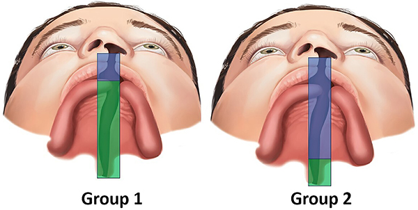
G*Power software (version 3.1.9.7; Heinrich-Heine-Universität Düsseldorf, Germany) was used for sample size calculation with α=5% and β=20%. In addition, a d-value of 0.86 (effect size) was considered for the parameter of cleft depth in the smaller segment, according to a previous study. The analysis indicated that the present study should have at least 23 participants/group. Thus, 28 individuals were allocated into each of two groups (Figure 1) according to the different surgical techniques. Group 1 (G1) included participants who had undergone the Millard technique (Figure 2) for cheiloplasty at three months of age and the von Langenbeck one-stage palatoplasty (Figure 3) at 12 months. Group 2 (G2) included participants who had undergone the Millard technique (Figure 2) for cheiloplasty at three months of age and simultaneous repair of the hard palate using the Hans Pichler technique; at 12 months of age, soft palate palatoplasty was performed using the Sommerlad technique. All participants had their palate molded at the pediatric dentistry clinic at the following stages: preoperative 1 (T1, at three months of age), preoperative 2 (T2, at 12 months of age), and postoperative (T3, at 24 months of age).
Group 1: Millard technique (cheiloplasty) at three months of age (blue) and von Langenbeck technique (one-stage palatoplasty) at 12 months (green). Group 2: Millard technique and Hans Pichler technique (hard palate repair) at three months (blue); Sommerlad technique (soft palate repair) at 12 months (green).
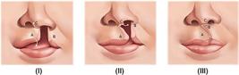
Millard Technique (cheiloplasty). Surgical flaps were made on the side without cleft for inferior rotation of the medial lip element (A); the medial advancement flap - side with cleft (B); and the base flap on the columella (C). I, Preoperative view; II, Transoperative view; III, Postoperative view.
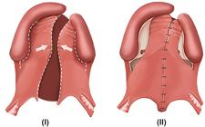
von Langenbeck technique (palatoplasty). I, Preoperative view: incisions were made on the margin of the cleft (from the alveolar region to the uvula), and relaxing incisions were made from the canine to the retromolar region. The incisions were made on the sides with and without cleft. II, Postoperative view.
Procedures
All impressions were sent to the dental laboratory of the rehabilitation center to obtain plaster dental models. After this stage, the models were scanned by a 3D scanner (R700™, 3Shape, Denmark) with proven accuracy in previous studies. The virtual models were exported in Standard Tesselation Language (STL) format and subsequently analyzed using Mirror¯ software (Canfield Scientific, USA) (15).
Digital morphometric analyses
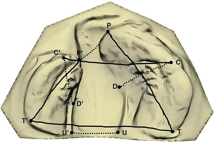
The software manually marked anatomical points according to published articles (14,16). Among these points, across-surface distances anterior (P-P′) and posterior (U-U′) cleft widths, intercanine (C-C′) and intertuberosity (T-T′) widths, lengths of the smallest (P′-T′) and largest (P-T) segments, and cleft depths in the smallest (C′-D′) and largest (C-D) segments were evaluated (16) (Figure 4). All analyses were automatically calculated by the software and quantified in mm.
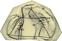
Across-surface distances evaluated in dental arches. Anterior (P-P′) and posterior (U-U′) cleft widths, intercanine (C-C′) and intertuberosity (T-T′) widths, lengths of the smaller (P′-T′) and larger (P-T) segments, and depths of the cleft in the smaller (C′-D′) and larger (C-D) segments.
Percent growth rate (GR)
The palatal growth was quantified in percentage and evaluated for all distances measured, according to the following formula: GR = (parameter T3 - parameter T1) × 100 / Parameter T1, where T1 = preoperative time 1 and T3 = postoperative time.
Statistical analysis
All across-surface distances were evaluated by Jamovi (computer software, version 2.3, Australia) and tested for normality (Shapiro-Wilk test). Fifty-six dental models (1/3 of the sample) were randomly selected for intra-rater (paired t-test) and inter-rater (Mann-Whitney test) agreement analysis. All parameters were quantified twice at an interval of 15 days (17). Paired t-test, Wilcoxon test, repeated measures ANOVA/Tukey test, and Friedman test/Dunn's multiple comparison test were used to compare within-group differences. Independent t-test and Mann-Whitney test were used to compare between-group percentage growth rates. Simple linear regression analysis and F-test were also applied. Descriptive data are reported as means±SD and median±interquartile range.
Results
Sample
One hundred and sixty-eight dental models were digitally evaluated, 84 from group 1, referring to 28 participants, and 84 from group 2 (28 participants). The impressions were taken at three different times, with the following mean ages in years: 0.35 (T1), 1.17 (T2), and 2.16 (T3).
Intra- and inter-rater reliability
There was no systematic error between the evaluated intra-rater (P=0.556, paired t-test) and inter-rater (P=0.491, Mann-Whitney test) distances, indicating that the measurements were reproducible.
Intragroup comparisons
In group 1, P-P′, U-U′, and C-C′ showed significant reductions after cheiloplasty (T2), unlike P′-T′, which increased (P<0.001, P=0.030, P<0.001, and P<0.001, respectively). Furthermore, the P-T and C′-D′ distances showed significant growth only after one-stage palatoplasty (T3) (P<0.001, P=0.003, in this order), while C-D increased at all stages (P<0.001) (Table 1).
Analysis between time points 1 (preoperative 1), 2 (preoperative 2), and 3 (postoperative) of the group treated with cheiloplasty and one-stage palatoplasty (G1).
In group 2, U-U′ and C-C′ showed significant reduction after cheiloplasty (T2) (P=0.001 and P<0.001, respectively). On the other hand, the distances P′-T′, P-T, C′-D′, and C-D showed significant growth after two-stage palatoplasty (T3) (P<0.001, for all) (Table 2).
Analysis between time points 1 (preoperative 1), 2 (preoperative 2) and 3 (postoperative) of the group treated with cheiloplasty and two-stage palatoplasty (G2).
Intergroup comparisons
There was no statistically significant difference in the comparison between groups at time 1 (P-P′, P=0.169; U-U′, P=0.782; C-C′, P=0.132; T-T′, P=0.281; C′-D′, P=0.826, independent t-test and P′-T′, P=0.248; P-T, P=0.571; C-D, P=0.187, Mann-Whitney test).
In general, the growth rate in group 2 was higher compared to group 1; however, only C′-D′ was significant (P=0.038) (Table 3).
Comparison of percentage growth rate between the group treated with cheiloplasty and one-stage palatoplasty (G1) and the group treated with cheiloplasty and two-stage palatoplasty (G2).
A simple linear regression analysis was performed in which the rehabilitation protocol was the independent variable and the measurements that presented normal distribution (T-T′ and C′-D′) were the dependent variables. The C′-D′ parameter showed a significant coefficient of determination (R2) of 0.076 (P=0.039), which means that the rehabilitation protocol explained 7.6% of the results of this distance. In other words, this model indicated that the rehabilitation protocol influenced the outcome (Table 4).
Simple linear regression between rehabilitation protocol (independent variable) and growth rate (dependent variable).
Discussion
In this study, one- and two-stage palatoplasty were evaluated in participants with unilateral cleft lip and palate operated on by a single plastic surgeon. Comparisons were made using dental models scanned with the across-surface distance method. This tool measures the shortest surface distance along a path defined by two anatomical points (16). Unfortunately, this tool needs to be used more in the analysis of dental arch development (16) and the face (18,19) of individuals with an orofacial cleft as more software tools make this anthropometric analysis tool available. However, its applicability should be disseminated to complement the knowledge of researchers and professionals who rehabilitate children with an orofacial cleft.
In group 1, both the intercanine measurement (C-C′) and the anterior (P-P′) and posterior (U-U′) cleft widths decreased after cheiloplasty (T2), indicating approximation between the alveolar segments in both the anterior and posterior portions of the dental arch. Although other authors have performed these analyses using the straight-line method, the data from this study corroborate the previous findings (11,12,20). In contrast, the lengths of the palatal segments, P′-T′ and P-T, and the cleft depths, C′-D′ and C-D, grew significantly from T1 to T3. Previous authors also reported total palate growth, measured as the distance from the inter-incisive point to the intertuberosity distance, after palatoplasty (13,21).
In group 2, the C-C′ distance and the posterior cleft width (U-U′) reduced after the first surgical step, i.e. cheiloplasty with anterior palatoplasty (T2). As in G1, group 2 also showed significant growth in palatal segment lengths (P′-T′ and P-T) and cleft depths (C′-D′ and C-D) between T1 and T3. These findings corroborate other data published in the scientific literature, in which the authors evaluated children with the same orofacial cleft phenotype but received different rehabilitation protocols (16).
Comparing the groups at T1, there was no statistically significant difference in the analyses before the surgical procedures. This finding is important, indicating no baseline discrepancies between groups. However, at T2, the growth rate of group 2 had higher growth percentages in five (C-C′, T-T′, P′-T′, C′-D′, and C-D) of the six parameters evaluated, but only C′-D′ showed a statistically significant difference. These data suggested that individuals who underwent two-stage palatal surgical repair showed better palatal growth, consistent with previous studies (22- 25). One systematic review on the effect of one- or two-stage palatoplasty on maxillofacial development was inconclusive due to contradictory results. However, the authors reinforce that the different surgical protocols and the age of the participants at assessment may have interfered with the inconclusive results (8).
It is well established in the literature that the development of dental arches of children with oral cleft can be influenced by various factors, such as genetics (9), treatment protocols (16), surgical techniques (17), age at surgeries (26), width of the cleft (9), in addition to the intrinsic characteristics of individuals. Although several studies (13,27,28) reinforce that the rehabilitation protocol influences palatal development, specific statistical analyses are not always applied to confirm this concept. This study used a simple linear regression analysis to evaluate the influence of the rehabilitation protocol on the quantitative parameters evaluated. As this analysis is conditioned to normally distributed data, only the T-T′ and C′-D′ parameters were assessed. The T-T′ distance was not significantly influenced by the rehabilitation protocol, but for the C′-D′ distance, the protocol explained 7.6% of the variation and this was statistically significant. These data reinforce that, in the evaluated model, the rehabilitation protocol affected palatal growth, especially the C′-D′ distance, corroborating the data from the between-group comparison of percentage growth rates.
The highlight of this study was that a single plastic surgeon operated on all participants; the involvement of more than one surgeon could bias the results (13). However, the existing limitation was the lack of classification for cleft width, i.e. wide, medium, or narrow. Therefore, this parameter could not be evaluated in the sample. Studies must be designed considering this vital factor, as wide clefts are challenging for surgeons and tend to lead to greater limitations in palatal development (9,10,14). Another point that should be considered is that, due to the absence of studies that evaluate the influence of the rehabilitation protocol from childhood to skeletal maturity, it is essential to perform comparative studies with other rehabilitation centers that have different rehabilitation protocols.
The evidence from this study allows us to conclude that, regardless of the surgical technique, dental arch development is influenced by the rehabilitation protocol. In the sample evaluated, individuals with a cleft palate operated on in two stages showed the most favorable palatal growth.
-
Funding
This study was supported by grants #2020/16690-0 and #2021/12424-6 from the São Paulo Research Foundation (FAPESP) and grant #310895/2020-0, from the National Council for Scientific and Technological Development (CNPq).
References
-
1 Freitas JAS, das Neves LT, de Almeida ALPF, Garib DG, Trindade-Suedam IK, Yaedú RYF, et al. Rehabilitative treatment of cleft lip and palate: experience of the Hospital for Rehabilitation of Craniofacial Anomalies/USP (HRAC/USP)--Part 1: overall aspects. J Appl Oral Sci 2012; 20: 9-15, doi: 10.1590/S1678-77572012000100003.
» https://doi.org/10.1590/S1678-77572012000100003 -
2 Tennison CW. The repair of the unilateral cleft lip by the stencil method. Plast Reconstr Surg (1946) 1952; 9: 115-120, doi: 10.1097/00006534-195202000-00005.
» https://doi.org/10.1097/00006534-195202000-00005 -
3 Randall P. A triangular flap operation for the primary repair of unilateral clefts of the lip. Plast Reconstr Surg Transplant Bull 1959; 23: 331-347, doi: 10.1097/00006534-195904000-00003.
» https://doi.org/10.1097/00006534-195904000-00003 -
4 Millard Jr DR. A radical rotation in single harelip. Am J Surg 1958; 95: 318-322, doi: 10.1016/0002-9610(58)90525-7.
» https://doi.org/10.1016/0002-9610(58)90525-7 - 5 Bosi VZ, Brandão GR, Yamashita RP. Speech resonance and surgical complications after primary palatoplasty with intravelar veloplasty in patients with cleft lip and palate. Braz J Plast Surg 2016; 31: 43-52.
-
6 Sommerlad BC. A technique for cleft palate repair. Plast Reconstr Surg 2003; 112: 1542-1548, doi: 10.1097/01.PRS.0000085599.84458.D2.
» https://doi.org/10.1097/01.PRS.0000085599.84458.D2 - 7 von Langenbeck BRK. Operation der angeborenem totalen spaltung des harten Gaumens nach einer neuen methode [in Germany]. Deutsh Klin 1861; 3: 231-232.
-
8 Reddy RR, Gosla Reddy S, Vaidhyanathan A, Bergé SJ, Kuijpers-Jagtman AM. Maxillofacial growth and speech outcome after one-stage or two-stage palatoplasty in unilateral cleft lip and palate. A systematic review. J Craniomaxillofac Surg 2017; 45: 995-1003, doi: 10.1016/j.jcms.2017.03.006.
» https://doi.org/10.1016/j.jcms.2017.03.006 -
9 Botticelli S, Küseler A, Marcusson A, Mølsted K, Nørholt SE, Cattaneo PM, et al. Do infant cleft dimensions have an influence on occlusal relations? A subgroup analysis within an RCT of primary surgery in patients with unilateral cleft lip and palate. Cleft Palate Craniofac J 2020; 57: 378-388, doi: 10.1177/1055665619875320.
» https://doi.org/10.1177/1055665619875320 -
10 Meazzini MC, Tortora C, Morabito A, Garattini G, Brusati R. Factors that affect variability in impairment of maxillary growth in patients with cleft lip and palate treated using the same surgical protocol. J Plast Surg Hand Surg 2011; 45: 188-193, doi: 10.3109/2000656X.2011.583493.
» https://doi.org/10.3109/2000656X.2011.583493 -
11 Shetty V, Agrawal RK, Sailer HF. Long-term effect of presurgical nasoalveolar molding on growth of maxillary arch in unilateral cleft lip and palate: randomized controlled trial. Int J Oral Maxillofac Surg 2017; 46: 977-987, doi: 10.1016/j.ijom.2017.03.006.
» https://doi.org/10.1016/j.ijom.2017.03.006 -
12 Jaklová LK, Hoffmannová E, Dupej J, Borský J, Jurovčík M, Černý M, et al. Palatal growth changes in newborns with unilateral and bilateral cleft lip and palate from birth until 12 months after early neonatal cheiloplasty using morphometric assessment. Clin Oral Investig 2021; 25: 3809-3821, doi: 10.1007/s00784-020-03711-9.
» https://doi.org/10.1007/s00784-020-03711-9 -
13 Ambrosio ECP, Sforza C, De Menezes M, Carrara CFC, Machado MAAM, Oliveira TM. Post-surgical effects on the maxillary segments of children with oral clefts: new three-dimensional anthro-pometric analysis. J Craniomaxillofac Surg 2018; 46: 1511-1514, doi: 10.1016/j.jcms.2018.06.017.
» https://doi.org/10.1016/j.jcms.2018.06.017 -
14 Ambrosio ECP, Pucciarelli MGR, Sforza C, Soares S, Menezes M, Carrara CFC, et al. Correlation between cleft width and dental arch symmetry in cleft lip and palate: a longitudinal study. Braz Oral Res 2023; 37: e021, doi: 10.1590/1807-3107bor-2023.vol37.0021.
» https://doi.org/10.1590/1807-3107bor-2023.vol37.0021 -
15 Ambrosio ECP, Camargo PB, Jorge PK, Carrara CFC, Machado MAAM, Oliveira TM. Digital workflow in the analysis of orofacial cleft in children: case series with 5 years of follow-up. Arch Health Invest 2023; 12: 6-11, doi: 10.21270/archi.v12i1.6049.
» https://doi.org/10.21270/archi.v12i1.6049 -
16 Cerón-Zapata AM, López-Palácio AM, Rodrigues-Ardila MJ, Berrio-Gutiérrez LM, De Menezes M, Sforza C. 3D evaluation of maxillary arches in unilateral cleft lip and palate patients treated with nasoalveolar moulding vs. Holtz's plate. J Oral Rehabil 2016; 43: 111-118, doi: 10.1111/joor.12350.
» https://doi.org/10.1111/joor.12350 -
17 Ambrosio ECP, Sartori IC, Jorge PK, Carrara CFC, Valarelli FP, Machado MAAM, et al. Six-year post-surgical evaluation in the treatment protocols in the dental arches of children with oral cleft: longitudinal study. J Appl Oral Sci 2022; 30: e20220120, doi: 10.1590/1678-7757-2022-0120.
» https://doi.org/10.1590/1678-7757-2022-0120 -
18 Alazzawi O, Morioka D, Miyabe M, Tosa Y, Ohkubo F, Yoshimoto S. Nasolabial growth in individuals with unilateral cleft lip and palate: a preliminary study of longitudinal observation using three-dimensional stereophotogrammetry. J Craniofac Surg 2017; 28: e449-e451, doi: 10.1097/SCS.0000000000003651.
» https://doi.org/10.1097/SCS.0000000000003651 -
19 Morioka D, Mandrano N, Fujimoto H, Koga Y, Sato N, Tosa Y, et al. Longitudinal follow-up of individuals with cleft lip using three-dimensional stereophotogrammetry. J Craniofac Surg 2018; 29: 1261-1265, doi: 10.1097/SCS.0000000000004434.
» https://doi.org/10.1097/SCS.0000000000004434 -
20 Saad MS, Fata M, Farouk A, Habib AMA, Gad M, Tayel MB, et al. Early progressive maxillary changes with nasoalveolar molding: randomized controlled clinical trial. JDR Clin Trans Res 2020; 5: 319-331, doi: 10.1177/2380084419887336.
» https://doi.org/10.1177/2380084419887336 -
21 Mello BZF, Ambrosio ECP, Jorge PK, de Menezes M, Carrara CFC, Soares S, et al. Analysis of dental arch in children with oral cleft before and after the primary surgeries. J Craniofac Surg 2019; 30: 2456-2458, doi: 10.1097/SCS.0000000000005775.
» https://doi.org/10.1097/SCS.0000000000005775 -
22 Yamanishi T, Nishio J, Kohara H, Hirano Y, Sako M, Yamanishi Y, et al. Effect on maxillary arch development of early 2-stage palatoplasty by modified furlow technique and conventional 1-stage palatoplasty in children with complete unilateral cleft lip and palate. J Oral Maxillofac Surg 2009; 67: 2210-2216, doi: 10.1016/j.joms.2009.04.038.
» https://doi.org/10.1016/j.joms.2009.04.038 -
23 Yamanishi T, Nishio J, Sako M, Kohara H, Hirano Y, Yamanishi Y, et al. Early two-stage double opposing Z-plasty or one-stage push-back palatoplasty?: comparisons in maxillary development and speech outcome at 4 years of age. Ann Plast Surg 2011; 66: 148-153, doi: 10.1097/SAP.0b013e3181d6e426.
» https://doi.org/10.1097/SAP.0b013e3181d6e426 -
24 Gundlach KKH, Bardach J, Filippow D, Stahl-de Castrillon F, Lenz JH. Two-stage palatoplasty, is it still a valuable treatment protocol for patients with a cleft of lip, alveolus, and palate? J Craniomaxillofac Surg 2013; 41: 62-70, doi: 10.1016/j.jcms.2012.05.013.
» https://doi.org/10.1016/j.jcms.2012.05.013 -
25 Mikoya T, Shibukawa T, Susami T, Sato Y, Tengan T, Katashima H, et al. Dental arch relationship outcomes in one- and two-stage palatoplasty for Japanese patients with complete unilateral cleft lip and palate. Cleft Palate Craniofac J 2015; 52: 277-286, doi: 10.1597/13-285.
» https://doi.org/10.1597/13-285 -
26 Kotova M, Urbanova W, Sukop A, Peterkova R, Peterka M, Petrova T. Dentoalveolar arch dimensions in UCLP boys after neonatal cheiloplasty or after lip surgery at the age of 3 or 6 months. Cleft Palate Craniofac J 2019; 56: 1020-1025, doi: 10.1177/1055665618824835.
» https://doi.org/10.1177/1055665618824835 -
27 Tome W, Yashiro K, Otsuki K, Kogo M, Yamashiro T. Influence of different palatoplasties on the facial morphology of early mixed dentition stage children with unilateral cleft lip and palate. Cleft Palate Craniofac J 2016; 53: e28-e33, doi: 10.1597/14-191.
» https://doi.org/10.1597/14-191 -
28 Xu X, Cao C, Zheng Q, Shi B. The influence of four different treatment protocols on maxillofacial growth in patients with unilateral complete cleft lip, palate, and alveolus. Plast Reconstr Surg 2019; 144: 180-186, doi: 10.1097/PRS.0000000000005711.
» https://doi.org/10.1097/PRS.0000000000005711