
 Effects of prolonged exposure to diclofenac sodium on strains of Staphylococcus aureus
Abstract
Staphylococcus aureus is a pathogen commonly involved in the etiology of nosocomial and community-acquired infections. This microorganism adheres and forms biofilm on biotic and abiotic surfaces, which is a significant complication for infection control. More recently, non-antibiotic medications, such as nonsteroidal anti-inflammatory agents (NSAIDs), have been studied as an alternative for treating infections caused by multidrug-resistant pathogens and infections associated with biofilms. This study assessed the effects of prolonged exposure of S. aureus strains to the NSAID diclofenac sodium regarding biofilm production, direct adhesion, hydrophobicity, susceptibility to antimicrobials, hemolysis, and biochemical profile. Some strains exposed to diclofenac showed increased direct adhesion to hydrophobic and hydrophilic surfaces and increased cell surface hydrophobicity. Two strains, one reference and one clinical, were subjected to repeated and prolonged passages in a culture medium with a diclofenac gradient. In one of them, the drug suppressed biofilm production in the assay with and without subinhibitory concentrations (sub-MICs) from NSAID. Additional repeated passages of this strain in a medium without diclofenac partially recovered the original biofilm-positive phenotype, but only in culture with sub-MICs of the drug. The original characteristics of the strains regarding susceptibility or resistance to antimicrobials and biochemical profile were not affected by prolonged exposure to diclofenac. However, the clinical isolate showed suppressed hemolytic activity.
Keywords:
Staphylococcus aureus; diclofenac sodium; resistance; biofilm; drug repositioning
Resumo
Staphylococcus aureus é um patógeno comumente envolvido na etiologia de infecções nosocomiais e adquiridas na comunidade. Este microrganismo adere e forma biofilme em superfícies bióticas e abióticas, o que é uma complicação significativa para o controle de infecções. Mais recentemente, medicamentos denominados “não antibióticos”, como agentes anti-inflamatórios não esteroides (AINEs), têm sido estudados como uma alternativa para o tratamento de infecções causadas por patógenos multirresistentes e infecções associadas a biofilmes. Este estudo avaliou os efeitos da exposição prolongada de amostras de S. aureus ao AINE diclofenaco de sódio em relação à produção de biofilme, adesão direta, hidrofobicidade, suscetibilidade a antimicrobianos, hemólise e perfil bioquímico. Algumas amostras expostas ao diclofenaco mostraram maior adesão direta a superfícies hidrofóbicas e hidrofílicas e maior hidrofobicidade da superfície celular. Duas amostras, uma de referência e uma clínica, foram submetidas a passagens repetidas e prolongadas em um meio de cultura com gradiente de diclofenaco. Em uma delas, o fármaco suprimiu a produção de biofilme no ensaio com e sem concentrações subinibitórias (sub-MICs) do AINE. Passagens repetidas adicionais dessa amostra em um meio sem diclofenaco recuperaram parcialmente o fenótipo original positivo para biofilme, mas apenas em cultura com sub-MICs da droga. As características originais das amostras em relação à suscetibilidade ou resistência a antimicrobianos e perfil bioquímico não foram afetadas pela exposição prolongada ao diclofenaco. No entanto, o isolado clínico mostrou supressão da atividade hemolítica.
Palavras-chave:
Staphylococcus aureus; diclofenaco de sódio; resistência; biofilme; reposicionamento de fármacos
1. Introduction
The increasing of multidrug-resistant bacterial pathogens has highlighted the need for novel therapeutic strategies. One promising approach involves the use of non-antibiotic drugs—compounds not initially designed to target bacterial infections but later found to possess antimicrobial properties. Among these are nonsteroidal anti-inflammatory drugs (NSAIDs), with diclofenac (2-[2-[(2,6-dichlorophenyl)amino]phenyl]acetic acid) being one of the most widely prescribed (de Lima e Silva and Martins Silva, 2018; Barbarossa et al., 2022). Diclofenac exerts its anti-inflammatory, analgesic, and antipyretic effects by inhibiting prostaglandin synthesis through the inactivation of cyclooxygenase enzymes (Altman et al., 2015). In addition to these effects, diclofenac has demonstrated antimicrobial activity both in vitro (Alqahtani et al., 2019; Hegazy, 2016) and in vivo (Dutta et al., 2004; Dutta et al., 2007) against various bacterial pathogens.
The minimum inhibitory concentration (MIC) of diclofenac for S. aureus has been reported to range from 50 to >1000 µg/mL. These discrepancies may be attributed to the intrinsic physiological characteristics of the bacterial isolates or to differences in the experimental methodologies, such as how the drug is solubilized in the culture medium (de Lima e Silva and Martins Silva, 2018). Although these MIC values exceed typical therapeutic plasma concentrations, topical diclofenac formulations can reach significantly higher local concentrations (Bariguian Revel et al., 2020).
In addition to its antibacterial activity, diclofenac's effect on biofilm formation has been investigated (Queiroz et al., 2021; Xi et al., 2024; de Lima e Silva et al., 2021). Biofilms are microbial communities encased in a protective polymeric matrix, which enhances bacterial resistance to immune responses and antimicrobial agents. In S. aureus, biofilm formation can occur either dependently or independently of the icaADBC operon. The ica operon encodes enzymes for polysaccharide intercellular adhesin (PIA) production, while alternative ica-independent pathways for biofilm formation involve proteins and/or extracellular DNA (McCarthy et al., 2015). Regardless of these mechanisms, biofilm formation is initiated by strain adhesion to surfaces, which is mediated by factors such as degree of hydrophobicity, electrostatic interactions, van der Waals forces, and protein adhesion (Sharma et al., 2023). This study aims to evaluate the effect of prolonged exposure to diclofenac on S. aureus strains, focusing on its impact on direct adhesion, hydrophobicity, biofilm formation, antimicrobial susceptibility, biochemical profile, and hemolytic activity.
2. Materials and Methods
2.1. Bacterial strains and preparation of diclofenac solution
Two reference strains of S. aureus (ATCC 25923 and ATCC 4300), five clinical isolates, and five nasal swab isolates from healthy individuals were examined. The clinical and carrier strains selected for this study are part of a group previously characterized by phenotypic and molecular methods, as well as their antibacterial activity and biofilm production induced by diclofenac (de Lima e Silva, et al., 2021). The diclofenac sodium solution (Sigma) was prepared in methanol and sterilized by filtration.
2.2. Biofilm production by strains
Biofilm production in the 12 S. aureus strains was assessed using the microtiter plate assay (MTPA) (Stepanović et al., 2007). Briefly, overnight cultures (1:100 dilution) were added to Tryptone Soy Broth (TSB) containing diclofenac (6.25–800 μg/mL) and to a TSB control. The bacterial suspensions were inoculated into 96-well plates (200 μL/well) and incubated for 24 hours at 35°C. Following incubation, the wells were washed, fixed with methanol, and stained for 15 minutes with a 2% Hucker's crystal violet solution. After removing the dye, the plates were washed, and 200 μL of 95% ethanol was added for 30 minutes to extract the dye. The optical density (OD570nm) of the biofilm extracts was then measured. The cut-off OD value used to differentiate biofilm-producing from non-biofilm-producing isolates was determined according to Christensen et al. (1985). The classification of biofilm production as weak, moderate, or strong was based on Stepanović et al. (2007).
2.3. Determination of direct adherence of strains after prolonged growth in a diclofenac gradient medium
The effect of prolonged exposure to diclofenac (96 hours) on bacterial adhesion to glass, stainless steel, and polystyrene was evaluated using two reference strains, five clinical isolates, and five isolates from healthy individuals. The tests were conducted in Tryptic Soya Agar (TSA) containing a diclofenac concentration gradient, as described by O'Leary et al. (2004). The second layer of molten TSA, poured onto a plate containing the first slanted layer of the medium, contained 1.089 mg/mL of diclofenac. Once the medium solidified, 50 µL of the strains, diluted 1:100, were inoculated using a sterile cell spreader. The plates were incubated for 96 hours at 35°C, and then standardized suspensions were prepared in phosphate-buffered saline (PBS, pH 6.8) from bacterial growth collected from the region bordering the area of inhibition. Suspensions were also prepared with strains grown in control TSA. The suspensions were centrifuged (2,000 x g, 10 min, 4°C), washed with PBS, resuspended in 400 µL of PBS, and homogenized in a tube shaker. Subsequently, 20 µL of the bacterial suspensions were transferred in triplicate to each tested surface. After 10 minutes of contact, the suspensions were removed by suction using a vacuum device, and the surface was washed with running purified water. The surfaces were then treated with methanol (1 minute), washed with running purified water, covered with 1% Hucker's crystal violet solution (1 minute), and washed again with running purified water. After drying, the purple color retained in the areas exposed to the bacterial suspensions was compared between the test and control strains.
2.4. Evaluation of the hydrophobicity of strains after prolonged growth in a diclofenac gradient medium
Hydrophobicity was assessed using bacterial suspensions prepared as described for the direct adherence test. The strains examined were the same as those described above. Solutions of ammonium sulfate in PBS (pH 6.8) at concentrations of 0.5, 1.5, 3.0, and 6.0 M were prepared according to Krepsky et al. (2003). Fifty microliters of the bacterial suspensions were deposited on Kline plates, followed by the addition of the same volume of each PBS solution with ammonium sulfate, as well as PBS without salt. The PBS solutions containing ammonium sulfate were diluted to half their original concentration (0.25, 0.75, 1.5, and 3.0 M). The plates were gently agitated on an orbital shaker for 5 minutes, and the presence of bacterial aggregates was assessed. The lowest concentration of ammonium sulfate that resulted in visible bacterial clumping was scored as a numerical value of the bacterial surface hydrophobicity (SAT value). A positive result was considered for strains with SAT values ≤1.5 M. The autoaggregation of the strains (i.e., bacterial aggregation in an isotonic buffer) was also evaluated.
2.5. Repeated passages of S. aureus in TSA medium containing a diclofenac gradient
Two strains were selected for this experiment: S. aureus ATCC 25923 and SA03. The preparation of the plates with the diclofenac concentration gradient and the inoculation procedures were performed as described previously. Control cultures were preserved in TSB with 20% glycerol and stored frozen. The plates were incubated for 96 hours at 35°C. Standardized bacterial suspensions were prepared in 3 mL of PBS, using bacterial growth collected from the region adjacent to the area of inhibition. Suspensions were also prepared from strains grown in TSA control medium. The suspensions were homogenized, diluted 1:100 in PBS, and 50 μL aliquots were immediately inoculated onto freshly prepared plates, both with and without the diclofenac gradient. Passages were repeated every 96 hours of incubation until 16 consecutive passages were completed, generating cultures from passage 1 in the diclofenac gradient (P1) and from passage 1 in the control medium (PC1). Subsequently, cultures subjected to a series of passage 1 (P1 and PC1) underwent 16 additional passages in TSA without diclofenac, resulting in series of passage 2 cultures, identified as P2 and PC2, respectively. The purity of the cultures was checked by inoculating TSA plates at each stage. Cultures subjected to passages P1 and PC1 were tested for MIC (Minimum Inhibitory Concentration) and MBC (Minimum Bactericidal Concentration) of diclofenac, biofilm production, hemolytic activity, biochemical profile, and antimicrobial susceptibility. Cultures from P2 and PC2 were only tested for biofilm production. To provide comparison, strains ATCC 25923 and SA03 that were not subjected to passage tests in TSA medium, either with or without diclofenac, were also tested. These cultures were referred to as P0 (zero passage).
2.6. Determination of the MIC and MBC of diclofenac after successive passages in the TSA medium with and without diclofenac
The MIC of the strains was determined using the broth dilution method (Clinical and Laboratory Standards Institute - CLSI, 2013). The MBC was determined by subculturing 10 μL of medium from wells that exhibited no visible growth onto TSA plates. After incubation for 18 hours at 35°C, the MBC was defined as the minimum concentration of diclofenac that resulted in a ≥99.9% reduction in CFU/mL relative to the initial inoculum.
2.7. Biofilm production by strains subjected to repeated passages in TSA medium with and without diclofenac
Biofilm production assays were performed in microtiter plates as described above in Section 2, using TSB supplemented with diclofenac at final concentrations of 25, 50, 100, 200, 400, and 800 µg/mL. Strains grown in TSB medium without diclofenac were used as controls. Cultures subjected to passage series 1 and 2 (P1, P2, PC1, and PC2) were tested, along with strains that were not subjected to passages (P0).
2.8. Effect of prolonged exposure to diclofenac on hemolysis
Suspensions derived directly from the plates of strains SA03 (P1 and PC1) and ATCC 25923 (P1 and PC1) were prepared in TSB. These suspensions were then inoculated as 10 µL spots onto Blood Agar plates and incubated at 35°C. Readings were taken at 24, 48, and 72 hours to assess the formation of hemolytic halos around bacterial growth. For comparison, suspensions prepared in TSB from the respective cultures that were not subjected to serial passage tests were also inoculated.
2.9. Effect of prolonged exposure to diclofenac on biochemical behavior and susceptibility to antimicrobials
Strains subjected to 16 serial passages in TSA medium with and without diclofenac (P1 and PC1) were evaluated for their biochemical profile and antimicrobial susceptibility using the VITEK® 2 System. Strains SA03 and ATCC 25923 that were not subjected to serial passage experiments (P0) were also tested as controls. The automated system assessed a set of 45 tests for the identification of Gram-positive cocci and determination of the MIC for 15 antimicrobials. Additionally, the strains underwent the cefoxitin disc screening test (CLSI, 2013).
3. Results and Discussion
3.1. Effects of sub-MICs of diclofenac on biofilm formation
All five clinical strains were negative for biofilm formation on TSB. In three of these strains, diclofenac induced biofilm production. Four carrier isolates were negative for biofilm formation on TSB, and only one of these strains exhibited biofilm production when exposed to diclofenac. The fifth strain was positive for biofilm production on both TSB with and without diclofenac. The two reference strains were biofilm-positive on both TSB and TSB supplemented with diclofenac (Table 1). These results confirm previous findings (de Lima e Silva et al. 2021).
The effect of diclofenac in inducing biofilm formation in Staphylococcus has been poorly investigated. Some studies have reported an anti-biofilm effect of this NSAID on various bacterial pathogens (Xi et al., 2024; El-Baky and El-Gendy, 2016; Hegazy, 2016). However, de Lima e Silva et al. (2021) demonstrated that sub-MICs of diclofenac can induce non-polysaccharide biofilm production in S. aureus strains, suggesting that this drug activates a PIA-independent pathway.
3.2. Effects on direct adhesion and hydrophobicity after prolonged growth in a diclofenac gradient medium
Bacterial attachment is a critical step in biofilm formation, involving reversible interactions governed by physicochemical factors such as electrostatic and hydrophobic forces. To investigate bacterial adhesion, tests were conducted on glass, polystyrene, and 316L stainless steel surfaces after prolonged cultivation in TSA with a diclofenac gradient. Stainless steel is widely used in medical implants, whereas polystyrene, although not employed in medical devices, is an excellent material for promoting cell adhesion (Merritt et al., 1998; Li et al., 2003).
Table 1 shows the results of adherence. The reference strains ATCC 43300 and ATCC 25923 (both biofilm-positive in media with and without diclofenac) exhibited increased adherence to all materials. Strains B03 (biofilm-negative in TSB but induced by diclofenac) and B56 (biofilm-positive in both conditions) demonstrated increased adhesion to polystyrene and steel. Strain B43 (biofilm-negative in both conditions) showed increased adhesion solely to polystyrene. Clinical strains, including those whose biofilm production was induced by diclofenac, did not show significant changes in adherence compared to controls. Therefore, these results suggest that diclofenac can induce increased adhesion in certain S. aureus strains to both hydrophilic and hydrophobic surfaces. Hegazy (2016) found that sub-MIC diclofenac did not affect Proteus mirabilis adhesion to polystyrene but inhibited biofilm formation, suggesting that diclofenac acts to prevent the later stages of biofilm formation.
In contrast to biofilm-positive strains in TSB, among the strains that transitioned from biofilm-negative to biofilm-positive due to diclofenac, only B03 exhibited increased adherence after prolonged exposure to the drug. This suggests that diclofenac-induced adherence is not necessarily linked to biofilm formation. Strain B43, which was biofilm-negative in both conditions, also showed increased adhesion following diclofenac exposure, supporting this hypothesis.
The study of direct adhesion was further complemented by evaluating the drug’s effect on the strains' hydrophobicity. Hydrophobic interactions between the microbial surface and the contact site influence adhesion, and evidence suggests their involvement in a wide range of infectious processes (Krepsky et al., 2003; Cerca et al., 2005). Among the surfaces tested, polystyrene is hydrophobic with little to no surface charge, while stainless steel is hydrophilic, with positive or neutral surface charges. Glass surfaces are also hydrophilic but carry negative surface charges (Pagedar et al., 2010). Diclofenac increased hydrophobicity in strains ATCC 25923, ATCC 43300, and B56, which also exhibited a biofilm-positive phenotype in TSB and increased adhesion after exposure to diclofenac.
For reference strains, the increase in hydrophobicity was also accompanied by marked autoaggregation in PBS (Figure 1). Autoaggregation and hydrophobicity are regarded as independent traits; however, Rahman et al. (2008) found a correlation between them, suggesting that hydrophobicity may influence autoaggregation. On the other hand, Burel et al. (2021) demonstrated that S. aureus planktonic cells aggregate rapidly, partly driven by the bacteria's surface potential and hydrophobicity. For instance, the suspension of S. aureus with more aggregates was found to be 20 times more resistant to quaternary ammonium compounds than one with fewer aggregates.
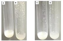
Autoaggregation of strains ATCC 43300 and ATCC 25923 in PBS after growth in the presence of diclofenac. A: ATCC 43300 strain not exposed to diclofenac; B: ATCC 43300 strain exposed to diclofenac; C: strain ATCC 25923 not exposed to diclofenac; D: ATCC 25923 strain exposed to diclofenac.
Thus, only the three strains primarily biofilm-positive showed an association between increased adhesion and hydrophobicity induced by diclofenac. Among the strains whose biofilm production was induced by diclofenac, only B03 exhibited an increase in adhesion, but no increase in hydrophobicity. Therefore, the results concerning biofilm, direct adhesion, and hydrophobicity suggest that the bacterial response to diclofenac seems to be more strain-specific. For example, in cases where diclofenac induced both increased hydrophobicity and adherence to both hydrophilic and hydrophobic surfaces, the ability to adhere to glass and steel (hydrophilic surfaces) may be attributed to the drug’s induction of slime production and/or the action of specific cell wall proteins, rather than solely the microorganism's surface hydrophobicity (Krepsky et al., 2003; Aboelnaga et al., 2024).
3.3. Effect of repeated passages in TSA medium with and without diclofenac gradient on susceptibility to diclofenac and biofilm production
Two strains, one reference (ATCC 25922) and one clinical (SA3, multidrug-resistant, identified as MRSA - data not shown), were subjected to repeated and prolonged passages in TSA with a diclofenac gradient. The serial exposure of bacteria to concentration gradients of antimicrobial agents is an effective method for selecting isolates that acquire resistance during exposure to the studied agent (Arsene et al., 2021).
After 16 passages of the two strains in diclofenac gradient medium, no significant differences in MIC or MBC were observed compared to the PC1 and P0 controls. This suggests a resistance barrier to diclofenac, even after prolonged exposure to the drug. Although the primary antibacterial mechanism of diclofenac appears to be the inhibition of DNA synthesis (Dastidar et al., 2000), other concurrently affected sites, such as the cell membrane (El-Baky and El-Gendy, 2016), may explain the absence of resistance emergence during exposure to this NSAID.
Table 2 displays the results regarding biofilm formation by the ATCC 25923 strain. Successive passages in TSA with diclofenac gradient (P1) and control medium (P2, PC1, and PC2) resulted in the loss of the strain’s original biofilm production capability in TSB without diclofenac in the MTP assay. Growth rates, as estimated by optical density in TSB without diclofenac, did not influence the results, as no significant differences were observed compared to the original strain (P0). Latimer et al. (2012) also reported the loss of S. aureus biofilm production after repeated exposure to triclosan, even in subsequent passages without the compound, while Arsene et al. (2021) observed increased biofilm production following prolonged exposure to sub-MICs of kanamycin and ampicillin but a decrease after exposure to silver nanoparticles.
Effects on biofilm production by repeated passages of S. aureus ATCC25922 in TSA medium with and without diclofenac gradient.
Sub-MICs of diclofenac in the MTP assay did not restore biofilm production in the P1 strain ATCC 25923. However, interestingly, after the second series of passages in TSA, this time without the drug (P2), it was observed that diclofenac induced weak biofilm production in almost all concentrations tested. Strain PC1 (control from the first series of 16 passages in TSA without diclofenac gradient) also restored biofilm production ability after exposure to sub-MICs of diclofenac. However, the levels of production ranged from moderate to strong. This phenotype induced by sub-MICs of the drug persisted after the second stage of 16 passages of the strain in TSA without the drug (PC2), with biofilm production levels similar to those of PC1 (Table 2).
The stressful conditions caused by high diclofenac concentrations (P1) and prolonged subcultures (P1 and PC1) were likely responsible for the suppression of the original biofilm-positive phenotype. These conditions may have triggered the phase variation phenomenon, which involves the reversible insertion of the IS256 element into the ica locus (Ziebuhr et al., 1999; Kiem et al., 2004) or insertions of this element into the rsbU gene of the sigB operon or the sarA locus, both global stress response regulators that influence ica expression (Conlon et al., 2004). The reversion to the original biofilm-positive phenotype observed in the P2 strain in the MTP assay with diclofenac suggests the potential of this drug to induce biofilm production. However, it is worth noting that this reversion was incomplete, as the levels of biofilm production induced by diclofenac were significantly lower than those expressed by the P0 strain (Table 2). Results for strain SA3 are not included in the table, as, unlike strain ATCC 25923, it did not produce biofilm under any condition. Thus, this strain demonstrated high stability in its original phenotype when exposed to diclofenac.
3.4. Effect of prolonged exposure to diclofenac on hemolysis, biochemical behavior and antibiotic susceptibility
Strain ATCC 25923-P1 (subjected to the diclofenac gradient) showed a significant reduction in hemolysis around bacterial growth compared to the results obtained for its control PC1 and for the culture of the P0 strain (Figure 2). Latimer et al. (2012) also observed inhibition of hemolysis after prolonged exposure of S. aureus to triclosan, in addition to inhibition of other virulence-related activities. They suggested that the staphylococcal accessory regulatory gene (sarA) could be implicated. Strain SA03 did not exhibit hemolysis under any culture condition but lost its yellow pigmentation after successive passages in the medium with diclofenac (data not shown). This pigment is considered a virulence factor for the microorganism, as mutants lacking the pigment are more susceptible to oxidative death and have compromised survival in the presence of neutrophils (Liu et al., 2005).

Strains subjected to repeated exposure to diclofenac did not show changes in the results of a set of biochemical tests. No alterations in the antimicrobial susceptibility profile were observed compared to controls. Zhang et al. (2021) also did not observe mutations for resistance to some antimicrobials after continuous passage of an MRSA strain in medium with high diclofenac concentrations. However, Riordan et al. (2011) reported that sub-MICs of diclofenac promoted changes in the expression of a broad range of genes and antimicrobial susceptibility in an MRSA strain. Therefore, diclofenac-induced changes at the genetic level, leading to broad phenotypic alterations, may depend on strain-specific responses.
4. Conclusion
Prolonged exposure of S. aureus to diclofenac affected adhesion and hydrophobicity in some strains but did not result in the development of resistance to this NSAID. Biofilm production was suppressed in originally biofilm-positive strains, but recovery of the original biofilm production phenotype partially occurred with exposure to sub-MICs of the drug. Biochemical and antimicrobial susceptibility profiles were not affected in the strains studied. However, the loss of hemolytic capacity in one of the strains suggests that virulence characteristics may be impacted.
Acknowledgments
This work was financed in part by Federal University of the State of Rio de Janeiro (UNIRIO).
References
-
ABOELNAGA, N., ELSAYED, S.W., ABDELSALAM, N.A., SALEM, S., SAIF, N.A., ELSAYED, M., AYMAN, S., NASR, M. and ELHADIDY, M., 2024. Deciphering the dynamics of methicillin-resistant Staphylococcus aureus biofilm formation: from molecular signaling to nanotherapeutic advances. Cell Communication and Signaling, vol. 22, pp. 188. http://doi.org/10.1186/s12964-024-01511-2 PMid:38519959.
» http://doi.org/10.1186/s12964-024-01511-2 -
ALQAHTANI, F.Y., ALEANIZY, F.S., TAHIR, E.E., ALQUADEIB, B.T., ALSARRA, I.A., ALANAZI, J.S. and ABDELHADY, H.G., 2019. Preparation, characterization, and antibacterial activity of diclofenac-loaded chitosan nanoparticles. Saudi Pharmaceutical Journal, vol. 27, no. 1, pp. 82-87. http://doi.org/10.1016/j.jsps.2018.08.001 PMid:30662310.
» http://doi.org/10.1016/j.jsps.2018.08.001 -
ALTMAN, R., BOSCH, B., BRUNE, K., PATRIGNANI, P. and YOUNG, C., 2015. Advances in NSAID development: evolution of diclofenac products using pharmaceutical technology. Drugs, vol. 75, no. 8, pp. 859-877. http://doi.org/10.1007/s40265-015-0392-z PMid:25963327.
» http://doi.org/10.1007/s40265-015-0392-z -
ARSENE, M.M.J., VIKTOROVNA, P.I., GRIGORIEVNA, V.E., DAVARES, A.K.L., SERGEEVNA, D.M. and NIKOLAEVNA, S.I., 2021. Prolonged exposure to antimicrobials induces changes in susceptibility to antibiotics, biofilm formation and pathogenicity in Staphylococcus aureus. Journal of Pharmaceutical Research International, vol. 33, no. 34B, pp. 140-151. http://doi.org/10.9734/jpri/2021/v33i34B31856
» http://doi.org/10.9734/jpri/2021/v33i34B31856 -
BARBAROSSA, A., ROSATO, A., CORBO, F., CLODOVEO, M. L., FRACCHIOLLA, G., CARRIERI, A. and CAROCCI, A., 2022. Non-antibiotic drug repositioning as an alternative antimicrobial approach.Antibiotics, vol. 11, no. 6, pp. 816. https://doi.org/10.3390/antibiotics11060816
» https://doi.org/10.3390/antibiotics11060816 -
BARIGUIAN REVEL, F., FAYET, M. and HAGEN, M., 2020. Topical diclofenac, an efficacious treatment for osteoarthritis: A narrative review. Rheumatology and Therapy, vol. 7, no. 2, pp. 217-236. http://doi.org/10.1007/s40744-020-00196-6 PMid:32086778.
» http://doi.org/10.1007/s40744-020-00196-6 -
BUREL, C., DREYFUS, R. and PUREVDORJ-GAGE, L., 2021. Physical mechanisms driving the reversible aggregation of Staphylococcus aureus and response to antimicrobials. Scientific Reports, vol. 11, no. 1, pp. 15048. http://doi.org/10.1038/s41598-021-94457-1 PMid:34294832.
» http://doi.org/10.1038/s41598-021-94457-1 -
CERCA, N., PIER, G.B., VILANOVA, M., OLIVEIRA, R. and AZEREDO, J., 2005. Quantitative analysis of adhesion and biofilm formation on hydrophilic and hydrophobic surfaces of clinical isolates of Staphylococcus epidermidis. Research in Microbiology, vol. 156, no. 4, pp. 506-514. http://doi.org/10.1016/j.resmic.2005.01.007 PMid:15862449.
» http://doi.org/10.1016/j.resmic.2005.01.007 -
CHRISTENSEN, G.D., SIMPSON, W.A., YOUNGER, J.J., BADDOUR, L.M., BARRETT, F.F., MELTON, D.M. and BEACHEY, E.H., 1985. Adherence of coagulase-negative staphylococci to plastic tissue culture plates: a quantitative model for the adherence of staphylococci to medical devices. Journal of Clinical Microbiology, vol. 22, no. 6, pp. 996-1006. http://doi.org/10.1128/jcm.22.6.996-1006.1985 PMid:3905855.
» http://doi.org/10.1128/jcm.22.6.996-1006.1985 - CLINICAL AND LABORATORY STANDARDS INSTITUTE – CLSI, 2013. VET01.A4 - Performance standards for antimicrobial disk and dilution susceptibility tests for bacteria isolated from animals. 3rd ed. Wayne: CLSI.
-
CONLON, K.M., HUMPHREYS, H. and O’GARA, J.P., 2004. Inactivations of rsbU and sarA by IS256 represent novel mechanisms of biofilm phenotypic variation in Staphylococcus epidermidis. Journal of Bacteriology, vol. 186, no. 18, pp. 6208-6219. http://doi.org/10.1128/JB.186.18.6208-6219.2004 PMid:15342591.
» http://doi.org/10.1128/JB.186.18.6208-6219.2004 -
DASTIDAR, S.G., GANGULY, K., CHAUDHURI, K. and CHAKRABARTY, A.N., 2000. The anti-bacterial action of diclofenac shown by inhibition of DNA synthesis. International Journal of Antimicrobial Agents, vol. 14, no. 3, pp. 249-251. http://doi.org/10.1016/S0924-8579(99)00159-4 PMid:10773497.
» http://doi.org/10.1016/S0924-8579(99)00159-4 -
DE LIMA E SILVA, A.A. and MARTINS SILVA, P., 2018. Non-antibiotic compounds: the activity of the NSAID diclofenac on bacteria- a review. International Journal of Current Microbiology and Applied Sciences, vol. 7, no. 12, pp. 340-351. http://doi.org/10.20546/ijcmas.2018.712.042
» http://doi.org/10.20546/ijcmas.2018.712.042 -
DE LIMA E SILVA, A.A., VIANA, A.S., SILVA, P.M., DE MATOS NOGUEIRA, E., SALGADO, L.T., TOMAZETTO DE CARVALHO, R. and SILVA FILHO, R.G., 2021. Diclofenac May Induce PIA-Independent Biofilm Formation inStaphylococcus aureusStrains. International Journal of Microbiology, vol. 2021, no. 1, pp. 8823775. http://doi.org/10.1155/2021/8823775
» http://doi.org/10.1155/2021/8823775 -
DUTTA, N.K., ANNADURAI, S., MAZUMDAR, K., DASTIDAR, S.G., KRISTIANSEN, J.E., MOLNAR, J., MARTINS, M. and AMARAL, L., 2007. Potential management of resistant microbial infections with a novel non-antibiotic: the anti-inflammatory drug diclofenac sodium. International Journal of Antimicrobial Agents, vol. 30, no. 3, pp. 242-249. http://doi.org/10.1016/j.ijantimicag.2007.04.018 PMid:17644318.
» http://doi.org/10.1016/j.ijantimicag.2007.04.018 - DUTTA, N.K., KUMAR, K.A., MAZUMDAR, K. and DASTIDAR, S.G., 2004. In vitro and in vivo antimycobacterial activity of antiinflammatory drug, diclofenac sodium. Indian Journal of Experimental Biology, vol. 42, no. 9, pp. 922-927. PMid:15462188.
-
EL-BAKY, R.M.A. and EL-GENDY, S.G., 2016. Effect of non-steroidal anti-inflammatory drugs and dexamethasone on the biofilm formation and expression of some adhesion-related genes ofCandida albicansandStaphylococcus aureus. African Journal of Microbiological Research, vol. 10, no. 20, pp. 694-707. http://doi.org/10.5897/AJMR2016.8013
» http://doi.org/10.5897/AJMR2016.8013 -
HEGAZY, W.A.H., 2016. Diclofenac inhibits virulence of Proteus mirabilis isolated from diabetic foot ulcer. African Journal of Microbiological Research, vol. 10, no. 21, pp. 733-743. http://doi.org/10.5897/AJMR2016.8043
» http://doi.org/10.5897/AJMR2016.8043 -
KIEM, S., OH, W.S., PECK, K.R., LEE, N.Y., LEE, J.Y., SONG, J.H., HWANG, E.S., KIM, E.C., CHA, C.Y. and CHOE, K.W., 2004. Phase variation of biofilm formation in Staphylococcus aureus by IS 256 insertion and its impact on the capacity adhering to polyurethane surface. Journal of Korean Medical Science, vol. 19, no. 6, pp. 779-782. http://doi.org/10.3346/jkms.2004.19.6.779 PMid:15608385.
» http://doi.org/10.3346/jkms.2004.19.6.779 -
KREPSKY, N., FERREIRA, R.B.R., NUNES, A.P.F., LINS, U.G.C., COSTA E SILVA FILHO, F., DE MATTOS-GUARALDI, A.L. and NETTO-DOSSANTOS, K.R., 2003. Cell surface hydrophobicity and slime production of Staphylococcus epidermidis Brazilian isolates. Current Microbiology, vol. 46, no. 4, pp. 280-286. http://doi.org/10.1007/s00284-002-3868-5 PMid:12732978.
» http://doi.org/10.1007/s00284-002-3868-5 -
LATIMER, J., FORBES, S. and MCBAIN, A.J., 2012. Attenuated virulence and biofilm formation in Staphylococcus aureus following sublethal exposure to triclosan. Antimicrobial Agents and Chemotherapy, vol. 56, no. 6, pp. 3092-3100. http://doi.org/10.1128/AAC.05904-11 PMid:22430975.
» http://doi.org/10.1128/AAC.05904-11 -
LI, X., YAN, Z. and XU, J., 2003. Quantitative variation of biofilms among strains in natural populations of Candida albicans. Microbiology (Reading, England), vol. 149, no. 2, pp. 353-362. http://doi.org/10.1099/mic.0.25932-0 PMid:12624197.
» http://doi.org/10.1099/mic.0.25932-0 -
LIU, G.Y., ESSEX, A., BUCHANAN, J.T., DATTA, V., HOFFMAN, H.M., BASTIAN, J.F., FIERER, J. and NIZET, V., 2005. Staphylococcus aureus golden pigment impairs neutrophil killing and promotes virulence through its antioxidant activity. The Journal of Experimental Medicine, vol. 202, no. 2, pp. 209-215. http://doi.org/10.1084/jem.20050846 PMid:16009720.
» http://doi.org/10.1084/jem.20050846 -
MCCARTHY, H., RUDKIN, J.K., BLACK, N.S., GALLAGHER, L., O’NEILL, E. and O’GARA, J.P., 2015. Methicillin resistance and the biofilm phenotype in Staphylococcus aureus. Frontiers in Cellular and Infection Microbiology, vol. 5, no. 1, pp. 1. http://doi.org/10.3389/fcimb.2015.00001 PMid:25674541.
» http://doi.org/10.3389/fcimb.2015.00001 -
MERRITT, K., GAIND, A. and ANDERSON, J.M., 1998. Detection of bacterial adherence on biomedical polymers. Journal of Biomedical Materials Research, vol. 39, no. 3, pp. 415-422. http://doi.org/10.1002/(SICI)1097-4636(19980305)39:3<415::AID-JBM10>3.0.CO;2-9 PMid:9468050.
» http://doi.org/10.1002/(SICI)1097-4636(19980305)39:3<415::AID-JBM10>3.0.CO;2-9 -
O’LEARY, J.O., LANGEVIN, M.J., PRICE, C.T., BLEVINS, J.S., SMELTZER, M.S. and GUSTAFSON, J.E., 2004. Effects of sarA inactivation on the intrinsic multidrug resistance mechanism of Staphylococcus aureus. FEMS Microbiology Letters, vol. 237, no. 2, pp. 297-302. http://doi.org/10.1111/j.1574-6968.2004.tb09710.x PMid:15321676.
» http://doi.org/10.1111/j.1574-6968.2004.tb09710.x -
PAGEDAR, A., SINGH, J. and BATISH, V.K., 2010. Surface hydrophobicity, nutritional contents affect Staphylococcus aureus biofilms and temperature influences its survival in preformed biofilms. Journal of Basic Microbiology, vol. 50, no. S1, suppl. 1, pp. S98-S106. http://doi.org/10.1002/jobm.201000034 PMid:20586075.
» http://doi.org/10.1002/jobm.201000034 -
QUEIROZ, H.A., DA SILVA, C.R., DE ANDRADE NETO, J.B., DO AV SÁ, L.G., DO NASCIMENTO, F.B., MORENO, L.S., BARROSO, F.D., DA SILVA, L.J., CÂNDIDO, T.M., DE OLIVEIRA, L.C., DE MESQUITA, J.R., DE MORAES, M.O., CAVALCANTI, B.C. and NOBRE JÚNIOR, H.V., 2021. Synergistic activity of diclofenac sodium with oxacillin against planktonic cells and biofilm of methicillin-resistantStaphylococcus aureusstrains. Future Microbiology, vol. 16, no. 6, pp. 375-387. http://doi.org/10.2217/fmb-2020-0095 PMid:33870731.
» http://doi.org/10.2217/fmb-2020-0095 -
RAHMAN, M., KIM, W.-S., KUMURA, H. and SHIMAZAKI, K.-I., 2008. Autoaggregation and surface hydrophobicity of bifidobacteria. World Journal of Microbiology & Biotechnology, vol. 24, no. 8, pp. 1593-1598. http://doi.org/10.1007/s11274-007-9650-x
» http://doi.org/10.1007/s11274-007-9650-x -
RIORDAN, J.T., DUPRE, J.M., CANTORE-MATYI, S.A., KUMAR-SINGH, A., SONG, Y., ZAMAN, S., HORAN, S., HELAL, N.S., NAGARAJAN, V., ELASRI, M.O., WILKINSON, B.J. and GUSTAFSON, J.E., 2011. Alterations in the transcriptome and antibiotic susceptibility of Staphylococcus aureus grown in the presence of diclofenac. Annals of Clinical Microbiology and Antimicrobials, vol. 10, no. 1, pp. 30. http://doi.org/10.1186/1476-0711-10-30 PMid:21774834.
» http://doi.org/10.1186/1476-0711-10-30 -
SHARMA, S., MOHLER, J., MAHAJAN, S.D., SCHWARTZ, S.A., BRUGGEMANN, L. and AALINKEEL, R., 2023. Microbial biofilm: a review on formation, infection, antibiotic resistance, control measures, and innovative treatment. Microorganisms, vol. 11, no. 6, pp. 1614. http://doi.org/10.3390/microorganisms11061614 PMid:39458421.
» http://doi.org/10.3390/microorganisms11061614 -
STEPANOVIĆ, S., VUKOVIĆ, D., HOLA, V., DI BONAVENTURA, G., DJUKIĆ, S., CIRKOVIĆ, I. and RUZICKA, F., 2007. Quantification of biofilm in microtiter plates: overview of testing conditions and practical recommendations for assessment of biofilm production by staphylococci. APMIS: Journal of Pathology, Microbiology and Immunology, vol. 115, no. 8, pp. 891-899. http://doi.org/10.1111/j.1600-0463.2007.apm_630.x PMid:17696944.
» http://doi.org/10.1111/j.1600-0463.2007.apm_630.x -
XI, H., LUO, Z., LIU, M.F., CHEN, Q., ZHU, Q., YUAN, L., SHENG, Y.Y. and ZHAO, R., 2024. Diclofenac sodium effectively inhibits the biofilm formation of Staphylococcus epidermidis. Archives of Microbiology, vol. 206, no. 7, pp. 289. http://doi.org/10.1007/s00203-024-04020-5 PMid:38847838.
» http://doi.org/10.1007/s00203-024-04020-5 -
ZHANG, S., QU, X., TANG, H., WANG, Y., YANG, H., YUAN, W. and YUE, B., 2021. Diclofenac resensitizes methicillin-resistantStaphylococcus aureustoβ-Lactams and prevents implant infections. Advanced Science, vol. 8, no. 13, pp. 2100681. http://doi.org/10.1002/advs.202100681 PMid:34258168.
» http://doi.org/10.1002/advs.202100681 -
ZIEBUHR, W., KRIMMER, V., RACHID, S., LÖSSNER, I., GÖTZ, F. and HACKER, J., 1999. A novel mechanism of phase variation of virulence in Staphylococcus epidermidis: evidence for control of the polysaccharide intercellular adhesin synthesis by alternating insertion and excision of the insertion sequence element IS256.Molecular Microbiology, vol. 32, no. 2, pp. 345-356. https://doi.org/10.1046/j.1365-2958.1999.01353.x PMid:10231490.
» https://doi.org/10.1046/j.1365-2958.1999.01353.x


