Metabolomic study and valuation of ethyl acetate extract of Uncaria gambir (W. Hunter) Roxb. as a natural antioxidant against lead acetate-induced free radicals
Abstract
A metabolomic study of Uncaria gambir W. Hunter (Roxb.) was conducted to characterize the bioactive compound profiles present in the roots, stems, leaves, and ethyl acetate extract, while the valuation of its ethyl acetate extract will be carried out through in vivo antioxidant assays against lead acetate-induced free radicals. The metabolomic workflow combined systematic sample preparation, UPLC-MS analysis, and downstream bioinformatic processing. Metabolites were extracted from 50 µg of freeze-dried material using a methanol-water solvent spiked with internal standards. Samples underwent homogenization, ultrasonication, and centrifugation, after which the supernatant was filtered prior to LC-MS injection. In vivo assessment of lead and U. gambir ethyl acetate extract was done to 25 mice were assigned to several treatments: lead acetate alone, or lead acetate combined withU. gambirethyl acetate extract at concentrations of 100 mg/L, 200 mg/L, and 300 mg/L for a period of 30 days. this study provides a valuable resource for determining the most suitable tissue types and processing conditions for maximizing the recovery of specific bioactive classes (e.g., catechins versus triterpenoids). This information is essential for ongoing efforts to standardize herbal preparations and to relate processing step with biological activity and product consistency. Measurements of MDA (malondialdehyde), SOD (superoxide dismutase), and CAT (catalase) revealed that the 100 mg/L and 200 mg/L doses enhanced endogenous SOD and CAT enzyme activity in the liver, kidneys, and testes. Furthermore, the 200 mg/L dose resulted in a 17.97% reduction in hepatocellular death. These findings suggest that the ethyl acetate extract ofU. gambirpossesses the ability to counteract lead acetate-induced free radicals and has the potential to be developed as a standardized herbal medicine candidate.
Keywords:
cell necrosis; free radical; health; metabolomic; superoxide dismutase
Resumo
Foi realizado um estudo metabolômico da Uncaria gambir W. Hunter (Roxb.) para caracterizar os perfis dos compostos bioativos presentes em raízes, caules, folhas e materiais processados, enquanto a avaliação de seu extrato de acetato de etila foi realizada por meio de ensaios antioxidantes in vivo contra radicais livres induzidos por acetato de chumbo. O fluxo de trabalho metabolômico combinou preparação sistemática de amostras, análise UPLC-MS e processamento bioinformático downstream. Os metabólitos foram extraídos de 50 µg de material liofilizado usando um solvente de metanol-água adicionado com padrões internos. As amostras foram submetidas a homogeneização, ultrassonização e centrifugação. Após estes processos, o sobrenadante foi filtrado antes da injeção em LC-MS. A avaliação in vivo do chumbo e do extrato de acetato de etila de U. gambir foi feita em 25 camundongos que foram designados a vários tratamentos: acetato de chumbo sozinho ou acetato de chumbo combinado com extrato de acetato de etila de U. gambir em concentrações de 100 mg/L, 200 mg/L e 300 mg/L, por um período de 30 dias. Este estudo fornece um recurso valioso para determinar os tipos de tecido e as condições de processamento mais adequados para maximizar a recuperação de classes bioativas específicas (por exemplo, catequinas versus triterpenoides). Essas informações são essenciais para os esforços contínuos de padronização das preparações à base de ervas e para relacionar a etapa de processamento com a atividade biológica e a consistência do produto. Medições de MDA (malondialdeído), SOD (superóxido dismutase) e CAT (catalase) revelaram que as doses de 100 mg/L e 200 mg/L aumentaram a atividade enzimática endógena da SOD e da CAT no fígado, nos rins e nos testículos. Além disso, a dose de 200 mg/L resultou em uma redução de 17,97% na morte hepatocelular. Esses achados sugerem que o extrato de acetato de etila de U. gambir possui a capacidade de neutralizar os radicais livres induzidos pelo acetato de chumbo e tem potencial para ser desenvolvido como um candidato a medicamento fitoterápico padronizado.
Palavras-chave:
necrose celular; radical livre; saúde; metabolômica; superóxido dismutase
1. Introduction
Uncaria gambir(W. Hunter) Roxb. is a plantation crop extensively utilized as a source of medicinal raw material, primarily obtained from the sap of its leaves or other plant parts. Its pharmacological potential is attributed to a diverse array of secondary metabolites, including alkaloids, flavonoids (e.g., gambirin), catechins (reaching up to 51%), tannins (22-40%), natural pigments, and various additional constituents that remain insufficiently characterized (Saad et al., 2020; Andasuryani et al., 2014). Traditionally, this plant has been widely used by local communities as a component of betel quid preparations, as a treatment for burns, headaches, diarrhea, dysentery, oral lesions (as a gargling agent), gastritis, and dermatological disorders, as well as a natural dye for textile processing (Dhalimi, 2006; Fauza, 2014).
Among the various Uncaria spp. species distributed across Sumatra, U. gambir cv. Cubadak is the only species that is currently cultivated. In West Sumatra, the local community extracts the leaves and twigs of this species using a hot-water extraction method, followed by drying to produce the processed leaf material. Currently, no studies have investigated the bioactive compounds present in the roots, stems, or fresh leaves of this species. However, the bioactive constituents of the processed extract have been characterized, and the ethyl acetate extract of U. gambir cv. Cubadak has been reported to contain the highest levels of phenolics, flavonoids, and catechin (Auliana et al., 2022; Marjoni et al., 2025; Munggari et al., 2022). A metabolomics analysis of different plant organs and processed materials of U. gambir is expected to reveal a diverse array of bioactive constituents. Moreover, this approach will help determine whether the bioactive compounds present in the roots, stems, leaves, and ethyl acetate extract differ from one another.
Previous studies have optimized various extraction solvents, and the ethyl acetate extract was found to exhibit the highest antioxidant activity (DPPH), along with the highest total phenolic, total flavonoid, and catechin contents (Manuhara et al., 2022, 2025). These secondary metabolites are well known for their capacity to scavege reactive oxygen species (ROS) and to protect cellular components from oxidative damage including heavy metals (Rudrapal et al., 2022; Bernatonienė and Kopustinskiene, 2018; Kim et al., 2020; Rao et al., 2025). To further elevate the valuation of U. gambir cv. Cubadak, in vivo antioxidant assays are required (Sharma and Singh, 2014; Andjelkovic et al., 2019; Sugiharto et al., 2022; Zubaidah et al., 2024) , in which mice exposed to lead-induced free radicals are treated with ethyl acetate extracts at various concentrations to assess their application as a protective agent against oxidative stress arising from environmental toxicants, particularly heavy metals that induce ROS overproduction.
Among heavy metals, lead is one of pervasive and hazardous environmental pollutants, primarily originatiing from industrial activities and persisting in the environment due to its non-biodegradable nature. It is absorbed into the body through respiration, the ingestion of contaminated food and dermal exposure. It then accumulates in organs such as the bone marrow, muscles, brain, kidneys, heart, spleen and liver. Excessive lead exposure induces oxidative stress due to the formation of reactive oxygen species (ROS) (Manuhara et al., 2020). ROS generated during normal metabolic processes, particularly within the respiratory chain, pose significant health risks. In mammals, energy essential for life is produced in the form of adenosine triphosphate (ATP) through the utilisation of oxygen. This process involves the electron transport chain located in the inner mitochondrial membrane, which produces both ATP and ROS (Bhatti et al., 2017). If not eliminated by endogenous antioxidants, excessive free radical production can trigger lipid peroxidation, affecting the integrity of the cell membrane and potentially damaging the cell. Liver cell swelling can result from lead exposure in mice over a period of 2-4 weeks. This condition is due to the swelling of intracellular organelles, particularly the mitochondria and the endoplasmic reticulum. Lead also inhibits the activity of antioxidant enzymes, including superoxide dismutase (SOD), catalase (CAT) and glutathione peroxidase (GPx) (Sugiharto et al., 2022). Furthermore, lead poisoning induces oxidative stress, as indicated by changes in haematological parameters and lipid peroxidation in the blood, liver, and kidneys, as well as decreased GPx-4 expression (Zubaidah et al., 2024). In this context, the present study aim to evaluate the capacity of U. gambir ethyl acetate extract to mitigate lead-induced oxidative damage at the biological level. Specifically, the study investigates whether secondary metabolites present in the extract can enhance endogenous antioxidant defenses and reduce oxidative injury in target organs following lead exposure.
Throughout evolution, humans have developed both endogenous and exogenous defense mechanisms to counteract ROS. Endogenous defence systems are primarily consist of enzymes that regulate intracellular ROS levels, including superoxide dismutase (SOD), catalase (CAT), glutathione S-transferase (GST) and glutathione peroxidase (GPx) (Forman and Zhang, 2021). Exogenous antioxidants, such as phenolic compounds (e.g., hydroxybenzoic acids and hydroxycinnamic acids), are obtained through diet and play a crucial role in mitigating ROS (Albarracin et al., 2012; Christodoulou et al., 2022; Pérez-Torres et al., 2021). These compounds are primarily found in plant-based foods, particularly fruits, vegetables and spices (Gorzynik-Debicka et al., 2018; Khan and Mukhtar, 2018; Podsędek, 2007; Rasines-Perea and Teissedre, 2017; Wahyuni et al., 2024). The ethyl acetate extract ofU. gambir, which exhibits strong antioxidant activity in in vitro assays, is expected to enhance endogenous SOD and CAT enzyme activities and reduce cellular damage in the hepatic tissue of mice.
Accordingly, this study was designed with two main objectives: (1) to comprehensively characterize and compare the secondary metabolite profiles of different Uncaria gambir organs and processing forms using UPLC-MS-based untargeted metabolomics, and (2) to evaluate the in vivo antioxidant efficacy of the ethyl acetate extract, selected based on its superior in vitro antioxidant activity, against lead acetate-induced oxidative stress in mice, as determined by malondialdehyde (MDA) levels, endogenous antioxidant enzyme activities (SOD and CAT), and histopathological alterations. By integrating untargeted metabolomic profiling with functional in vivo validation at the extract level, this study provides a holistic framework for linking phytochemical composition to biological efficacy and supports the rational development and standardization of U. gambir-based antioxidant preparations.
2. Material and Method
2.1. Metabolomic analysis
The metabolomic workflow combined systematic sample preparation, UPLC-MS analysis, and downstream bioinformatic processing. Metabolites were extracted from 50 µg of freeze-dried material using a methanol-water solvent spiked with internal standards. A total of 50 mg of each sample was weighed into a 1.5 mL Eppendorf tube for metabolite extraction. Cold methanol-water (7:3, v/v; −20 °C; 800 µL) containing internal standards (d3-leucine, 13C9-phenylalanine, d5-tryptophan, and 13C3-progesterone; 20 µL) was added, together with two stainless steel grinding beads. The mixture was mechanically disrupted at 50 Hz for 5 min using a tissue homogenizer followed by ultrasonic treatment at 4 °C for 30 min. Protein precipitation was achieved by incubation at −20 °C for 1 h. After centrifugation at 14,000 rpm for 15 min at 4 °C, 600 µL of the supernatant was filtered through a 0.22 µm membrane and collected for LC-MS measurement. QC samples were generated by pooling equal volumes (20 µL) from each extract to evaluate instrument performance and data consistency. Chromatographic separation was carried out on a Waters UPLC I-Class Plus system coupled with a Q Exactive mass spectrometer, using a Hypersil GOLD aQ column. A gradient mobile phase consisting of water with formic acid (A) and acetonitrile (B) was employed. Mass spectrometric acquisition was performed in dual-polarity mode, collecting high-resolution full-scan data along with targeted MS/MS fragmentation of selected precursor ions across a predetermined m/z range. Data processing and metabolite annotation were conducted in Compound Discoverer 3.3, utilizing several compound databases (BMDB, MZCloud, and ChemSpider). Annotation levels followed the Metabolomics Standards Initiative (MSI), with the majority of detected compounds assigned to Level 2 putatively identified metabolites supported by MS/MS spectral matching. The final data matrix, which included peak intensities and metabolite annotations, was further analyzed using MetaX. Processing steps included probabilistic quotient normalization, correction of batch-related variation, and stringent filtering to exclude metabolites with poor reproducibility. PCA was applied to assess data integrity and visualize clustering among sample groups. QC samples were used to evaluate system performance, with LOESS-based signal correction implemented to reduce batch effects.
2.2. Differential metabolite screening
Differential metabolite screening across sample groups was conducted using a combination of multivariate and univariate statistical methods within the metaX platform. Principal Component Analysis (PCA), an unsupervised multivariate tool, was applied to examine overall variance and visualize clustering trends among samples. For univariate comparisons, fold-change (FC) analysis and Student’s t-tests were used to identify metabolites that differed significantly between groups.
2.3. In vitro antioxidant activity assay (ABTS)
In vitro antioxidant activity of the samples was evaluated using the ABTS (2,2′-azinobis-(3-ethylbenzothiazoline)-6-sulfonic acid) assay, following the modified method of Dong et al. (2015). The generation of free radicals was initiate through the incubation of 7 mM ABTS solution with 2.45 mM potassium persulfate (K2S2O8) at room temperature for a period of 12 hours. The ABTS solution was then diluted with ethanol to obtain an absorbance of 0.7 at 734 nm. Standard BHA (butylated hydroxyanisole) solutions at various concentrations (5, 10, 25, 50, and 100 mg/L) were prepared by dissolving BHA in ethanol. Sample solutions were prepared at concentrations of 5, 10, 25, 50, 100, 250, and 500 mg/L. A total of 200 µL of the ABTS radical solution was added to 20 µL of each sample and BHA standard, followed by incubation at room temperature in the dark for 6 minutes. The absorbance was then measured at 734 nm. Antioxidant activity was determined based on the extraction yield nine extracts as follows: catechu-1 methanolic extract (G1M), catechu-2 methanolic extract (G2M), dried leaves methanolic extract (DM), catechu-1 ethyl acetate extract (G1E), catechu-2 ethyl acetate extract (G2E), dried leaves ethyl acetate extract (DE), catechu-1 n-hexane extract (G1H), catechu-2 n-hexane extract (G2H), dried leaves n-hexane extract (DH), fresh leaves methanolic extract (FL), fresh twigs methanolic extract (FS), and fresh root methanolic extract (FR). Ascorbic acid was utilised as a reference standard.
2.4. In vivo assessment of lead (Pb) acetate andGambirethyl acetate extract
A total of 25 healthy adult male mice (Mus musculus, Balb/C strain) were obtained from the Faculty of Pharmacy, Airlangga University. The animals were maintained in plastic cages, being fed with standard commercial mice chow, and provided with drinking water ad libitum. The use of animal subjects in this research does not violate of animal welfare and have been approved by Faculty of Dental Medicine Health Research Ethical Clearence, Airlangga University (certificate no. 0339/HRECC.FODM/IV/2024). Mice were randomly selected and subsequently divided into five treatment groups, and each group contained five mice (n=5), namely:
P1 : 0.25 mL of sterile distilled water (control);
P2 : 0.25 mL of lead (Pb) acetate at a concentration of 100 mg/L;
P3 : 0.25 mL of U. gambir ethyl acetate extract at a concentration of 100 mg/L and 0.25 mL of Pb acetate at a concentration of 100 mg/L;
P4 : 0.25 mL of U. gambir ethyl acetate extract at a concentration of 200 mg/L and 0.25 mL of Pb acetate at a concentration of 100 mg/L;
P5 : 0.25 mL of U. gambir ethyl acetate extract at a concentration of 300 mg/L and 0.25 mL of Pb acetate at a concentration of 100 mg/L.
Each morning, theU. gambirethyl acetate extract was given orally to the mice, followed two hours later by Pb acetate. Treatments were conducted daily for 30 days. At the end of the treatment period, the mice were euthanized following anesthesia with xylazine. For serum collection, blood samples were obtained via intracardiac puncture and centrifuged at 3,000 rpm at 10 °C for 10 minutes. The liver, kidneys, and testes were excised for subsequent MDA, SOD, and CAT analyses. Approximately 70 gram of each organ was rinsed with phosphate-buffered saline (PBS) to remove residual blood. The tissues were homogenized in PBS at a 1:10 (w/v) ratio using a homogenizer pre-cooled on ice, followed by sonication for 6 × 15 seconds and centrifugation at 2,000 rpm at 4 °C for 20 minutes. The results of liver, kidney, and testis homogenates were transferred into sterile 1.5-mL microtubes for further analysis.
2.5. MDA (malondialdehyde) assay
The measurement of MDA levels was performed according to the instructions of the BioAssay TBARS Assay Kit (DTBA-100). A total of 100 µL of blood serum or liver, kidney, and testis homogenates was mixed with 200 µL of 10% trichloroacetic acid (TCA) and incubated for 5 minutes on ice, followed by centrifugation at 14,000 rpm for 5 minutes. A 200-µL supernatant was transferred into a new tube. Subsequently, 200 µL of thiobarbituric acid (TBA) was added to each of the tubes containing the MDA standards and test samples. The mixture was homogenized and incubated at 100 °C for 60 minutes. Following a cooling period at room temperature, the solution was the solution was vortexed and centrifuged at 14,000 rpm for 5 minutes. A volume of 100 µL from each treatment mixture was transferred into a 96-well plate, and the absorbances were measured using a microplate reader Multiskan Go-Thermo scientific ƛ=535 nm. MDA levels were calculated using the following Formula 1:
2.6. SOD (superoxide dismutase) activity assay
The SOD assay was performed using a SOD ELISA Kit (E0290Mo) following the manufacturer’s instructions (BioAssay). A total of 50 μL of standard solution (without antibody) was added to the standard wells of a 96-well plate. Approximately 40 μL of serum or liver homogenate samples was added to the sample wells, followed by the addition of 10 μL of anti-SOD antibody. Subsequently, 50 μL of streptavidin-HRP was added to each sample and control well, and the plate was incubated for 60 minutes at 37 °C. The plate was then washed five times, with each wash lasting one minute, using 0.35 mL wash buffer. After washing, 50 μL of Substrate Solution A and B were added to each well, and the plate was incubated at 37 °C for 10 minutes in the dark. Then, approximately 50 μL of stop solution was added to each well, resulting in a yellow coloration. Absorbance was measured at λ = 450 nm using a Multiskan GO microplate reader (Thermo Scientific). The values thus obtained were then analysed using regression analysis of the optical density (OD) values and compared with the standard curve.
2.7. CAT (catalase) activity assay
The CAT assay was performed using a CAT ELISA Kit (E00776Mo) following the manufacturer’s instructions (BioAssay). A total of 50 μL of standard solution (without antibody) was added to the standard wells of a 96-well plate, while 40 μL of serum or liver homogenate was added to the sample wells, together with 10 μL of anti-CAT antibody. Subsequently, 50 μL of streptavidin-HRP was added to each sample and control well, and the plate was incubated at 37 °C for 60 minutes. The plate was then washed five times, each wash lasting 1 minute, using 0.35 mL wash buffer. After washing, 50 μL of Substrate Solution A and B were added to each well, followed by incubation at 37 °C for 10 minutes in the dark. Then, 50 μL of stop solution was added to each well until a yellow color developed. Absorbance was measured at λ = 450 nm using a microplate reader (Multiskan Go-Thermo scientific).
2.8. Histological of hepatic cell
Histopathological observations of the liver, kidneys, and testis were conducted by preparing histological sections. The sequential procedures for histological preparation included: dissection of mice, fixation, dehydration, clearing, infiltration, embedding, sectioning, affixing, staining with hematoxylin-eosin (H&E), labeling, microscopic examination at 400× magnification using a binocular microscope, and documentation. Histopathological evaluation of structural damage to the liver, kidneys, and testes was conducted microscopically by counting the number of normal cells and cells exhibiting varying degrees of damage, with the following criteria: Damage I:cellular swelling characterized by the presence of cytoplasmic granules, vacuolization, or hydropic degeneration, observable as enlarged vacuoles, swollen cells, and pale, homogeneous cytoplasm. Damage II:necrosis, indicated by cell shrinkage, nuclear condensation, or nuclear fragmentation. Cell damage quantification was performed using a microscope equipped with a graticule. The numbers of normal, swollen/hydropic, and necrotic cells were counted and expressed as cells/cm2. Each sample was examined to three times in different fields of view, and a total of 15 observations were conducted for each treatment group.
2.9. Statistical analysis
All data are presented as mean ± standard deviation (SD) from three replicates. Statistical analysis was performed using SPSS version 27 through one-way ANOVA followed by Duncan’s post hoc test at a significance level of 5%. A p-value < 0.05 was considered statistically significant.
3. Results
3.1. Metabolite profile of Uncaria gambir organs
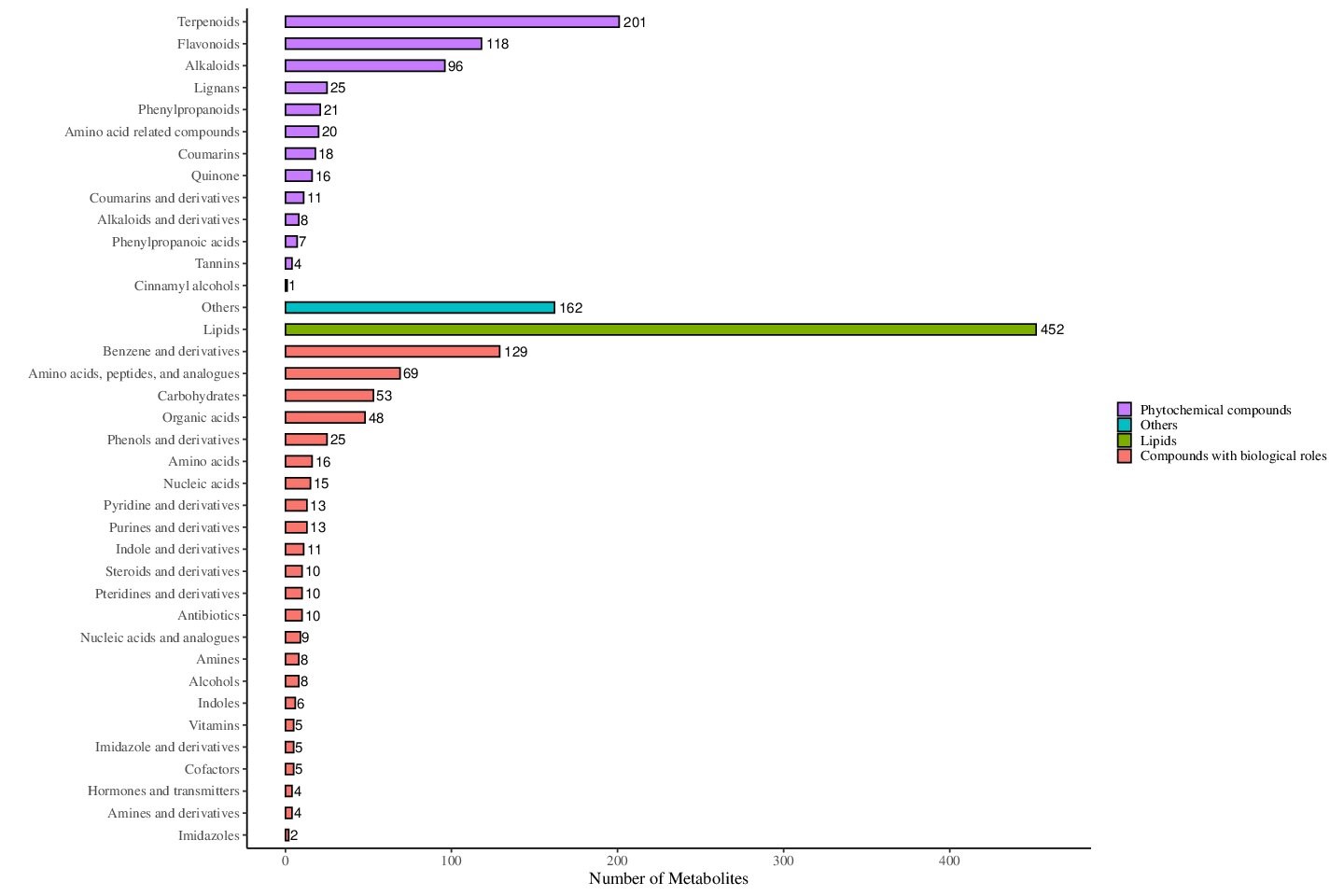
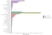
Liquid chromatography-tandem mass spectrometry (LC-MS/MS)-based metabolomic analysis was conducted to characterize the distribution of bioactive compounds in U. gambir leaf, stem, and root tissues. The analysis identified 5.681 distinct metabolites in accross all tissue types (as shown in Figure 1). The metabolome of U. gambir exhibited extensive chemical diversity with compounds distributed across multiple metabolite classes. Compound classified as lipids emerged as the predominant metabolite family, with 452 unique compounds. Secondary metabolites were also abundant and well-represented by terpenoids (201 compounds), followed by flavonoids (118 compounds), and Alkaloids (96 compounds). This analysis also revealed substantial populations of benzene and its derivates (129 compounds), amino acids and peptides (69 compounds), and carbohydrates (53 compounds). Additional secondary metabolite families included lignans (25 compounds), phenylpropanoids (21 compounds), coumarins (18 compounds), and quinone (16 compounds). Among the secondary metabolites, catechin, D-(-)-quinic acid, rauwolcine, palmitic acid, 6-Methylquinoline, and L-(-)-Malic acid were identified as the most abundant compounds, indicating their major contribution to the metabolic profile. Minor but significant contributions from tannins, cinnamyl alcohol, and imidazole derivates contributed to the metabolic landscape, demonstrating the complex phytochemical composition of U. gambir (as shown in Figure 1).
3.2. Metabolite composition across organ types and preparation methods
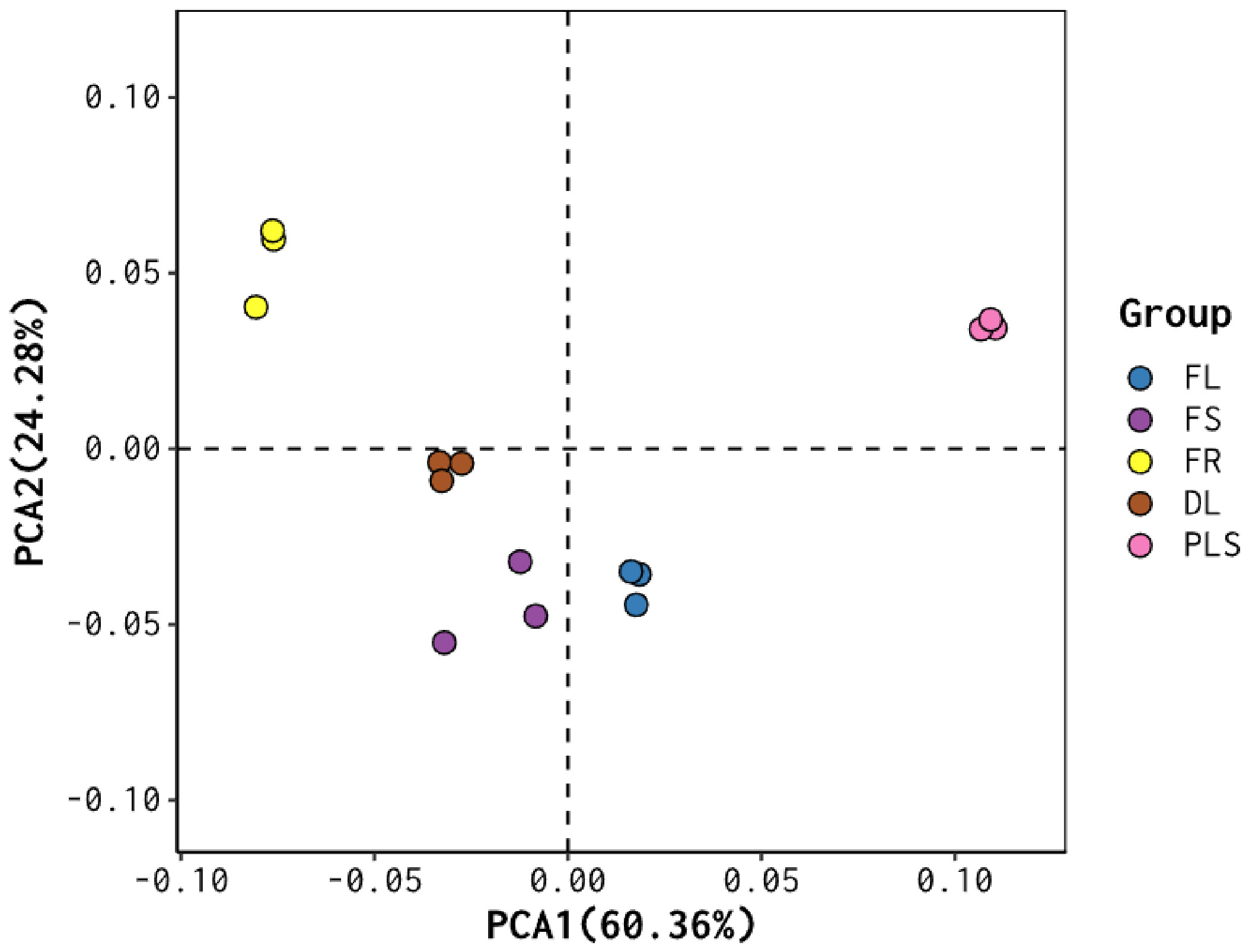
The PCA plot (Figure 2) showed a clear separation of U. gambir samples according to both organ type and preparation method, including fresh root (FR), fresh shoot (FS), fresh leaf (FL), dried leaf (DL), and processed leaf (PLS) or ethyl acetate leaves extract. Notably, FR formed a distinct cluster, indicating that roots possess a unique metabolite profile that differentiates them from the other sample groups. Likewise. PLS samples also showed clear separation from the other groups, indicating that the heat and water-based processing used to produce the concentrated extract significantly altered the metabolite composition of the leaves. Furthermore, FL, DL, and FS samples clustered relatively close to one another, although a noticeable separation among these groups was still evident (Figure 2). Overall, the PCA plot highlighted substantial differences in metabolite composition between organ types, as well as the strong influence of sample processing on metabolite profiles. While air-drying appeared to have only a minor effect on leaf metabolites, heat-based extraction produced marked alterations in metabolite composition.
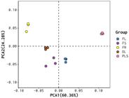
Principal Component Analysis (PCA) of Uncaria gambir metabolites from different organ types and processing methods. FR: fresh root; FS: fresh stem; FL: fresh leaf; DL: dried leaf; PLS: processed leaf.
3.3. Differential metabolite composition across organ types
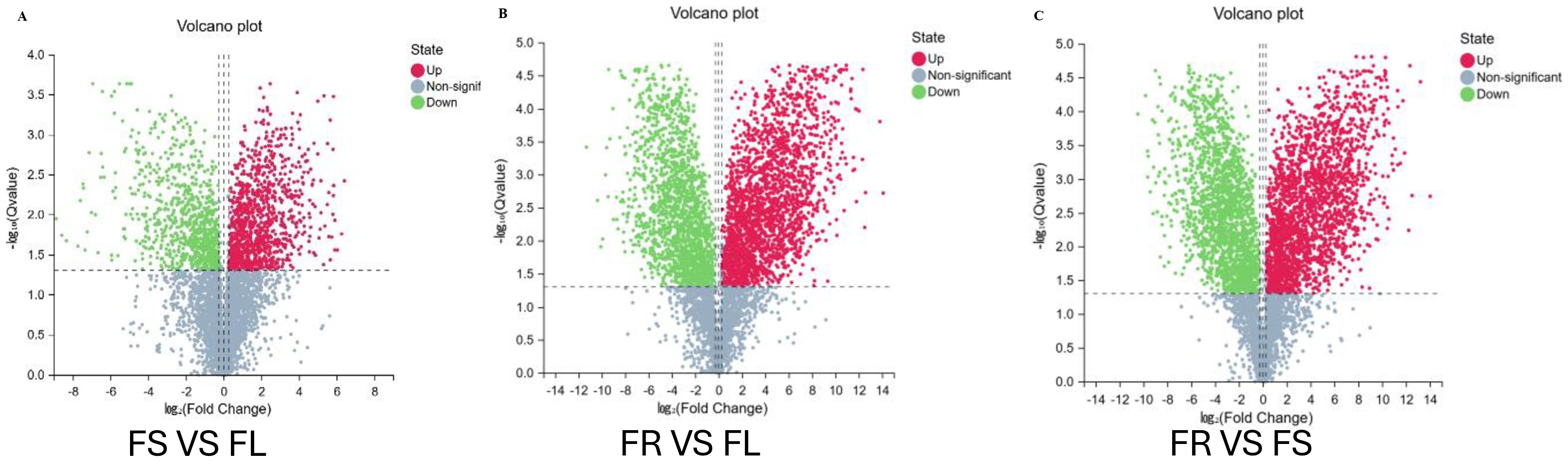
Expanding on the PCA findings, which revealed distinct metabolite composition differences between organ types, we further performed differential metabolite analysis across these groups. A total of 2,007 metabolites showed significant differences in abundance between fresh shoot (FS) and fresh leaf (FL). Of these, 1,114 metabolites were upregulated in FS, whereas 893 were downregulated (Figure 3A). Notable upregulated metabolites in FS included Etoposide, Phenyl-2-propenoyl, 4-fluorophenyl, and Geissoschizine, while major downregulated metabolites included Isoschaftoside, Caudatin, and Robinin.
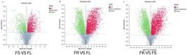
Analysis of differential metabolites between Uncaria gambir organ types. FS: fresh stem; FL: fresh leaf; FR: fresh root.
Comparison between fresh root (FR) and fresh leaf (FL) revealed 3,894 differentially expressed metabolites, with 2,201 upregulated in FR and 1,693 downregulated (Figure 3B). Key upregulated metabolites in FR included Oleanoic acid, Clitoriacetal, and Xanthurenic acid, while Deacetylisoipecoside, Phylloflavan, and Kaempferol were among the major downregulated compounds. Lastly, the comparison between FR and FS identified 4,045 metabolites with significant differential expression, of which 2,208 were upregulated and 1,837 were downregulated in FR (Figure 3C). Prominent upregulated metabolites included Eupachloroxin, Oleanoic acid, and Deoxyloganetin, whereas Methyl rosmarinate, Methylquinoline, and Mulberrin were among the major downregulated metabolites. These results emphasize the notable shifts in metabolite expression patterns across different U. gambir organs, underscoring the organ-specific specialization of metabolic pathways and the distinct biochemical functions associated with each tissue type.
3.4. Impact of processing methods on differential metabolite composition
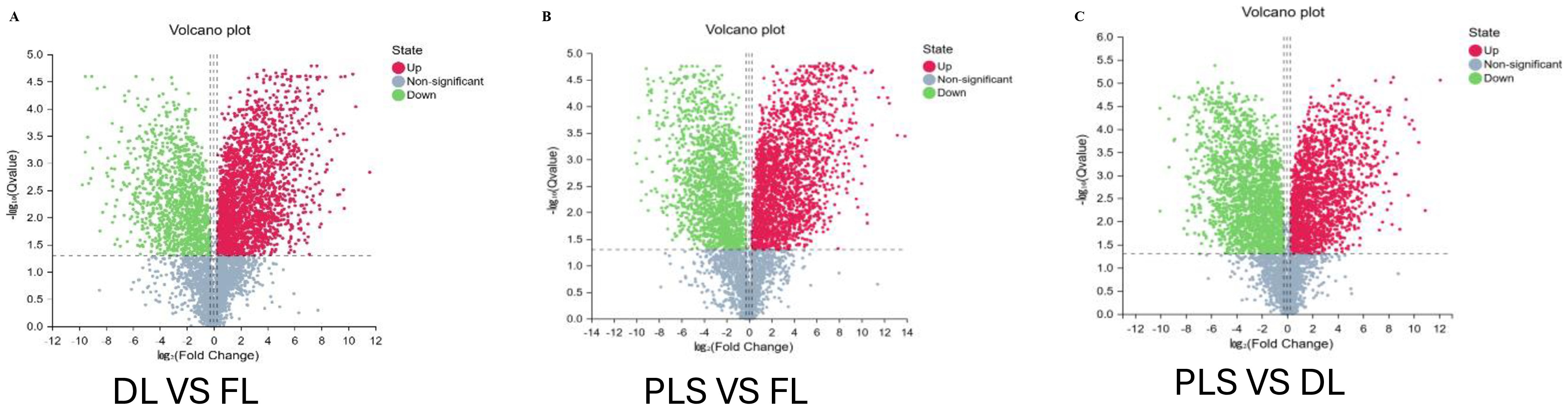
Besides the differences observed between organ types, the PCA analysis also revealed clear variations in metabolite composition among leaf samples subjected to different processing methods. Fresh leaf samples (FL) clustered distinctly from both air-dried leaves (DL) and heat-processed concentrated leaf extracts (PLS). A total of 3,762 metabolites were differentially expressed between FL and DL, with 2,511 upregulated and 1,251 downregulated in DL (Figure 4A). Notable upregulated metabolites included Dianthramine, Oleanoic acid, and various diterpenoids, whereas Calocarpin, Phylloflavan, and Propylparaben were among the major downregulated compounds.
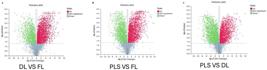
Differential Metabolite Analysis of Uncaria gambir Leaf Under Different Processing Methods. DL: dried leaf; FL: fresh leaf; PLS: processed leaf.
Comparison between PLS and FL identified 4,104 differentially expressed metabolites, of which 2,315 were upregulated and 1,789 downregulated in PLS DL (Figure 4B). Key upregulated metabolites included Norcimifugin, Amarogentin, and Baicalin, while Propylparaben, Arctigenin, and Flavonol represented major downregulated metabolites. Between PLS and DL, 4,117 metabolites showed significant differential expression, with 1,865 upregulated and 2,252 downregulated in PLS (Figure 4C). Important upregulated metabolites included Amarogentin, Norcimifugin, and Daidzein, whereas Myristic acid, Montanol, and several diterpenoids were prominently downregulated. These results demonstrate that different processing methods markedly alter the leaf metabolite profile, with heat-based extraction causing the most substantial changes compared to air-drying and fresh tissue.
3.5. Antioxidant activity ofU. gambir cv. Cubadak
The results of the antioxidant activity assay using the ABTS method are presented in Table 1. The antioxidant activity measurements of variousU. gambircv. Cubadak extracts showed that all extract types exhibited IC50 values below 50 µg/mL, indicating very strong antioxidant activity. Among the extracts, the catechu-2 ethyl acetate extract (G2 E) showed the strongest antioxidant activity, with an IC50 value of 8.09 µg/mL, which was even higher than ascorbic acid (10.00 µg/mL). Antioxidant activity analysis using the DPPH method also demonstrated that the catechu-2 ethyl acetate extract had the strongest antioxidant activity, with an IC50 value of 8.82 µg/mL (Manuhara et al., 2025). The use of the ABTS method to measure antioxidant activity was intended to confirm the results obtained using the DPPH method. According to Knez et al. (2025), the DPPH assay exhibits the highest degree of variability among antioxidant assessment approaches, involving methodological differences and unit inconsistencies that do not accurately reflect true antioxidant activity. Their study demonstrated that any variation in these factors can substantially increase uncertainty levels, with relative standard deviations reaching up to 11%, depending on the compound being tested (Shimamura et al., 2014). Nevertheless, the result of antioxidant potency assays have generally shown a positive correlation, supporting the rationale for using multiple methods to obtain a more reliable assessment (Chaves et al., 2020). In the context of radical-based techniques, the ABTS assay has been shown to exhibit higher repeatability and stability compared with DPPH, which is highly prone to environmental factors (Knez et al., 2025).The choice of extraction solvent had a significant effect on the antioxidant activity of the samples. Extracts obtained usingn-hexane displayed substantially lower antioxidant activity compared with those extracted with ethyl acetate or methanol (31.19-33.67 µg/mL). Furthermore, The antioxidant activity assays conducted on differentU. gambirsample types revealed that fresh materials (FL, FS, FR) exhibited lower antioxidant activity compared with the other sample categories. Nevertheless, allU. gambirextracts, regardless of sample type or extraction solvent, consistently demonstrated strong to very strong antioxidant activity. Based on these findings, the ethyl acetate extract ofU. gambirwas selected for subsequent in vivo evaluation to assess its protective antioxidant potential against Pb acetate-induced oxidative stress in mice. To further elucidate the chemical basis underlying the antioxidant activity, correlation analysis was conducted between IC50 values and the metabolites detected in U. gambir extracts (Table 2).
Antioxidant activity of various U. gambir cv. Cubadak extracts from different sample types and solvents based on the ABTS method.
Correlation analysis between ABTS IC50 values and the relative abundance of selected secondary metabolites revealed several strong and statistically significant negative relationships (Table 2). Catechin exhibited the strongest negative correlation with IC50 (r = −0.9977), indicating that higher catechin abundance was strongly associated with lower IC50 values and thus higher antioxidant activity. Significant negative correlations were also observed for naringenin chalcone (r = −0.9433), procyanidin B1 (r = −0.9355), chlorogenic acid (r = −0.9153), and coumarin (r = −0.9603). In contrast, other detected metabolites, including organic acids (e.g., citric acid, malic acid), fatty acids, alkaloids, and several phenolic acids, showed weak or non-significant correlations with IC50 values. These results indicate that only specific phenolic and flavonoid compounds are closely linked to the antioxidant potency of U. gambir extracts.
3.6. Effects of U. gambir cv Cubadak ethyl acetate extract on MDA levels and endogenous enzyme activity in liver, kidney, and testis homogenates
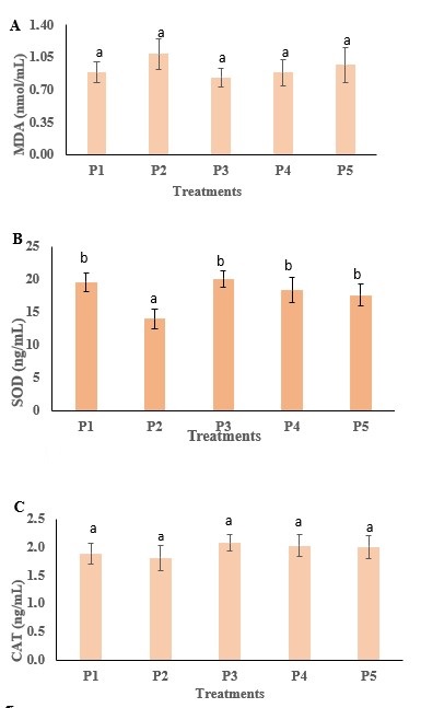
The effects of the ethyl acetate extract ofU. gambircv. Cubadak on MDA levels and endogenous antioxidant enzyme activity (SOD and CAT) were evaluated in liver, kidney, and testis homogenates of mice. Oxidative stress induced by heavy metal accumulation can be detected by an increase in MDA levels and a decrease in the activity of antioxidant enzymes such as SOD and CAT. Reactive oxygen species (ROS) stimulate the elevation of MDA levels through enhanced lipid peroxidation, whereas SOD and CAT function to reduce ROS formation and mitigate oxidative stress resulting from excessive lipid peroxidation. The in vivo antioxidant effects of theU. gambirethyl acetate extract on MDA, SOD, and CAT activity in liver homogenates (see Figure 5).
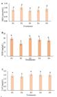
Levels of MDA (A), SOD (B), and CAT (C) in liver homogenates of mice subjected to different treatments with U. gambir cv. Cubadak ethyl acetate extract. P1: positive control (sterile distilled water), P2: negative control (Pb acetate only), P3: U. gambir ethyl acetate extract 100 mg/L + Pb acetate 100 mg/L, P4: U. gambir ethyl acetate extract 200 mg/L + Pb acetate 100 mg/L, P5: U. gambir ethyl acetate extract 300 mg/L + Pb acetate 100 mg/L.
MDA levels in the liver of mice showed an increase, with the highest level observed in the Pb acetate-only group (P2). However, this increase was not statistically significant (p< 0.05) when compared with the other treatment groups and the control (P1, P3, P4, and P5). This indicates that reactive oxygen species are capable of elevating MDA levels through enhanced lipid peroxidation. The SOD measurements showed a significant increase (p< 0.05) in hepatic SOD activity in the control group (P1) and in all groups receiving Pb acetate together withU. gambirextract (P3, P4, and P5), compared with the Pb acetate-only group (P2). In contrast, the CAT levels in the liver did not differ significantly (p> 0.05) between the control and treatment groups.
SOD plays an essential role in reducing the formation of ROS and mitigating oxidative stress resulting from elevated lipid peroxidation. SOD functions by catalyzing the dismutation of superoxide radicals (O2•−) into molecular oxygen (O2) or hydrogen peroxide (H2O2), whereas CAT decreases ROS levels by facilitating the conversion of H2O2 into water (H2O). The activities of these antioxidant enzymes are capable of reducing lipid peroxidation by approximately 70-90% (Ighodaro and Akinloye, 2018; Seifried et al., 2017; Sheng et al., 2014; Zulaikhah, 2017).
Under normal conditions, the elevated production of ROS is counterbalanced by the presence of enzymatic antioxidants (SOD, CAT, GPx) and non-enzymatic antioxidants (GSH, vitamins C and E). In mammals, three types of SOD enzymes are distinguished based on their metal cofactors: SOD-1 (Cu/Zn SOD), SOD-2 (Mn SOD), dan SOD-3 (extracellular SOD) (Kim et al., 2018). One approach to counteracting oxidative stress caused by heavy metals is to increase the intake of essential bioelements (Ca, Fe, P, K, Se, Mn, Zn), which function as cofactors for endogenous antioxidant enzymes, along with dietary supplementation of antioxidant compounds such as vitamins C and E, as well as phenolic and flavonoid compounds that can chelate Fe ions in the intestine (Anantharaju et al., 2016; Genchi et al., 2020; Kumar et al., 2020).
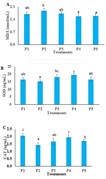
Measurements of MDA, SOD, and CAT levels were also performed on kidney homogenates. The results for renal MDA, SOD, and CAT across the different treatment groups are presented in Figure 6. In the kidneys of mice, oral galvage of theU. gambirethyl acetate extract exerted a significant effect on MDA, SOD, and CAT levels (p< 0.05). The highest MDA levels were observed in the negative control group (P2), in which mice received only Pb acetate. In contrast, treatment withU. gambirethyl acetate extract at concentrations of 200 mg/L and 300 mg/L resulted in a significant reduction in MDA levels (p< 0.05). At 100 mg/L, however, MDA levels were comparable to those of the control group that did not receive Pb acetate. The highest SOD and CAT activities in kidney tissue were obtained from the group treated withU. gambirethyl acetate extract at 200 mg/L.
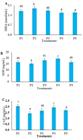
MDA (A), SOD (B), and CAT (C) levels in kidney homogenates of mice subjected to different treatments with U. gambir ethyl acetate extract. P1: positive control (sterile distilled water), P2: negative control (Pb acetate only), P3: U. gambir ethyl acetate extract 100 mg/L + Pb acetate 100 mg/L, P4: U. gambir ethyl acetate extract 200mg/L + Pb acetate 100 mg/L, P5: U. gambir ethyl acetate extract 300 mg/L + Pb acetate 100 mg/L.
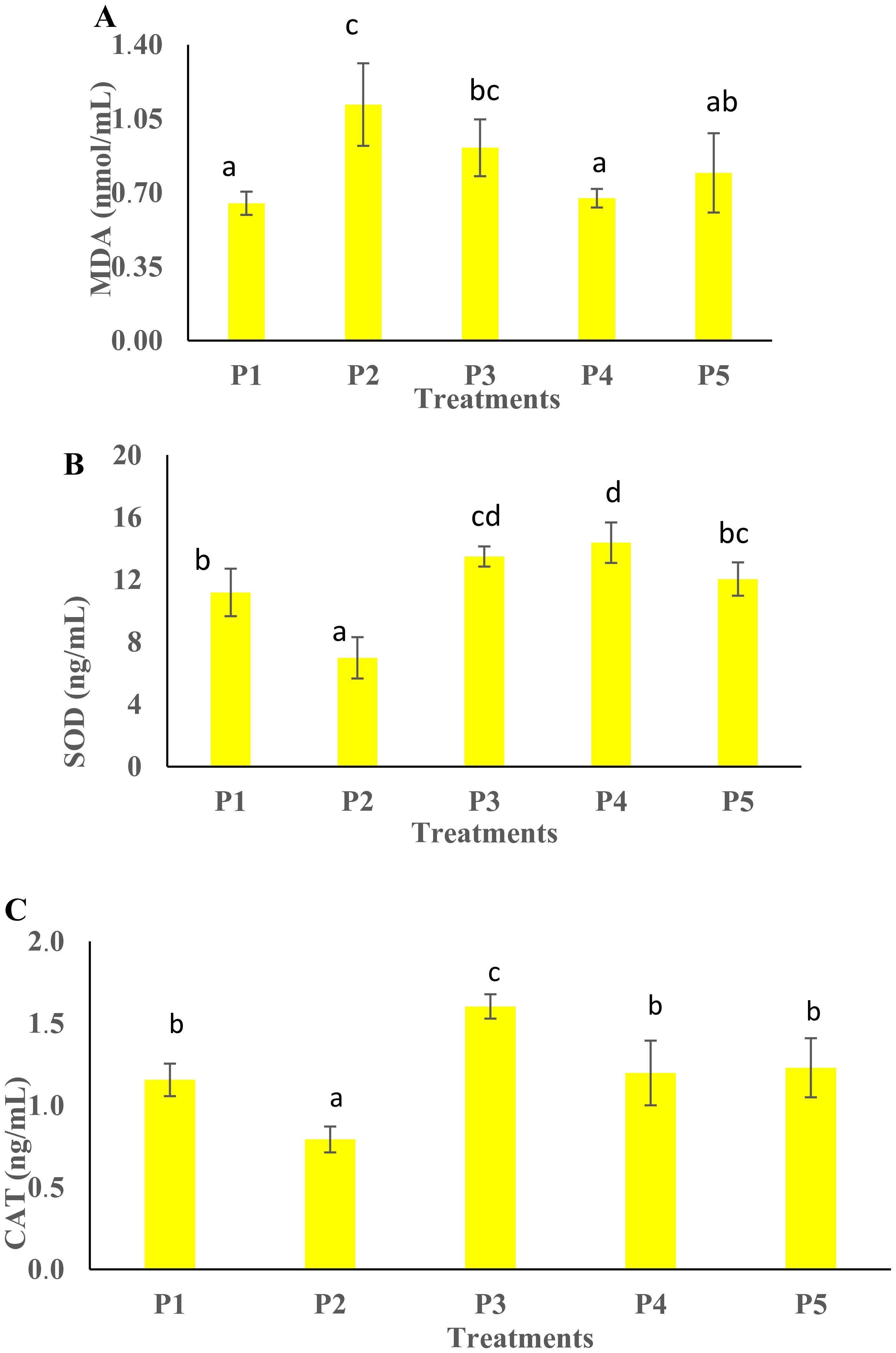
MDA levels in the testis of mice were significantly different from the control in all treatment groups, except for the group receivingU. gambirethyl acetate extract at 200 mg/L (P4), which did not differ significantly (p< 0.05) (Figure 7A). In this group, oral galgave of theU. gambirextract reduced MDA levels to nearly the same level as the positive control (no Pb acetate exposure). In contrast, MDA levels in P3 and P5 were higher than the control, although still lower than those in the negative control (P2). The highest SOD activity was observed in the P4 group (200 mg/LU. gambirethyl acetate extract), whereas P3 and P5 also showed elevated SOD levels compared with the negative control. The negative control exhibited the lowest SOD activity (Figure 7B). These findings indicate that theU. gambirethyl acetate extract is capable of enhancing antioxidant enzyme activity to counteract Pb acetate-induced free radicals. For CAT activity, groups P4 and P5 did not differ significantly from the positive control (no Pb acetate), whereas P3 showed the highest CAT level (Figure 7C). This suggests that administration of theU. gambirethyl acetate extract can increase endogenous antioxidant enzyme levels in the testis to combat Pb acetate-induced oxidative stress.
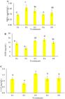
MDA (A), SOD (B), and CAT (C) levels in testis homogenates of mice subjected to different treatments with U. gambir ethyl acetate extract. P1: positive control (sterile distilled water), P2: negative control (Pb acetate only), P3: U. gambir ethyl acetate extract 100 mg/L + Pb acetate 100 mg/L, P4: U. gambir ethyl acetate extract 200 mg/L + Pb acetate 100 mg/L, P5: U. gambir ethyl acetate extract 300 mg/L + Pb acetate 100 mg\L.
3.7. Effects of U. gambir cv. Cubadak ethyl acetate extract on liver histology in mice
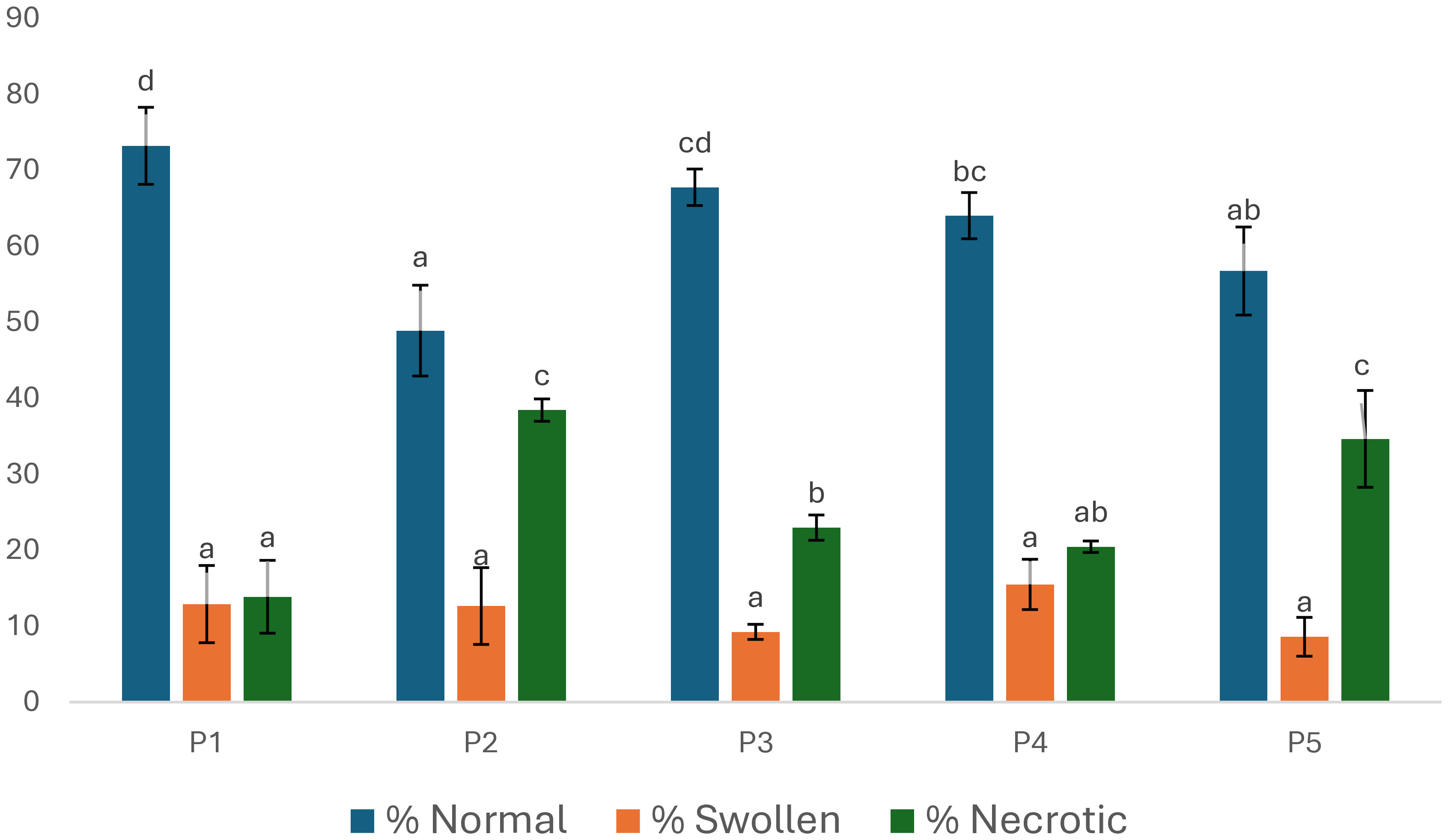
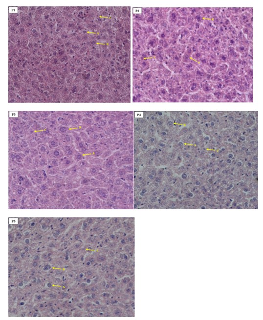
The histological observations of mice liver tissue, showing the percentages of normal cells, swollen cells, and necrotic cells (see Figure 8). Representative liver histology images from mice treated with different concentrations of U. gambir ethyl acetate extract (see Figure 9).
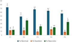
Percentage of normal, swollen, and necrotic liver cells in mice under different treatment conditions. P1: positive control (sterile distilled water), P2: negative control (Pb acetate only), P3: U. gambir ethyl acetate extract 100 mg/L + Pb acetate 100 mg/L, P4: U. gambir ethyl acetate extract 200 mg/L + Pb acetate 100 mg/L, P5: U. gambir ethyl acetate extract 300 mg/L + Pb acetate 100 mg/L.
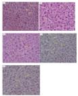
Histological features of mice liver under different treatments with Pb acetate and U. gambir ethyl acetate extract, stained with hematoxylin and eosin. A: normal cells; B: swollen cells; C: necrotic cells. P1: positive control (sterile distilled water), P2: negative control (Pb acetate only), P3: U. gambir ethyl acetate extract 100 mg/L + Pb acetate 100 mg/L, P4: U. gambir ethyl acetate extract 200 mg/L + Pb acetate 100 mg/L, P5: U. gambir ethyl acetate extract 300 mg/L + Pb acetate 100 mg/L. Magnification: 400×.
Microscopic examination of hepatic cells showed that oral administration of theU. gambirethyl acetate extract at different concentrations did not have a significant effect on the occurrence of cellular swelling (statistical analysis,p> 0.05). However, it had a significant effect on the formation of necrotic liver cells (p< 0.05) (Figure 8). The highest level of cell death was observed in the group receiving Pb acetate alone, The highest level of cell death occurred in the group that received Pb acetate alone, whereas lower levels of cell death were observed in the control group (sterile distilled water) as well as in P3 and P4, which received 100 mg/L and 200 mg/L ofU. gambirextract in combination with 100 mg/L Pb acetate, respectively. In the control group, cell death reached 13.86%, while in the Pb acetate group, cell death increased markedly to 38.44%. These findings indicate that Pb acetate induces the formation of free radicals, leading to damage of cellular membranes and leading to cell death (necrosis).
Oral administration ofU. gambirethyl acetate extract at 100 mg/L (P3) in mice exposed to Pb acetate reduced cell death by 15.46%, whereas treatment with 200 mg/L (P4) reduced cell death by 17.97%. These findings are supported by the significant increase in hepatic SOD activity observed in P3 and P4 (Figure 6) compared with P2 (Pb acetate only). Moreover, oral administration ofU. gambirethyl acetate extract at 200 mg/L achieved a greater reduction in cell death than methanolic extracts ofGynura procumbensat 300 mg/L, which decreased cell death by less than 10% (Sugiharto et al., 2022). Similarly, treatment with a methanolic extract combination ofZingiber officinaleandBoesenbergia rotunda(1:1) was able to reduce cell death by only 6.77% (Sugiharto et al., 2022). The administration of a 300 mg/L U. gambir extract dose did not yield a positive response in the livers of rats. A similar observation was made in the administration of Gynura procumbens extract, where a 300 mg/L dose did not yield a positive response (Sugiharto et al., 2022). The administration of herbal formulas containing gambier and Caesalpiniasappan extracts at doses of 300 mg and 1,200 mg/kgBW also resulted in the induction of lesions in the liver, kidneys, and heart of male and female mice, particularly at higher doses (Armenia et al., 2021).
4. Discussion
Our comprehensive untargeted metabolomic survey extends previous phytochemical studies on Uncaria gambir, which have largely relied on targeted approaches such as the isolation and quantification of catechins, tannins, and selected flavonoids, as well as pharmacological evaluations of plant extracts. Earlier reviews consistently describe U. gambir as rich in flavonoids, phenolics (particularly catechins), alkaloids, and terpenoids, emphasizing its traditional medicinal applications and antioxidant and anti-inflammatory activities. In contrast, our dataset comprising 5,681 metabolites detected across multiple organs and processing methods greatly expands this foundation by revealing far broader chemical diversity, including numerous lipids, terpenoids, flavonoids, and alkaloids.
The organ-specific metabolite patterns observed here align with findings from other medicinal plants, in which roots typically accumulate triterpenoids and other defense or storage-related metabolites, whereas aerial tissues are enriched in flavonoids and phenolic compounds associated with photoprotection, signaling, and environmental responsiveness.
Regarding processing effects, our observation that air-drying caused relatively lower alterations in leaf metabolite profiles, while heat-based extraction (PLS) led to extensive compositional changes, is consistent with metabolomics studies in other species. These studies have shown that drying, especially thermal extraction, can selectively concentrate heat-stable metabolites while degrading or transforming heat-labile compounds. In agreement with this pattern, we found strong enrichment of thermostable metabolites such as derivatives of amarogentin, norcimifugin, and baicalin in PLS samples, accompanied by the depletion of several flavonols and other heat-sensitive compounds. Amarogentin, a secoiridoid glycoside detected in the metabolomics profile, has been reported to exhibit diverse protective and regulatory biological activities. Previous studies indicate that amarogentin is involved in metabolic and vascular regulation through activation of AMP-activated protein kinase (AMPK) signaling, which contributes to improved glucose and lipid metabolism. In addition, amarogentin has been associated with neuroprotective and anti-inflammatory effects, mediated through the modulation of oxidative stress– and inflammation-related pathways. The presence and upregulation of this secoiridoid glycoside may therefore contribute to the observed bioactivity by supporting antioxidant capacity and cellular protection under stress conditions. (Potunuru et al., 2019; Song and Zhou. 2022; Singh et al., 2025). Norcimifugin, classified as a chromone-type phenolic compound, and dianthramine, an alkaloid, were detected in the metabolomics analysis. As specific functional studies on these individual metabolites remain limited, their biological relevance can be inferred at the metabolite-class level. Chromone-derived phenolic compounds in plants are generally associated with antioxidant and anti-inflammatory properties and are known to contribute to chemical defense, often exhibiting antimicrobial and cytotoxic activities in vitro. The upregulation of such compounds is commonly linked to enhanced resistance against environmental stressors and pathogenic challenges (Lewandowski et al., 2020; Gao et al., 2025; Liu et al., 2025)
Altogether, this study provides a valuable resource for determining the most suitable tissue types and processing conditions for maximizing the recovery of specific bioactive classes (e.g., catechins versus triterpenoids). This information is essential for ongoing efforts to standardize herbal preparations and to relate processing step with biological activity and product consistency.
Lead (Pb) exposure in the body has been demonstrated to induce the formation of ROS. In instances where ROS production exceeds the threshold that can be neutralised by endogenous antioxidants, an imbalance between oxidants and antioxidants ensues, giving rise to oxidative stress. This condition is reflected by an increase in MDA levels. The elevated MDA concentration observed in the negative control group (P2) suggests a correlation between high MDA levels and the degree of hepatocellular death. Similar findings were reported in mice exposed to cadmium (Cd) (Sugiharto et al., 2022). Cd accumulation causes severe organ damage and induces rigidity of the cell membrane, leading to increased extracellular fluid influx into the cell, cellular swelling, and cell death, particularly in the liver and kidneys (Kerek et al., 2018; Khalesi et al., 2017). Lead toxicity could induce oxidative stress and caused lipid peroxidation in cell membrane. Thereby increasing cells damage, especially in liver and kidney. Similar to present study, Zidi I (2014) reported administration of lead acetate 2 g/L for 35 days increased hypertrophy of hepatic cells and accompanied by an increase in AST and ALT levels. Administration of lead acetate 20 mg/kg BW for 3 weeks increased necrotic of hepatic cells, MDA levels, AST (aspartate aminotransferase), and ALT (alanine aminotransfer ase). levels as indicators of damage in liver cells (Yuniarti et al., 2021). The strong and significant negative correlations between IC50 values and the abundance of catechin, naringenin chalcone, procyanidin B1, chlorogenic acid, and coumarin clearly indicate that these compounds play a major role in determining the antioxidant activity of U. gambir extracts. Because lower IC50 values correspond to higher antioxidant capacity, the observed inverse relationships suggest that increased concentrations of these metabolites directly enhance radical-scavenging efficiency in the ABTS assay (Frediansyah et al., 2021)
Catechin emerged as the most influential contributor, consistent with previous reports identifying catechins as the dominant antioxidants in U. gambir (Manuhara et al., 2025). Similarly, procyanidin B1, a condensed tannin dimer composed of catechin units, is well known for its high radical-scavenging capacity, supporting its significant association with low IC50 values (Gao et al., 2021). Naringenin chalcone and chlorogenic acid are phenolic compounds that act as efficient antioxidants through both direct radical scavenging and metal-chelating mechanisms (Abbasi-Parizad et al., 2020). Their significant negative correlations with IC50 values indicate that, although present at lower abundance than catechin, they contribute synergistically to the overall antioxidant capacity of the extract. Coumarin also showed a strong inverse correlation with IC50, suggesting a meaningful role in antioxidant activity, potentially through stabilization of radical intermediates and modulation of redox reactions (Mohammadnia et al., 2025). Overall, the correlation analysis supports the conclusion that the high antioxidant activity of U. gambir ethyl acetate extract is primarily driven by a specific group of phenolic and flavonoid compounds rather than by the total metabolite pool. These findings provide a clear chemical basis for the strong antioxidant activity observed in vitro and support the use of catechin-rich U. gambir extracts as candidates for standardized antioxidant formulations.
5. Conclusion
Based on metabolite composition across organ types and preparation methods this research obtained substantial differences in metabolite composition between organ types, as well as the strong influence of sample processing on metabolite profiles. While air-drying appeared to have only a minor effect on leaf metabolites, heat-based extraction produced marked alterations in metabolite composition. Different processing methods markedly alter the leaf metabolite profile, with heat-based extraction causing the most substantial changes compared to air-drying and fresh tissue. This study provides a valuable resource for determining the most suitable tissue types and processing conditions for maximizing the recovery of specific bioactive classes.
Oral administration of theU. gambirethyl acetate extract to mice exposed to lead acetate was able to decrease MDA levels in the kidney and testis, although no reduction was observed in the liver. The extract effectively increased SOD activity in the liver, kidney, and testis, while CAT activity increased in the kidney and testis. Histological analysis of the liver revealed that treatment with 100 mg/L and 200 mg/L of the ethyl acetate extract reduced hepatocellular death by 15.46% and 17.97%, respectively. The findings suggest that the U. gambir ethyl acetate extract possesses the potential to counteract lead acetate-induced oxidative stress, indicating its suitability as a candidate for development as a standardized herbal therapeutic agent.
Acknowledgements
This study was funded by Universitas Airlangga through the Riset Kolaborasi Indonesia scheme under the contract number 1685/B/UN3.LPPM/PT.01.03/2025.
Data Availability Statement
The data that support the findings of this study are available on request from the corresponding author.
References
-
ABBASI-PARIZAD, P., DE NISI, P., ADANI, F., PEPÉ SCIARRIA, T., SQUILLACE, P., SCARAFONI, A., IAMETTI, S. and SCAGLIA, B., 2020. Antioxidant and anti-inflammatory activities of the crude extracts of raw and fermented tomato pomace and their correlations with aglycate-polyphenols. Antioxidants, vol. 9, no. 2, pp. 179. https://doi.org/10.3390/antiox9020179 PMid:32098217.
» https://doi.org/10.3390/antiox9020179 -
ALBARRACIN, S.L., STAB, B., CASAS, Z., SUTACHAN, J.J., SAMUDIO, I., GONZALEZ, J., GONZALO, L., CAPANI, F., MORALES, L. and BARRETO, G.E., 2012. Effects of natural antioxidants in neurodegenerative disease. Nutritional Neuroscience, vol. 15, no. 1, pp. 1-9. https://doi.org/10.1179/1476830511Y.0000000028 PMid:22305647.
» https://doi.org/10.1179/1476830511Y.0000000028 -
ANANTHARAJU, P.G., GOWDA, P.C., VIMALAMBIKE, M.G. and MADHUNAPANTULA, S.V., 2016. An overview on the role of dietary phenolics for the treatment of cancers. Nutrition Journal, vol. 15, no. 1, pp. 1-16. https://doi.org/10.1186/s12937-016-0217-2
» https://doi.org/10.1186/s12937-016-0217-2 -
ANDASURYANI, A., PURWANTO, Y.A., BUDIASTRA, I.W. and SYAMSU, K., 2014. Determination of catechin content in gambir powder from dried gambir leaves quickly using FT NIR PLS model. International Journal on Advanced Science, Engineering and Information Technology, vol. 4, no. 5, pp. 303-307. https://doi.org/10.18517/ijaseit.4.5.423
» https://doi.org/10.18517/ijaseit.4.5.423 -
ANDJELKOVIC, M., DJORDJEVIC, A.B., ANTONIJEVIC, E., ANTONIJEVIC, B., STANIC, M., KOTUR-STEVULJEVIC, J., SPASOJEVIC-KALIMANOVSKA, V., JOVANOVIC, M., BORICIC, N., WALLACE, D. and BULAT, Z., 2019. Toxic effect of acute cadmium and lead exposure in rat blood, liver, and kidney. International Journal of Environmental Research and Public Health, vol. 16, no. 2, pp. 274. https://doi.org/10.3390/ijerph16020274 PMid:30669347.
» https://doi.org/10.3390/ijerph16020274 -
ARMENIA, A., PERMATASARI, D., SINAMAR, L.P., ESTERA, K. and AHMADIN, A., 2021. The impact of sub acute administration of purified gambier (Uncaria gambir roxb.) to the liver and kidney functions and its reversibility on rats. Pharmacognosy Journal, vol. 13, no. 1, pp. 44-51. https://doi.org/10.5530/pj.2021.13.7
» https://doi.org/10.5530/pj.2021.13.7 -
AULIANA, F.R., IFORA, I. and FAUZIAH, F., 2022. Phytochemical and anti-inflammatory of uncaria gambir: a review. Asian Journal of Pharmaceutical Research and Development, vol. 10, no. 1, pp. 79-83. https://doi.org/10.22270/ajprd.v10i1.1077
» https://doi.org/10.22270/ajprd.v10i1.1077 -
BERNATONIENĖ, J. and KOPUSTINSKIENE, D., 2018. The role of catechins in cellular responses to oxidative stress. Molecules: a Journal of Synthetic Chemistry and Natural Product Chemistry, vol. 23, no. 4, pp. 965. https://doi.org/10.3390/molecules23040965
» https://doi.org/10.3390/molecules23040965 -
BHATTI, J.S., BHATTI, G.K. and REDDY, P.H., 2017. Mitochondrial dysfunction and oxidative stress in metabolic disorders: a step towards mitochondria based therapeutic strategies. Biochimica et Biophysica Acta. Molecular Basis of Disease, vol. 1863, no. 5, pp. 1066-1077. https://doi.org/10.1016/j.bbadis.2016.11.010 PMid:27836629.
» https://doi.org/10.1016/j.bbadis.2016.11.010 -
CHAVES, N., SANTIAGO, A. and ALÍAS, J.C., 2020. Quantification of the antioxidant activity of plant extracts: analysis of sensitivity and hierarchization based on the method used. Antioxidants, vol. 9, no. 1, pp. 76. https://doi.org/10.3390/antiox9010076 PMid:31952329.
» https://doi.org/10.3390/antiox9010076 -
CHRISTODOULOU, M.C., ORELLANA PALACIOS, J.C., HESAMI, G., JAFARZADEH, S., LORENZO, J.M., DOMÍNGUEZ, R., MORENO, A. and HADIDI, M., 2022. Spectrophotometric methods for measurement of antioxidant activity in food and pharmaceuticals. Antioxidants, vol. 11, no. 11, pp. 2213. https://doi.org/10.3390/antiox11112213 PMid:36358583.
» https://doi.org/10.3390/antiox11112213 - DHALIMI, A., 2006. Permasalahan gambir (Uncaria gambir L.) di Sumatera Barat dan alternatif pemecahannya. Perspektif, vol. 5, no. 1, pp. 46-59.
-
DONG, J.-W., CAI, L., XING, Y., YU, J. and DING, Z.-T., 2015. Re-evaluation of ABTS*+ assay for total antioxidant capacity of natural products. Natural Product Communications, vol. 10, no. 12, pp. 2169-2172. https://doi.org/10.1177/1934578X1501001239 PMid:26882692.
» https://doi.org/10.1177/1934578X1501001239 -
FAUZA, H., 2014. Gambier: Indonesia leading commodities in the past. International Journal on Advanced Science, Engineering and Information Technology, vol. 4, no. 6, pp. 455. https://doi.org/10.18517/ijaseit.4.6.463
» https://doi.org/10.18517/ijaseit.4.6.463 -
FORMAN, H.J. and ZHANG, H., 2021. Targeting oxidative stress in disease: promise and limitations of antioxidant therapy. Nature Reviews. Drug Discovery, vol. 20, no. 9, pp. 689-709. https://doi.org/10.1038/s41573-021-00233-1 PMid:34194012.
» https://doi.org/10.1038/s41573-021-00233-1 -
FREDIANSYAH, A., ROMADHONI, F., SURYANI., NURHAYATI, R. and WIBOWO, A.T., 2021. Fermentation of jamaican cherries juice using lactobacillus plantarum elevates antioxidant potential and inhibitory activity against type ii diabetes-related enzymes. Molecules, vol. 26, no. 10, pp. 2868. https://doi.org/10.3390/molecules26102868
» https://doi.org/10.3390/molecules26102868 -
GAO, R., CHEN, F., CHEN, L. and MA, H., 2025. DNA methylation changes in Medicago sativa under salt-alkaline stress and the function of 5-azacytidine in enhancing stress tolerance. BMC Plant Biology, vol. 25, no. 1, pp. 1419. https://doi.org/10.1186/s12870-025-07424-7 PMid:41120937.
» https://doi.org/10.1186/s12870-025-07424-7 -
GAO, W., YU, T., LI, G., SHU, W., JIN, Y., ZHANG, M. and YU, X., 2021. Antioxidant activity and anti-apoptotic effect of the small molecule procyanidin B1 in early mouse embryonic development produced by somatic cell nuclear transfer. Molecules, vol. 26, no. 20, pp. 6150. https://doi.org/10.3390/molecules26206150 PMid:34684730.
» https://doi.org/10.3390/molecules26206150 -
GENCHI, G., SINICROPI, M.S., GRAZIANTONO, L., CAROCCI, A. and ALESSIA, C., 2020. Review: the effects of cadmium toxicity. International Journal of Environmental Research and Public Health, vol. 17, no. 11, pp. 1-24. https://doi.org/10.3390/ijerph17113782
» https://doi.org/10.3390/ijerph17113782 -
GORZYNIK-DEBICKA, M., PRZYCHODZEN, P., CAPPELLO, F., KUBAN-JANKOWSKA, A., MARINO GAMMAZZA, A., KNAP, N., WOZNIAK, M. and GORSKA-PONIKOWSKA, M., 2018. Potential health benefits of olive oil and plant polyphenols. International Journal of Molecular Sciences, vol. 19, no. 3, pp. 686. https://doi.org/10.3390/ijms19030686 PMid:29495598.
» https://doi.org/10.3390/ijms19030686 -
IGHODARO, O.M. and AKINLOYE, O.A., 2018. First line defence antioxidants-superoxide dismutase (SOD), catalase (CAT) and glutathione peroxidase (GPX): their fundamental role in the entire antioxidant defence grid. Alexandria Journal of Medicine, vol. 54, no. 4, pp. 287-293. https://doi.org/10.1016/j.ajme.2017.09.001
» https://doi.org/10.1016/j.ajme.2017.09.001 -
KEREK, E., HASSANIN, M. and PRENNER, E.J., 2018. Inorganic mercury and cadmium induce rigidity in eukaryotic lipid extracts while mercury also ruptures red blood cells. Biochimica et Biophysica Acta. Biomembranes, vol. 1860, no. 3, pp. 710-717. https://doi.org/10.1016/j.bbamem.2017.12.014 PMid:29269315.
» https://doi.org/10.1016/j.bbamem.2017.12.014 -
KHALESI, K., ABEDI, Z., BEHROUZI, S. and KOHESTAN ESKANDARI, S., 2017. Haematological, blood biochemical and histopathological effects of sublethal cadmium and lead concentrations in common carp. Bulgarian Journal of Veterinary Medicine, vol. 20, no. 2, pp. 141-150. https://doi.org/10.15547/bjvm.965
» https://doi.org/10.15547/bjvm.965 -
KHAN, N. and MUKHTAR, H., 2018. Tea polyphenols in promotion of human health. Nutrients, vol. 11, no. 1, pp. 39. https://doi.org/10.3390/nu11010039 PMid:30585192.
» https://doi.org/10.3390/nu11010039 -
KIM, K.S., LIM, H.-J., LIM, J.S., SON, J.Y., LEE, J., LEE, B.M., CHANG, S.-C. and KIM, H.S., 2018. Curcumin ameliorates cadmium-induced nephrotoxicity in Sprague-Dawley rats. Food and Chemical Toxicology: an International Journal Published for the British Industrial Biological Research Association, vol. 114, pp. 34-40. https://doi.org/10.1016/j.fct.2018.02.007 PMid:29421648.
» https://doi.org/10.1016/j.fct.2018.02.007 -
KIM, T., LEEM, E., LEE, J. and KIM, S., 2020. Control of reactive oxygen species for the prevention of parkinson’s disease: the possible application of flavonoids. Antioxidants, vol. 9, no. 7, pp. 583. https://doi.org/10.3390/antiox9070583 PMid:32635299.
» https://doi.org/10.3390/antiox9070583 -
KNEZ, E., KADAC-CZAPSKA, K. and GREMBECKA, M., 2025. Evaluation of spectrophotometric methods for assessing antioxidant potential in plant food samples: a critical Approach. Applied Sciences, vol. 15, no. 11, pp. 5925. https://doi.org/10.3390/app15115925
» https://doi.org/10.3390/app15115925 -
KUMAR, A., KHUSHBOO, P.R. and SHARMA, B., 2020. Modulation of superoxide dismutase activity by mercury, lead, and arsenic. Biological Trace Element Research, vol. 196, no. 2, pp. 654-661. https://doi.org/10.1007/s12011-019-01957-3
» https://doi.org/10.1007/s12011-019-01957-3 -
LEWANDOWSKI, W., LEWANDOWSKA, H., GOLONKO, A., ŚWIDERSKI, G., ŚWISŁOCKA, R. and KALINOWSKA, M., 2020. Correlations between molecular structure and biological activity in “logical series” of dietary chromone derivatives. PLoS One, vol. 15, no. 8, e0229477. https://doi.org/10.1371/journal.pone.0229477 PMid:32822343.
» https://doi.org/10.1371/journal.pone.0229477 -
LIU, X., WANG, B., ZHAO, Y., CHU, M., YU, H., GAO, D., WANG, J., LI, Z., LIU, S., LI, Y., WEI, Y., WEI, J. and XU, J., 2025. Physiological and transcriptional responses of sorghum seedlings under alkali stress. Plants, vol. 14, no. 19, pp. 1. https://doi.org/10.3390/plants14193106 PMid:41095246.
» https://doi.org/10.3390/plants14193106 -
MANUHARA, Y.S.W., SUGIHARTO, S., KRISTANTI, A.N., AMINAH, N.S., WIBOWO, A.T., WARDANA, A.P., PUTRO, Y.K. and SUGIARSO, D., 2022. Antioxidant activities, total phenol, flavonoid, and mineral content in the rhizome of various Indonesian herbal plants. Rasayan Journal of Chemistry, vol. 15, no. 4, pp. 2724-2730. https://doi.org/10.31788/RJC.2022.1548024
» https://doi.org/10.31788/RJC.2022.1548024 - MANUHARA, Y.S.W., SYAMSUARDI, SUPRAPTO, S., ZUBAIDAH, U., WIBOWO, A.T., AMINAH, N.A., KRISTANTI, A.N., SUGIHARTO and SUGIARSO, D., 2025. Exploring the medicinal potency of Uncaria gambir (W. Hunter) Roxb. cv. cubadak: antioxidant activity, catechin content, metabolite profiling, and antimicrobial testing as a basis for potential therapeutic use. Research Journal of Pharmacy and Technology In press.
- MANUHARA, Y.S.W., WIBOWO, A.T., WINARNI, D., ISLAMATASYA, U. and KHASANAH, U.W.N., 2020. The comparison toxicity effects of lead and cadmium exposure on hematological parameters and organs of mice. Ecology. Environmental Conservation, vol. 26, no. 4, pp. 1842-1846.
- MARJONI, R., TRIJUANDA, S.E. and EVANI, N.P., 2025. Analysis of tannin and catechin levels in ethyl acetate extract of gambir twigs (Uncaria gambir Roxb.) using UV-Vis spectrophotometry. Science Midwifery, vol. 13, no. 3, pp. 735-744.
-
MOHAMMADNIA, M., EMAMGHOLIPOUR, Z., PEYTAM, F., NIKBAKHTZADEH, M., HOSSEINDOOST, S., ALSAEED, S.B., SEHATI, F., SHAHBA, M., BIJANZADEH, H.R., GULCAN, H.O., FIROOZPOUR, L., GHASEMI, F., ASHABI, G. and FOROUMADI, A., 2025. Coumarin-Chalcone derivatives as promising antioxidant agents targeting ischemia/reperfusion injury through Nrf2 pathway activation. Bioorganic Chemistry, vol. 164, pp. 108790. https://doi.org/10.1016/j.bioorg.2025.108790 PMid:40743708.
» https://doi.org/10.1016/j.bioorg.2025.108790 -
MUNGGARI, I.P., KURNIA, D., DEAWATI, Y. and JULAEHA, E., 2022. Current research of phytochemical, medicinal and non-medicinal uses of Uncaria gambir Roxb.: a review. Molecules (Basel, Switzerland), vol. 27, no. 19, pp. 6551. https://doi.org/10.3390/molecules27196551 PMid:36235088.
» https://doi.org/10.3390/molecules27196551 -
PÉREZ-TORRES, I., CASTREJÓN-TÉLLEZ, V., SOTO, M.E., RUBIO-RUIZ, M.E., MANZANO-PECH, L. and GUARNER-LANS, V., 2021. Oxidative stress, plant natural antioxidants, and obesity. International Journal of Molecular Sciences, vol. 22, no. 4, pp. 1786. https://doi.org/10.3390/ijms22041786 PMid:33670130.
» https://doi.org/10.3390/ijms22041786 -
PODSĘDEK, A., 2007. Natural antioxidants and antioxidant capacity of Brassica vegetables: a review. Lebensmittel-Wissenschaft + Technologie, vol. 40, no. 1, pp. 1-11. https://doi.org/10.1016/j.lwt.2005.07.023
» https://doi.org/10.1016/j.lwt.2005.07.023 -
POTUNURU, U., PRIYA, V., VARSHA, M., MEHTA, N., CHANDEL, S., MANOJ, N., RAMAN, T., RAMAR, M., GROMIHA, M. and DIXIT, M., 2019. Amarogentin, a secoiridoid glycoside, activates AMP- activated protein kinase (AMPK) to exert beneficial vasculo-metabolic effects. Biochimica et Biophysica Acta. General Subjects, vol. 1863, no. 8, pp. 1270-1282. https://doi.org/10.1016/j.bbagen.2019.05.008 PMid:31125678.
» https://doi.org/10.1016/j.bbagen.2019.05.008 -
RAO, M., DUAN, M., ZHOU, C., JIAO, J., CHENG, P., YANG, L., WEI, W., SHEN, Q., JI, P., YANG, Y., CONTEH, O., YAN, D., YUAN, H., RAUF, A., AI, J. and ZHENG, B., 2025. Antioxidant defense system in plants: reactive oxygen species production, signaling, and scavenging during abiotic stress-induced oxidative damage. Horticulturae, vol. 11, no. 5, pp. 477. https://doi.org/10.3390/horticulturae11050477
» https://doi.org/10.3390/horticulturae11050477 -
RASINES-PEREA, Z. and TEISSEDRE, P.-L., 2017. Grape polyphenols’ effects in human cardiovascular diseases and diabetes. Molecules, vol. 22, no. 1, pp. 68. https://doi.org/10.3390/molecules22010068 PMid:28045444.
» https://doi.org/10.3390/molecules22010068 -
RUDRAPAL, M., KHAIRNAR, S., KHAN, J., DUKHYIL, A., ANSARI, M., ALOMARY, M., ALSHABRMI, F., PALAI, S., DEB, P. and DEVI, R., 2022. Dietary polyphenols and their role in oxidative stress-induced human diseases: insights into protective effects, antioxidant potentials and mechanism(s) of action. Frontiers in Pharmacology, vol. 13, pp. 13. https://doi.org/10.3389/fphar.2022.806470 PMid:35237163.
» https://doi.org/10.3389/fphar.2022.806470 -
SAAD, M.F.M., GOH, H.H., RAJIKAN, R., YUSOF, T.R.T., BAHARUM, S.N. and BUNAWAN, H., 2020. Uncaria gambir (W. Hunter) Roxb: from phytochemical composition to pharmacological importance. Tropical Journal of Pharmaceutical Research, vol. 19, no. 8, pp. 1767-1773. https://doi.org/10.4314/tjpr.v19i8.28
» https://doi.org/10.4314/tjpr.v19i8.28 -
SEIFRIED, R.M., HARRISON, E. and SEIFRIED, H.E., 2017. Antioxidants in health and disease. In: A.M. COULSTON, C. BOUSHEY, M.G. FERRUZZI and L.M. DELAHANTY, eds. Nutrition in the prevention and treatment of disease London: Elsevier, pp. 321-346. https://doi.org/10.1016/B978-0-12-802928-2.00016-3
» https://doi.org/10.1016/B978-0-12-802928-2.00016-3 -
SHARMA, S. and SINGH, B., 2014 [viewed 24 November 2025]. Effects of acute and chronic lead exposure on kidney lipid peroxidation and antioxidant enzyme activities in Balb-C mice (Mus musculus). International Journal of Science and Research [online], vol. 3, no. 9, pp. 2012-2015. Available from: http://www.ijsr.net/archive/v3i9/U0VQMTQ0MjU=.pdf
» http://www.ijsr.net/archive/v3i9/U0VQMTQ0MjU=.pdf -
SHENG, Y., ABREU, I.A., CABELLI, D.E., MARONEY, M.J., MILLER, A.-F., TEIXEIRA, M. and VALENTINE, J.S., 2014. Superoxide dismutases and superoxide reductases. Chemical Reviews, vol. 114, no. 7, pp. 3854-3918. https://doi.org/10.1021/cr4005296 PMid:24684599.
» https://doi.org/10.1021/cr4005296 -
SHIMAMURA, T., SUMIKURA, Y., YAMAZAKI, T., TADA, A., KASHIWAGI, T., ISHIKAWA, H., MATSUI, T., SUGIMOTO, N., AKIYAMA, H. and UKEDA, H., 2014. Applicability of the DPPH assay for evaluating the antioxidant capacity of food additives - inter-laboratory evaluation study. Analytical Sciences, vol. 30, no. 7, pp. 717-721. https://doi.org/10.2116/analsci.30.717 PMid:25007929.
» https://doi.org/10.2116/analsci.30.717 -
SINGH, S., VARSHNEY, M. and SHARMA, H., 2025. Amarogentin, natural bitter terpenoids: research update with pharmacological potential, patent and toxicity aspects. Current Topics in Medicinal Chemistry, vol. 25. https://doi.org/10.2174/0115680266392073250808110715 PMid:40849776.
» https://doi.org/10.2174/0115680266392073250808110715 -
SONG, B. and ZHOU, W., 2022. Amarogentin has protective effects against sepsis-induced brain injury via modulating the AMPK/SIRT1/NF-κB pathway. Brain Research Bulletin, vol. 189, pp. 44-56. https://doi.org/10.1016/j.brainresbull.2022.08.018 PMid:35985610.
» https://doi.org/10.1016/j.brainresbull.2022.08.018 -
SUGIHARTO, S., TRI WIBOWO, A., ZUBAIDAH, U., DWI SAVITRI, A., FAUKIB, M.S., SAFIRA SALSABILA, N. and WULAN MANUHARA, Y.S., 2022. Biological properties of Gynura procumbens leaves extract to MDA levels and antioxidant activities in liver of mice. Research Journal of Pharmacy and Technology, vol. 15, pp. 5829-5834. https://doi.org/10.52711/0974-360X.2022.00984
» https://doi.org/10.52711/0974-360X.2022.00984 -
WAHYUNI, D.K., KHARISMA, V.D., MURTADLO, A.A.A., RAHMAWATI, C.T., SYUKRIYA, A.J., PRASONGSUK, S., SUBRAMANIAM, S., WIBOWO, A.T. and PURNOBASUKI, H., 2024. The antioxidant and antimicrobial activity of ethanolic extract in roots, stems, and leaves of three commercial Cymbopogon species. BMC Complementary Medicine and Therapies, vol. 24, no. 1, pp. 272. https://doi.org/10.1186/s12906-024-04573-4 PMid:39026301.
» https://doi.org/10.1186/s12906-024-04573-4 -
YUNIARTI, W.M., KRISMAHARANI, N., CIPTANINGSIH, P., CELIA, K., MA’RUF, A. and LUKISWANTO, B.S., 2021. The protective effect of Ocimum sanctum leaf extract against lead acetate-induced nephrotoxicity and hepatotoxicity in mice (Mus musculus). Veterinary World, vol. 14, no. 1, pp. 250-258. https://doi.org/10.14202/vetworld.2021.250-258 PMid:33642811.
» https://doi.org/10.14202/vetworld.2021.250-258 -
ZIDI, I.S.A., 2014. Hepatotoxic effects of lead acetate in rats: histopathological and cytotoxic studies. Journal of Cytology & Histology, vol. 5, no. 5, pp. 1000256. https://doi.org/10.4172/2157-7099.1000256
» https://doi.org/10.4172/2157-7099.1000256 -
ZUBAIDAH, U., SUGIHARTO, SIREGAR, M.I.P., ISLAMATASYA, U., NISA, N., WIBOWO, A.T. and MANUHARA, Y.S.W., 2024. Gynura procumbens adventitious root ameliorates oxidative stress and has cytotoxic activity against cancer. BIO Integration, vol. 5, no. 1, pp. 1-9. https://doi.org/10.15212/bioi-2024-0020
» https://doi.org/10.15212/bioi-2024-0020 -
ZULAIKHAH, S.T., 2017. The role of antioxidant to prevent free radicals in the body. Sains Medika, vol. 8, no. 1, pp. 39. https://doi.org/10.26532/sainsmed.v8i1.1012
» https://doi.org/10.26532/sainsmed.v8i1.1012
Edited by
-
Editor:
Takako Matsumura Tundisi