Biosynthesis of iron nanoparticles by Westerdykella dispersa and their application in synthetic dye decolorization
Abstract
This study reports, for the first time, the green synthesis of iron nanoparticles (FeNPs) using Westerdykella dispersa (AmlDm3), a fungal species not previously associated with FeNP production. The isolate, recovered from wastewater-contaminated sites in Dammam, Saudi Arabia, was identified through morphological examination and ITS-based phylogenetic analysis. UV–Vis spectroscopy confirmed nanoparticle formation with a characteristic absorption peak at 240 nm. SEM analysis revealed predominantly spherical nanoparticles, while zeta potential measurements indicated moderate colloidal stability (–20.2 mV). FTIR spectra demonstrated the presence of Fe–O, COO−, C–O, and amide functional groups, suggesting stabilization by fungal biomolecules. EDX analysis further verified the elemental composition of iron-based nanoparticles. The synthesized FeNPs exhibited rapid catalytic performance, achieving 95.09 ± 0.08% decolorization of malachite green within 5 min at 0.2 mg/mL, with a maximum removal efficiency of 96.72 ± 0.03% after 24 h. Methyl violet and methylene blue showed decolorization efficiencies up to ~85%, whereas safranin reached ~69%. Effective dye removal was also maintained under mixed-dye conditions and at low nanoparticle dosages. No antimicrobial activity was observed. Overall, W. dispersa-mediated FeNPs represent a rapid, efficient, and sustainable nanobiotechnological platform for environmentally friendly wastewater remediation.
Keywords:
iron nanoparticles; fungal-mediated synthesis; dye decolorization; environmental remediation; Westerdykella dispersa
Resumo
Este estudo relata, pela primeira vez, a síntese verde de nanopartículas de ferro (FeNPs) utilizando o fungo Westerdykella dispersa (AmlDm3), uma espécie fúngica que ainda não havia sido associada à produção de FeNPs. O isolado, recuperado de locais contaminados por águas residuais em Dammam, Arábia Saudita, foi identificado por meio de exame morfológico e análise filogenética baseada na região ITS. A espectroscopia UV-Vis confirmou a formação das nanopartículas com um pico característico de absorção em 240 nm. A análise por microscopia eletrônica de varredura (SEM) revelou nanopartículas predominantemente esféricas, enquanto as medições de potencial zeta indicaram estabilidade coloidal moderada (–20,2 mV). Os espectros de FTIR demonstraram a presença de grupos funcionais Fe–O, COO−, C–O e amida, sugerindo estabilização por biomoléculas fúngicas. A análise por EDX confirmou adicionalmente a composição elementar das nanopartículas à base de ferro. As FeNPs sintetizadas apresentaram rápido desempenho catalítico, alcançando 95,09 ± 0,08% de descoloração do verde malaquita em 5 min na concentração de 0,2 mg/mL, com eficiência máxima de remoção de 96,72 ± 0,03% após 24 h. O violeta de metila e o azul de metileno apresentaram eficiências de descoloração de até ~85%, enquanto a safranina atingiu ~69%. A remoção eficiente de corantes também foi mantida em condições de mistura de corantes e em baixas dosagens de nanopartículas. Não foi observada atividade antimicrobiana. De modo geral, as FeNPs mediadas por W. dispersa representam uma plataforma nanobiotecnológica rápida, eficiente e sustentável para a remediação ambientalmente amigável de águas residuais.
Palavras-chave:
nanopartículas de ferro; síntese mediada por fungos; descoloração de corantes; remediação ambiental; Westerdykella dispersa
1. Introduction
Nanotechnology is an emerging interdisciplinary field focused on the design and manipulation of materials at the atomic, molecular, and supramolecular levels typically within the 1 - 100 nm scale (Koul et al., 2021). This field has gained remarkable global attention due to its vast potential in applications spanning medicine, energy, agriculture, environmental protection, and electronics (Srivastava and Bhargava, 2021).
Nanoparticle synthesis encompasses a variety of physical and chemical techniques, each with distinct strengths and limitations. Physical methods, such as ball milling and pulsed laser ablation, rely on mechanical and high-energy processes to break down bulk materials into nanoscale particles, often yielding high purity but at high energy and financial costs (Dhand et al., 2015). Chemical methods, like the sol-gel and co-precipitation techniques, use controlled reactions to produce nanoparticles with precise size and composition but may involve hazardous chemicals and complex procedures (Patil et al., 2021). To address the environmental and safety concerns associated with traditional methods, researchers have increasingly turned to green synthesis approaches, which use natural, eco-friendly resources and processes to produce nanoparticles more sustainably and cost-effectively (Mehta et al., 2021).
Fungi have proven particularly valuable in this process due to their ability to secrete abundant biomolecules like enzymes and proteins that effectively reduce and stabilize nanoparticles. This method has gained special relevance for iron nanoparticle synthesis, given their wide applications in biomedical imaging, biosensors, magnetic recording media, and ferrofluids. The fungal-mediated green synthesis approach addresses key limitations of conventional methods by eliminating toxic chemicals while maintaining nanoparticle stability and functionality, making it an attractive sustainable alternative for industrial and medical applications (Sidhu and Kaushal, 2023; Muzahid et al., 2023).
Among the various metallic nanoparticles, iron-based nanoparticles (FeNPs) have received special attention due to their exceptional physicochemical and catalytic properties, including high surface reactivity, magnetic behavior, and redox potential, which make them well-suited for environmental remediation applications. Compared with noble metals such as silver, gold, and platinum, iron is inexpensive, biocompatible, and abundant in nature, making it a sustainable choice for large-scale remediation systems (Adeleye et al., 2020; Kumar et al., 2024).
Industrial expansion and intensive anthropogenic activities have resulted in the continuous discharge of persistent organic and inorganic pollutants into aquatic systems, including synthetic dyes, which are resistant to biodegradation due to their complex aromatic structures, and high stability, posing significant ecological and human health risks (Saied et al., 2022). Although conventional treatments can degrade dyes, they may generate secondary by-products limiting their environmental sustainability (Mathur et al., 2021). In response to this limitation, iron-based nanoparticles have attracted considerable attention in environmental remediation owing to their large surface area, catalytic and redox activity, and magnetic characteristics, which enable the removal of diverse organic and inorganic pollutants through adsorption, photocatalysis, and redox-mediated pathways (Kumar et al., 2023). Mycosynthesized iron oxide nanoparticles have demonstrated effective dye degradation, achieving up to 97% crystal violet removal under optimized conditions (Saied et al., 2022), while fungal-mediated FeNPs have also shown enhanced methylene blue decolorization, particularly in the presence of H2O2 (Mathur et al., 2021).
Beyond dye remediation, biogenic iron oxide nanoparticles have been successfully employed for the adsorption of toxic heavy metals such as Pb(II), Ni(II), Cu(II), and Zn(II), with removal efficiencies exceeding 90% under optimized experimental parameters (Mahanty et al., 2020). Additionally, extracellularly synthesized FeNPs have demonstrated catalytic reduction of nitroaromatic pollutants such as p-nitrophenol, further confirming their multifunctional applicability in industrial wastewater treatment systems (Kumar et al., 2024). Collectively, these findings highlight the environmental relevance of iron nanoparticles as versatile and sustainable tools for nano remediation strategies targeting complex industrial effluents
While numerous fungal genera, such as Purpureocillium lilacinum (Hammad et al., 2022) and Fusarium proliferatum (Schuster and Ting, 2022), have been investigated for iron nanoparticle biosynthesis, many others remain largely underexplored. Among these, Westerdykella dispersa (AmlDm3) is a filamentous fungus belonging to the phylum Ascomycota, class Sordariomycetes, order Pleosporales, and family Sporormiaceae (Goh et al., 2021). This species, typically isolated from marine and mangrove-associated environments, exhibits remarkable chemical diversity and a rich secondary metabolism. Previous studies have identified its production of cytochalasans, tyrosine-derived alkaloids such as gymnastatin Z, and polyketide-derived tetrahydropyrans compounds known for antibacterial, antifungal, and cytotoxic activities (Xu et al., 2017).
Given its unique metabolic richness combined with its secretion of diverse bioactive molecules, W. dispersa (AmlDm3), represents a promising yet largely unexplored candidate for green nanoparticle synthesis. In this study we investigate the biosynthesis, characterization, and functional evaluation of iron nanoparticles synthesized by W. dispersa (AmlDm3), selected from a broader set of fungal isolates in the Eastern Province of Saudi Arabia for its novelty and previously unreported potential for FeNP production. The work further investigates the dye-decolorization and antibacterial properties of biosynthesized nanoparticles, advancing fungal-mediated green nanotechnology.
2. Material and method
2.1. Isolation of fungal strain
Wastewater samples (code: Dm) were collected in March 2024 from the inlet basin of a sewage pumping station in the Second Industrial City, Dammam, Eastern Province, Saudi Arabia. Samples were collected in sterile 250 mL bottles and transported to the Microbiology Laboratory at the Basic and Applied Scientific Research Center (BASRC), Imam Abdulrahman Bin Faisal University.
The samples were cultured on malt extract agar (MEA; Sigma-Aldrich) and incubated at 28 °C for seven days. Pure fungal colonies were obtained by successive sub-culturing, and ten morphologically distinct isolates with stable growth were selected for subsequent evaluation of iron nanoparticle biosynthesis.
2.2. Preliminary fungal cultivation and screening for nanoparticle production
The Fungal cultivation and extract preparation were performed according to Hammad et al. (2022) with minor modifications. The ten purified fungal isolates were cultivated in 300 mL of malt extract broth (MEB) at 28 °C for seven days under shaking conditions (80 rpm). The fungal biomass was harvested by vacuum filtration, washed with deionized water, and resuspended in 100 mL of deionized water, followed by incubation for two additional days to allow the release of extracellular metabolites. The biomass was then removed by filtration, and the aqueous fungal extracts were collected for nanoparticle synthesis.
Preliminary screening for iron nanoparticle biosynthesis was carried out by reacting the fungal extracts with iron salt precursors following Ramadan Gouda et al. (2020) and Schuster and Ting (2022). Ferric chloride (FeCl3, 5 mM) and ferrous sulfate heptahydrate (FeSO4·7H2O, 1 mM) were evaluated using a 2:1 (v/v) extract-to-precursor ratio, with reactions incubated at 28 °C for 72 h under shaking conditions. Both boiled and non-boiled extracts were tested to assess the contribution of heat-stable biomolecules. Control reactions containing iron salts without fungal extract were included.
Nanoparticle formation was preliminarily confirmed by a visible color change from colorless to orange-brown. Based on the screening results, the most efficient fungal isolate and iron precursor were selected for large-scale synthesis and detailed physicochemical characterization.
2.3. Characterization and identification of fungal isolates
2.3.1. Morphological identification
Fungal isolates showing a positive color change during preliminary FeNP biosynthesis screening were subjected to morphological identification using the slide culture technique following Matthapan et al. (2018). Cultures were incubated to allow the development of diagnostic reproductive structures, stained with Lactophenol Cotton Blue, and examined under a light microscope (40×). Key morphological features, including hyphal septation, conidiophore structure, and spore morphology, were recorded to support preliminary taxonomic identification.
2.3.2. Molecular identification
Molecular identification was performed for the FeNP-producing fungal isolates. Genomic DNA was extracted using the QIAamp DNA Mini Kit (QIAGEN, Germany) with minor modifications. DNA quality and concentration were assessed spectrophotometrically.
The internal transcribed spacer (ITS) region was amplified using universal primers ITS1 and ITS4. PCR products were verified by agarose gel electrophoresis and subjected to Sanger sequencing. Species-level identification was achieved by comparing the obtained sequences with those in the NCBI BLAST database. Phylogenetic relationships were inferred using the neighbor-joining method and visualized using iTOL. All molecular analyses were conducted at King Saud University Medical City, Riyadh, Saudi Arabia.
2.4. Large-scale production of iron nanoparticles
Large-scale synthesis of iron nanoparticles (FeNPs) was carried out using strain Dm3 (Westerdykella dispersa (AmlDm3)), following the procedure described above with an increased culture volume from 300 mL to 1000 mL. Ferrous sulfate heptahydrate (FeSO4·7H2O) and non-boiled fungal extracts were used based on preliminary screening results.
After synthesis, the FeNPs were collected by centrifugation at 10,000 rpm for 15 min, washed three times with ethanol, re-dispersed by ultrasonication, and dried at 40 °C for further analyses.
2.5. Chemical characterization of iron nanoparticles
The synthesized iron nanoparticles (FeNPs) were characterized using multiple analytical techniques. UV–Visible spectroscopy (Shimadzu UV-1900, Japan) was used to confirm nanoparticle formation by recording absorbance spectra in the 200 - 600 nm range (Ingle et al., 2023; Al-Harbi and Abd-Elrahman 2024). Particle size distribution and zeta potential were measured using dynamic light scattering (DLS) and electrophoretic light scattering (ELS) on a Zetasizer Nano ZEN3600 (Malvern Instruments, UK), providing insight into nanoparticle stability and surface charge (Tarafdar and Raliya 2013; Saha et al., 2023). Fourier transform infrared (FTIR) spectroscopy (Shimadzu IRSpirit, Japan) equipped with an ATR accessory was used to detect functional groups involved in nanoparticle capping and stabilization (Saha et al., 2023). Morphological and elemental characterization was performed using scanning electron microscopy (TESCAN VEGA3, Czech Republic) coupled with energy-dispersive X-ray spectroscopy (EDX; EDAX Z2-i7 Analyzer). Prior to SEM analysis, samples were coated with a thin layer of chromium using a Quorum Q150T ES Plus sputter coater (Mathur et al., 2021).
2.6. Application of biosynthesized iron nanoparticles (FeNPs)
2.6.1. Individual dye decolorization assay
The decolorization efficiency of W. dispersa (AmlDm3)–derived iron nanoparticles (FeNPs) was evaluated against four synthetic dyes: methylene blue, malachite green, methyl violet, and safranin (10 mg/L), following a previously reported dye decolorization protocol with minor modifications Kumar et al. (2024). Based on preliminary optimization, FeNPs were tested at final concentrations of 0.1 and 0.2 mg/mL in a total reaction volume of 2 mL at room temperature.
Absorbance measurements were recorded at 5, 15, and 25 min, and after 24 h using a UV–Vis spectrophotometer (PerkinElmer Lambda 365, USA) at the respective λmax values of each dye (665, 617, 584, and 525 nm). Decolorization efficiency (%) was calculated as (Equation 1):
where A0 and Aₜ represent the initial and final absorbance values, respectively.
2.6.2. Mixed-dye decolorization assay
A mixed-dye solution was prepared by combining equal volumes of methylene blue, malachite green, methyl violet, and safranin (each at 10 mg/L) to simulate multi-dye wastewater systems. FeNPs were evaluated at concentrations of 0.1 and 0.2 mg/mL in a total volume of 2 mL and incubated at room temperature for 24 h.
Decolorization efficiency was assessed by recording UV–Vis absorbance spectra in the range of 400–700 nm.
2.6.3. Antimicrobial activity assay
The antimicrobial activity of W. dispersa (AmlDm3)-derived iron nanoparticles (FeNPs) was evaluated using the agar well diffusion method following Hammad et al. (2022). Six reference microbial strains were tested, including Klebsiella oxytoca ATCC 700324, Escherichia coli ATCC 25922, Staphylococcus aureus ATCC 29213, Staphylococcus epidermidis ATCC 12228, Candida tropicalis ATCC 13803, and Candida albicans ATCC 14053.
Microbial suspensions were spread onto agar plates, and wells (6 mm diameter) were filled with colloidal FeNPs suspensions at concentrations of 0.1, 0.2, and 0.3 mg/mL. The FeNPs powder was also tested for comparison. Deionized water served as a negative control, while ciprofloxacin, vancomycin, and nystatin were used as positive controls for Gram-negative bacteria, Gram-positive bacteria, and unicellular fungi, respectively. Plates were incubated at 37 °C for 24 h, and antimicrobial activity was assessed by measuring inhibition zone diameters.
3. Results
3.1. Isolation of fungal strains
A total of ten morphologically distinct fungal isolates were successfully recovered from the wastewater samples and selected for subsequent screening of their iron nanoparticle biosynthesis potential.
3.2. Preliminary screening for nanoparticle biosynthesis
Preliminary screening for iron nanoparticle biosynthesis was based on visible color changes as a qualitative indicator of FeNP formation. Fungal extracts were evaluated using two iron precursors (FeSO4·7H2O and FeCl3) and both boiled and non-boiled extracts. Among the tested isolates, three strains (Dm3, Dm6, and Dm2) showed a visible color change when reacted with ferrous sulfate (FeSO4·7H2O, 1 mM) using non-boiled extracts (Figure 1). The intensity of color development varied among the isolates, ranging from pale yellow to deep orange-brown.

Visual screening of fungal extracts for iron nanoparticle biosynthesis using non-boiled extracts and ferrous sulfate (FeSO4·7H2O, 1 mM). The appearance of an orange–brown color indicates positive nanoparticle formation.
Isolate Dm3 showed the most intense and rapid color transition, whereas Dm6 and Dm2 displayed weaker or delayed responses. No distinguishable color change beyond that of the controls was observed when ferric chloride was used as the precursor or when boiled fungal extracts were applied.
3.3. Characterization and identification of fungal isolates
The fungal isolates that demonstrated potential for iron nanoparticle biosynthesis were characterized using both morphological and molecular approaches.
3.3.1. Morphological characterization
Morphological characterization was conducted for the three isolates showing positive color responses during preliminary screening (Dm3, Dm6, and Dm2). Colony morphology on malt extract agar and microscopic features stained with Lactophenol Cotton Blue (×400) revealed distinct macroscopic and microscopic characteristics, enabling clear differentiation among the isolates (Figure 2).

Macroscopic colony morphology and microscopic features of the three nanoparticle-producing fungal isolates (Dm3, Dm6, and Dm2). Microscopic observations were conducted using lactophenol cotton blue staining at ×400 magnification.
3.3.2. Molecular identification
Molecular identification of the nanoparticle-producing isolates (Dm3, Dm6, and Dm2) was performed by amplification and sequencing of the ITS region of rRNA. BLAST analysis showed 100% query coverage and 100% sequence identity with reference sequences in the GenBank database. Accordingly, the isolates were identified as Westerdykella dispersa (AmlDm3), Alternaria sp. (Dm6), and Curvularia nicotiae (Dm2), with accession numbers listed in Table 1. Phylogenetic analysis further confirmed these identifications, showing distinct clustering within their respective genera (Figure 3).

Phylogenetic relationships among nanoparticle-producing fungal isolates inferred from ITS rRNA gene sequences. The isolates clustered within their respective genera (Alternaria, Westerdykella, and Curvularia) with high sequence similarity to reference strains obtained from GenBank.
3.4. Large-scale production and yield of FeNPs
For large-scale production, strain (W. dispersa AmlDm3) was cultured in 1000 mL medium under optimized conditions. After seven days of incubation, the harvested fungal biomass reached approximately 9 g (wet weight). Following iron nanoparticle synthesis and purification, the total amount of dried FeNPs obtained was approximately 1 g.
3.5. Chemical characterization of iron nanoparticles
3.5.1. UV–Vis spectroscopic analysis of iron nanoparticles
UV–Vis spectrophotometry confirmed the biosynthesis of iron nanoparticles by W. dispersa (AmlDm3), showing a distinct absorption peak at 240 nm, whereas no characteristic peak was observed in the control sample (Figure 4).

UV–Vis absorption spectra of iron nanoparticles synthesized by W. dispersa (AmlDm3). The FeNPs showed a characteristic absorption peak at 240 nm, while no corresponding peak was observed in the control.
3.5.2. Zeta potential analysis
Zeta potential measurements revealed a surface charge of –20.2 mV for the synthesized FeNPs, indicating moderate colloidal stability (Figure 5)

Zeta potential distribution of iron nanoparticles synthesized by W. dispersa (AmlDm3), indicating a negative surface charge and good colloidal stability.
3.5.3. Fourier-Transform Infrared (FTIR) analysis
FTIR analysis showed a characteristic Fe–O stretching band at 467 cm−1, along with absorption bands corresponding to C–O, COO−, and amide functional groups, indicating the involvement of fungal biomolecules in nanoparticle stabilization (Figure 6).

FTIR spectrum of iron oxide nanoparticles synthesized using the fungal extract of W. dispersa (AmlDm3), indicating the presence of functional groups involved in nanoparticle reduction and stabilization.
3.5.4. Scanning Electron Microscopy (SEM) analysis
SEM micrographs showed well-defined iron nanoparticles with minimal agglomeration and relatively uniform morphology, suggesting effective stabilization during biosynthesis (Figure 7).

SEM images of iron nanoparticles synthesized by W. dispersa (AmlDm3), revealing aggregated particles with relatively uniform morphology and nanoscale size (~100 nm).
3.5.5. Energy Dispersive X-ray (EDX) analysis
EDX analysis confirmed the elemental composition of the synthesized nanoparticles, with iron as the major constituent, along with oxygen, carbon, and trace amounts of phosphorus (Figure 8; Table 2).

EDX spectrum of iron oxide nanoparticles synthesized using the extract of W. dispersa (AmlDm3), confirming the presence of iron and oxygen as the main constituent elements.
Elemental composition of iron nanoparticles synthesized using W. dispersa (AmlDm3) extract determined by EDX analysis.
3.6. Dye decolorization
3.6.1. Individual dye decolorization assay
Iron nanoparticles synthesized by W. dispersa (AmlDm3) showed rapid and efficient decolorization of all tested dyes at both nanoparticle concentrations (0.1 and 0.2 mg/mL). Visual inspection after 24 h confirmed effective dye removal compared with untreated controls (Figure 9). Quantitative analysis showed clear time- and concentration-dependent decolorization trends (Table 3, Figure 10).

Visual decolorization of synthetic dyes (methyl violet, methylene blue, safranin, and malachite green) after 24 h of treatment with FeNPs synthesized by W. dispersa (AmlDm3) at two concentrations (0.1 and 0.2 mg/mL), compared with untreated controls.
Decolorization efficiency (%) of synthetic dyes by W. dispersa (AmlDm3)-mediated Fe-NPs at two concentrations (0.1 and 0.2 mg/mL).

Decolorization efficiency (%) of synthetic dyes after 24 h of treatment with FeNPs synthesized by W. dispersa (AmlDm3) at two concentrations (0.1 and 0.2 mg/mL).
Among the dyes, malachite green showed the highest susceptibility, with removal efficiencies exceeding 95% within the first 5 min and reaching up to 96.7% after 24 h. Methyl violet and methylene blue also showed rapid decolorization, achieving up to 84.8% and 85.0% removal after 24 h, respectively. In contrast, safranin displayed slower removal kinetics, reaching a maximum of 69.5% after 24 h.
Overall, W. dispersa (AmlDm3)-derived FeNPs demonstrated strong catalytic activity toward all dyes, particularly malachite green and methyl violet, even at relatively low nanopaterial concentrations.
3.6.2. Mixed-dye decolorization assay
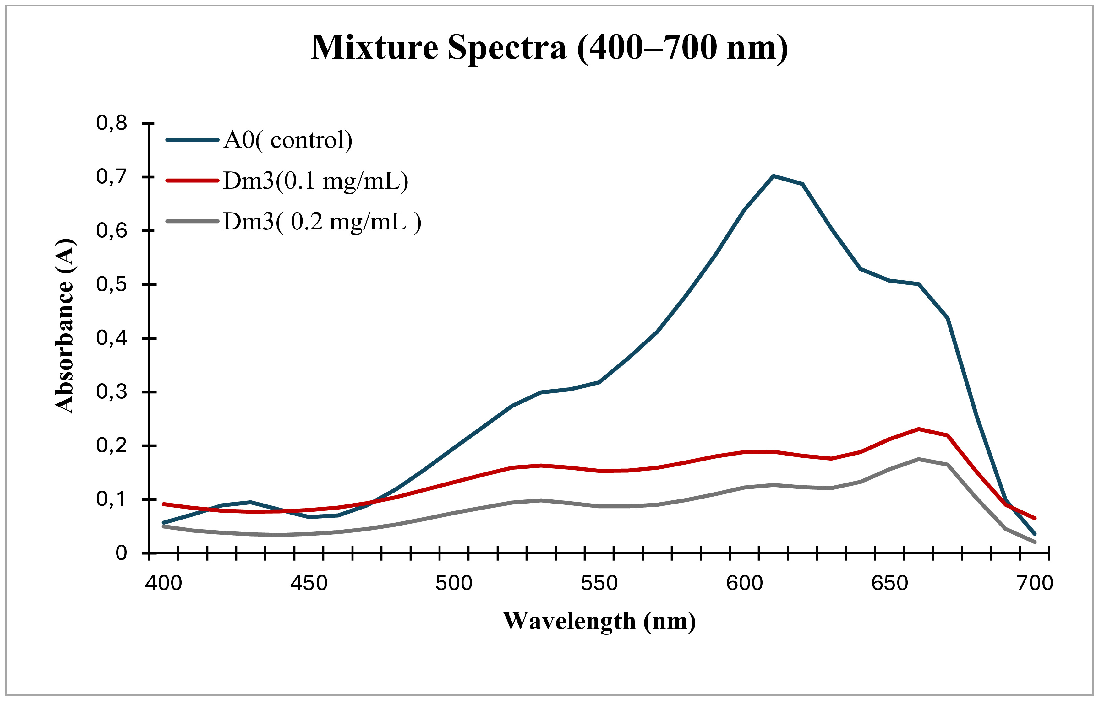
To evaluate the performance of W. dispersa (AmlDm3)-derived FeNPs under conditions resembling real wastewater, a mixed-dye solution containing methylene blue, malachite green, methyl violet, and safranin was prepared at a final reaction volume of 2 mL. After 24 h of treatment with FeNPs at 0.2 mg/mL, a pronounced reduction in color intensity was observed compared with the deep-blue untreated control (Figure 11).

Visual observation of mixed-dye decolorization after 24 h of treatment with FeNPs synthesized by W. dispersa (AmlDm3) at two concentrations (0.1 and 0.2 mg/mL), compared with the untreated control.
Spectrophotometric analysis showed a substantial decrease in absorbance across the visible range (400 - 700 nm), indicating simultaneous degradation of multiple chromophoric compounds within the mixture (Figure 12). The decolorization profile remained comparable across the tested FeNP amounts, with only a slight increase in efficiency at the higher applied volume, suggesting that W. dispersa (AmlDm3)-mediated FeNPs maintain strong catalytic performance even at relatively low nanoparticle dosages.

UV–Vis absorption spectra (400–700 nm) of a mixed-dye system showing a marked decrease in absorbance intensity after 24 h of treatment with FeNPs synthesized by W. dispersa (AmlDm3) at two concentrations (0.1 and 0.2 mg/mL), indicating effective dye degradation.
3.6.3. Antimicrobial activity assay
The antimicrobial activity of W. dispersa (AmlDm3)-derived FeNPs was evaluated against six clinically relevant microorganisms, including Gram-positive bacteria (Staphylococcus aureus, Staphylococcus epidermidis), Gram-negative bacteria (Escherichia coli, Klebsiella oxytoca), and unicellular fungi (Candida albicans and Candida tropicalis). At all tested concentrations (0.1–0.3 mg/mL), and in both colloidal and powder forms, the FeNPs produced no measurable zones of inhibition against any of the tested strains. In contrast, the positive controls showed clear inhibition zones, confirming the assay's validity. (Figure 13).

Agar well diffusion assay evaluating the antimicrobial potential of FeNPs synthesized by Westerdykella dispersa (AmlDm3) against Gram-negative bacteria, Gram-positive bacteria, and yeast strains. No inhibition zones were observed around wells containing FeNPs, indicating the absence of antimicrobial activity, whereas the positive controls produced clear inhibition halos.
4. Discussion
Fungal isolates were obtained from wastewater-impacted environments at a sewage pumping station in Dammam, Eastern Province, where continuous exposure to industrial effluents likely promotes the presence of metal-tolerant and pollutant-adapted fungi. Similar observations have been reported by Mahanty et al. (2019), who noted that fungi from contaminated habitats often exhibit enhanced redox activity, making them suitable candidates for nanoparticle biosynthesis. Among the screened isolates, W. dispersa (AmlDm3) showed the most pronounced visual response during preliminary assays, as evidenced by the rapid orange-brown color change upon exposure to ferrous sulfate. This color transition is commonly associated with Fe2+ reduction and the initiation of iron nanoparticle formation and is widely recognized as a primary indicator of biogenic FeNP synthesis (Ramadan Gouda et al., 2020; Mathur et al., 2021). The intensity and stability of the developed color suggest strong bioreductive activity and the involvement of extracellular metabolites in nanoparticle formation and stabilization.
UV–Vis analysis of FeNPs synthesized by Westerdykella dispersa (AmlDm3) showed a distinct absorption peak at 242 nm, which was absent in the control, confirming nanoparticle formation. Absorption bands below 300 nm are characteristic of biogenic iron nanoparticles and are commonly associated with metabolite-mediated iron reduction. Similar UV features have been reported for fungal-derived FeNPs, including Alternaria alternata and other iron-tolerant fungi (Mohamed et al., 2015; Gouda et al., 2020).
Zeta potential analysis showed a surface charge of –20.2 mV, indicating moderate colloidal stability of the synthesized nanoparticles. Although values beyond ±30 mV are generally associated with higher stability, the observed charge suggests sufficient electrostatic repulsion to limit aggregation. This stability is likely enhanced by fungal biomolecules acting as natural capping agents, a phenomenon commonly reported in fungal-mediated nanoparticle synthesis and associated with proteins and polysaccharides present in the fungal filtrate (Hammad et al. (2022); Ingle et al., 2023).
FTIR analysis of FeNPs synthesized using W. dispersa (AmlDm3) extract showed functional groups indicative of fungal biomolecules involved in nanoparticle formation and stabilization. The Fe–O stretching band at 467 cm−1 confirmed iron oxide formation, while the bands associated with C–O and carboxylate (COO−) groups suggest the presence of polysaccharides and organic acids in the synthesis medium. Similar functional groups have been widely reported as stabilizing moieties in biologically synthesized nanoparticles, where carbohydrates and proteins act as surface-capping ligands rather than metal-specific indicators (Manivasagan et al., 2015; Ahmed, 2017).
SEM analysis showed predominantly spherical FeNPs with an average size of ~100 nm. Similar particle sizes have been reported for biogenic FeNPs synthesized using apple peel extract (50–100 nm) and other plant-mediated systems (Devatha et al., 2016; Ting and Chin, 2020), supporting the role of biomolecular capping in controlling particle growth.
EDX analysis confirmed that iron and oxygen were the predominant elements in the biosynthesized FeNPs, accompanied by a notable carbon signal, indicating the presence of organic capping layers on the nanoparticle surface. Comparable elemental compositions have been reported for biogenic iron oxide nanoparticles, where Fe and O typically account for ~50–55% and ~20–30% of the total elemental content, respectively, while carbon signals are attributed to residual biological molecules involved in surface stabilization (Yassin et al., 2023; Kamal et al., 2023). The minor phosphorus peak observed in the present study may arise from phosphate-containing fungal metabolites or trace components of the growth medium adsorbed onto the nanoparticle surface.
The FeNPs biosynthesized by W. dispersa (AmlDm3) showed strong catalytic activity toward all tested dyes, with decolorization efficiency varying according to dye structure. Malachite green displayed the highest susceptibility, achieving rapid and near-complete removal (>95%). Previous studies on fungal-mediated FeNPs have reported variable decolorization efficiencies for triphenylmethane dyes, with malachite green removal ranging from ~18–35% in adsorption-dominated systems to values exceeding 90% under enhanced catalytic or Fenton-like conditions (Schuster and Ting, 2022; Kumar et al., 2024). The pronounced response of malachite green observed in the present study may be attributed to its relatively lower structural stability and higher accessibility of the chromophoric center compared to other triphenylmethane dyes.
Methyl violet and methylene blue exhibited comparatively slower decolorization kinetics, reaching approximately 85% and 82% removal, respectively. This trend aligns with earlier reports identifying methyl violet as one of the least susceptible triphenylmethane dyes during degradation due to its higher resonance stabilization (Chen et al., 2010). Although effective methylene blue removal has been reported in FeNP-mediated systems, high efficiencies often require extended reaction times or auxiliary oxidants (Mathur et al., 2021; Jayeola et al., 2022). Safranin showed moderate decolorization (~70%), which is in close agreement with the 66% removal efficiency reported for phenazine dyes using fungal-derived FeNPs (Hammad et al., 2022).
Evaluation of FeNP performance in a mixed-dye system further demonstrated their robustness under conditions simulating real wastewater. Despite the increased complexity and competitive interactions among dyes, W. dispersa (AmlDm3)-mediated FeNPs maintained substantial decolorization efficiency at low nanoparticle dosage. Comparable performance across different FeNP volumes suggests near-saturation of active sites under the tested conditions.
Overall, these results highlight the strong and consistent catalytic performance of W. dispersa (AmlDm3)-derived FeNPs in both single- and mixed-dye systems, supporting their potential as an efficient and sustainable nanocatalyst for complex wastewater remediation.
The absence of antimicrobial activity observed in this study contrasts with previous reports on biogenic iron nanoparticles that showed inhibitory effects against microbial strains (Mohamed et al., 2015; Hammad et al., 2022). Such discrepancies can be attributed to differences in nanoparticle size, surface properties, and capping efficiency. Larger and well-stabilized nanoparticles generally display reduced biological reactivity, as reported by Gupta et al. (2023). In the present study, the FeNPs synthesized by W. dispersa (AmlDm3) were strongly capped by fungal metabolites, which likely limited their interaction with microbial cells and explain the lack of antimicrobial activity.
Overall, the findings demonstrate that W. dispersa (AmlDm3) represents an effective and previously unexplored fungal platform for sustainable iron nanoparticle biosynthesis. The resulting FeNPs showed favorable physicochemical characteristics and strong catalytic performance in both single- and mixed-dye systems, supporting their potential application as environmentally safe nanocatalysts for wastewater remediation
Acknowledgements
The authors acknowledge the Biology Department, College of Science, Imam Abdulrahman Bin Faisal University, the Basic and Applied Scientific Research Center (BASRC), and King Saud University Medical City, Riyadh, Saudi Arabia, for providing laboratory facilities and molecular identification support.
Data Availability Statement
The dataset analyzed or produced in this study is available from the corresponding author upon reasonable request.
References:
-
ADELEYE, T.M., KAREEM, S.O. and KEKERE-EKUN, A.A., 2020. Optimization studies on biosynthesis of iron nanoparticles using Rhizopus stolonifer. IOP Conference Series. Materials Science and Engineering, vol. 805, no. 1, pp. 012037. http://doi.org/10.1088/1757-899X/805/1/012037
» http://doi.org/10.1088/1757-899X/805/1/012037 -
AHMED, T., 2017. Elucidation of emerging nanomaterials impacts on antibiotic resistance against soil and aquatic microflora. In: J. DUTTA and T. BANERJEE, eds. Emerging nanomaterials for agriculture, food and environmental safety Singapore: Springer, pp. 259–281. http://doi.org/10.1007/978-3-319-66260-2_14
» http://doi.org/10.1007/978-3-319-66260-2_14 -
AL-HARBI, N. and ABD-ELRAHMAN, N.K., 2024. Physical methods for preparation of nanomaterials, their characterization and applications: a review. Journal of Umm Al-Qura University for Applied Sciences, vol. 11, no. 2, pp. 356-377. http://doi.org/10.1007/s43994-024-00165-7
» http://doi.org/10.1007/s43994-024-00165-7 -
CHEN, C.H., CHANG, C.F. and LIU, S.M., 2010. Partial degradation mechanisms of malachite green and methyl violet B by Shewanella decolorationis NTOU1 under anaerobic conditions. Journal of Hazardous Materials, vol. 177, no. 1-3, pp. 281-289. http://doi.org/10.1016/j.jhazmat.2009.12.030 PMid:20060225.
» http://doi.org/10.1016/j.jhazmat.2009.12.030 -
DEVATHA, C.P., THALLA, A.K. and KATTE, S.Y., 2016. Green synthesis of iron nanoparticles using different leaf extracts for treatment of domestic wastewater. Journal of Cleaner Production, vol. 139, pp. 1425-1435. http://doi.org/10.1016/j.jclepro.2016.09.019
» http://doi.org/10.1016/j.jclepro.2016.09.019 -
DHAND, C., DWIVEDI, N., LOH, X.J., JIE YING, A., VERMA, N.K., BEUERMAN, R.W., LAKSHMINARAYANAN, R. and RAMAKRISHNA, S., 2015. Methods and strategies for the synthesis of diverse nanoparticles and their applications: a comprehensive overview. RSC Advances, vol. 5, no. 127, pp. 105003-105037. http://doi.org/10.1039/C5RA19388E
» http://doi.org/10.1039/C5RA19388E - GOH, J., MUN, H.Y. and OH, Y., 2021. Seven previously unrecorded fungal species isolated from freshwater ecosystems in Korea. Korean Journal of Mycology, vol. 49, pp. 183-197.
-
GOUDA, A.R., SIDKEY, N.M., SHAWKY, H.A. and ABDEL-HADY, Y.A., 2020. Biosynthesis, characterization and antimicrobial activity of iron oxide nanoparticles synthesized by fungi. Azhar Journal of Pharmaceutical Sciences, vol. 62, no. 2, pp. 164-179. http://doi.org/10.21608/ajps.2020.118382
» http://doi.org/10.21608/ajps.2020.118382 -
GUPTA, I., GHUGE, A., DAHM, H. and RAI, M., 2023. Toxicity of mycosynthesised nanoparticles. In: I. GUPTA, A. GHUGE, H. DAHM and M. RAI, eds. Mycosynthesis of nanomaterials: perspectives and challenges Abingdon: Routledge/Taylor & Francis, pp. 283–296. http://doi.org/10.1201/9781003327387-17
» http://doi.org/10.1201/9781003327387-17 -
HAMMAD, E.N., SALEM, S.S., MOHAMED, A.A. and EL-DOUGDOUG, W., 2022. Environmental impacts of ecofriendly iron oxide nanoparticles on dyes removal and antibacterial activity. Applied Biochemistry and Biotechnology, vol. 194, no. 12, pp. 6053-6067. http://doi.org/10.1007/s12010-022-04105-1 PMid:35881227.
» http://doi.org/10.1007/s12010-022-04105-1 -
INGLE, P., KAMBLE, K., GOLIŃSKA, P., RAI, M. and GADE, A., 2023. Techniques for characterization of biologically synthesized nanoparticles by fungi. In: I. GUPTA, A. GHUGE, H. DAHM and M. RAI, eds. Mycosynthesis of nanomaterials: perspectives and challenges Abingdon: Routledge/Taylor & Francis, pp. 233–253. http://doi.org/10.1201/9781003327387-14
» http://doi.org/10.1201/9781003327387-14 -
JAYEOLA, J.N., BELLO, I.A., JULIUS, F.A. and ADIO, S.W., 2022 [viewed 14 January 2026]. Oxidative degradation of methylene blue dye from wastewater by Fenton process. International Journal of Applied Chemical and Biological Sciences [online], vol. 3, no. 2, pp. 34-43. Available from: https://identifier.visnav.in/1.0001/ijacbs-22c-03003/
» https://identifier.visnav.in/1.0001/ijacbs-22c-03003/ -
KAMAL, A., SABA, M. and FAROOQ, M., 2023. Biocompatible formulations based on mycosynthesized iron oxide nanoparticles: Fabrication, characterization, and biological investigation. Journal of Basic Microbiology, vol. 63, no. 2, pp. 156-167. http://doi.org/10.1002/jobm.202200560 PMid:36529705.
» http://doi.org/10.1002/jobm.202200560 -
KOUL, B., POONIA, A.K., YADAV, D. and JIN, J.-O., 2021. Microbe-mediated biosynthesis of nanoparticles: applications and future prospects. Biomolecules, vol. 11, no. 6, pp. 886. http://doi.org/10.3390/biom11060886 PMid:34203733.
» http://doi.org/10.3390/biom11060886 -
KUMAR, V., KAUSHIK, N.K., TIWARI, S.K., SINGH, D. and SINGH, B., 2023. Green synthesis of iron nanoparticles: sources and multifarious biotechnological applications. International Journal of Biological Macromolecules, vol. 253, no. Pt 4, pp. 127017. http://doi.org/10.1016/j.ijbiomac.2023.127017 PMid:37742902.
» http://doi.org/10.1016/j.ijbiomac.2023.127017 -
KUMAR, V., SINGH, D. and SINGH, B., 2024. Mycogenic synthesis of iron nanoparticles using thermophilic mould Myceliophthora thermophila and their applicability in environmental remediation. Biocatalysis and Agricultural Biotechnology, vol. 56, pp. 103020. http://doi.org/10.1016/j.bcab.2024.103020
» http://doi.org/10.1016/j.bcab.2024.103020 -
MAHANTY, S., CHATTERJEE, S., GHOSH, S., TUDU, P., GAINE, T., BAKSHI, M., DAS, S., DAS, P., BHATTACHARYYA, S., BANDYOPADHYAY, S. and CHAUDHURI, P., 2019. Green synthesis of iron oxide nanoparticles mediated by filamentous fungi isolated from Sundarban mangrove ecosystem, India. BioNanoScience, vol. 9, no. 3, pp. 637-651. http://doi.org/10.1007/s12668-019-00644-w
» http://doi.org/10.1007/s12668-019-00644-w -
MAHANTY, S., CHATTERJEE, S., GHOSH, S., TUDU, P., GAINE, T., BAKSHI, M., DAS, S., DAS, P., BHATTACHARYYA, S., BANDYOPADHYAY, S. and CHAUDHURI, P., 2020. Synergistic approach towards the sustainable management of heavy metals in wastewater using mycosynthesized iron oxide nanoparticles: biofabrication, adsorptive dynamics and chemometric modeling study. Journal of Water Process Engineering, vol. 37, pp. 101426. http://doi.org/10.1016/j.jwpe.2020.101426
» http://doi.org/10.1016/j.jwpe.2020.101426 -
MANIVASAGAN, P., KANG, K.-H., KIM, D.G. and KIM, S.-K., 2015. Production of polysaccharide-based bioflocculant for the synthesis of silver nanoparticles by Streptomyces sp. International Journal of Biological Macromolecules, vol. 77, pp. 159-167. http://doi.org/10.1016/j.ijbiomac.2015.03.022 PMid:25799882.
» http://doi.org/10.1016/j.ijbiomac.2015.03.022 -
MATHUR, P., SAINI, S., PAUL, E., SHARMA, C. and MEHTANI, P., 2021. Endophytic fungi mediated synthesis of iron nanoparticles: characterization and application in methylene blue decolorization. Current Research in Green and Sustainable Chemistry, vol. 4, pp. 100220. http://doi.org/10.1016/j.crgsc.2020.100053
» http://doi.org/10.1016/j.crgsc.2020.100053 - MATTHAPAN, L., PRASONG, W., LEEYAPHAN, C., BUNYARATAVEJ, S. and LERTRUJIWANIT, K., 2018. A novel technique of aluminum multiplier slide culture for fungal identification. Siriraj Medical Journal, vol. 70, pp. 438-441..
-
MEHTA, M., SHARMA, M., PATHANIA, K., JENA, P.K. and BHUSHAN, I., 2021. Degradation of synthetic dyes using nanoparticles: a mini-review. Environmental Science and Pollution Research International, vol. 28, no. 36, pp. 49434-49446. http://doi.org/10.1007/s11356-021-15470-5 PMid:34350572.
» http://doi.org/10.1007/s11356-021-15470-5 -
MOHAMED, Y.M., AZZAM, A.M., AMIN, B.H. and SAFWAT, N.A., 2015. Mycosynthesis of iron nanoparticles by Alternaria alternata and its antibacterial activity. African Journal of Biotechnology, vol. 14, no. 14, pp. 1234-1241. http://doi.org/10.5897/AJB2014.14286
» http://doi.org/10.5897/AJB2014.14286 -
MUZAHID, A.N.M., ARAF, Y., MAHMUD, N.U., SARKER, A., AKTER, F., CHOWDHURY, M.T.I., SHIDDIKY, M.J.A., SOHRAWARDY, H., CHAKRABORTY, M. and ISLAM, T., 2023. Potentials of mycosynthesized nanomaterials for efficient remediation of environmental contaminants. In: K.A. ABD-ELSALAM, ed. Fungal cell factories for sustainable nanomaterials production and agricultural applications Amsterdam: Elsevier, pp. 693–724. http://doi.org/10.1016/B978-0-323-99922-9.00015-5
» http://doi.org/10.1016/B978-0-323-99922-9.00015-5 -
PATIL, N., BHASKAR, R., VYAVHARE, V., DHADGE, R., KHAIRE, V. and PATIL, Y., 2021. Overview on methods of synthesis of nanoparticles. International Journal of Current Pharmaceutical Research, vol. 13, no. 2, pp. 11-16. http://doi.org/10.22159/ijcpr.2021v13i2.41556
» http://doi.org/10.22159/ijcpr.2021v13i2.41556 -
SAHA, I., KARMAKAR, P. and BHATTACHARYA, D., 2023. Fungi-mediated fabrication of copper nanoparticles and copper oxide nanoparticles, physical characterization and antimicrobial activity. In: M. RAI and P. GOLINSKA, eds. Mycosynthesis of nanomaterials: perspectives and challenges Boca Raton: CRC Press, pp. 112-125.. http://doi.org/10.1201/9781003327387-7
» http://doi.org/10.1201/9781003327387-7 -
SAIED, E., SALEM, S.S., AL-ASKAR, A.A., ELKADY, F.M., ARISHI, A.A. and HASHEM, A.H., 2022. Mycosynthesis of hematite (α-Fe2O3) nanoparticles using Aspergillus niger AH1 and their photocatalytic activity against crystal violet dye. Bioengineering, vol. 9, pp. 397. http://doi.org/10.3390/bioengineering9080397 PMid:36004922.
» http://doi.org/10.3390/bioengineering9080397 -
SCHUSTER, S. and TING, A.S.Y., 2022. Decolourisation of triphenylmethane dyes by biogenically synthesised iron nanoparticles from fungal extract. Mycology, vol. 13, no. 1, pp. 56-67. http://doi.org/10.1080/21501203.2021.1948928 PMid:35186413.
» http://doi.org/10.1080/21501203.2021.1948928 -
SIDHU, A.K. and KAUSHAL, P., 2023. Biosynthesis of iron oxide nanoparticles using fungi. In: M. RAI and P. GOLINSKA, eds. Mycosynthesis of nanomaterials: perspectives and challenges. Boca Raton: CRC Press, pp. 81-98.. http://doi.org/10.1201/9781003327387-5
» http://doi.org/10.1201/9781003327387-5 -
SRIVASTAVA, S. and BHARGAVA, A., 2021. Green nanoparticles: the future of nanobiotechnology Singapore: Springer, vol. 2, pp. 1-352. http://doi.org/10.1007/978-981-16-7106-7
» http://doi.org/10.1007/978-981-16-7106-7 -
TARAFDAR, J.C. and RALIYA, R., 2013. Rapid, low-cost, and ecofriendly approach for iron nanoparticle synthesis using Aspergillus oryzae TFR9. Journal of Nanoparticles, vol. 2013, pp. 1-4. http://doi.org/10.1155/2013/141274
» http://doi.org/10.1155/2013/141274 -
TING, A.S.Y. and CHIN, J.E., 2020. biogenic synthesis of iron nanoparticles from apple peel extracts for decolorization of malachite green dye. Water, Air, and Soil Pollution, vol. 231, no. 6, pp. 1. http://doi.org/10.1007/s11270-020-04658-z
» http://doi.org/10.1007/s11270-020-04658-z -
XU, D., PANG, X.-J., ZHAO, T., XU, L.-L. and YANG, X.-L., 2017. New alkenylated tetrahydropyran derivatives from the marine sediment-derived fungus Westerdykella dispersa and their bioactivities. Fitoterapia, vol. 122, pp. 45-51. http://doi.org/10.1016/j.fitote.2017.08.010 PMid:28842357.
» http://doi.org/10.1016/j.fitote.2017.08.010 -
YASSIN, M.T., AL-OTHMAN, M.R., ALI, A.M., AL-QURAINY, F.H. and AL-HARBI, N.S., 2023. Green synthesis, characterization, and antifungal efficiency of biogenic iron oxide nanoparticles. Applied Sciences, vol. 13, no. 17, pp. 9942. http://doi.org/10.3390/app13179942
» http://doi.org/10.3390/app13179942
Edited by
-
Editor:
Takako Matsumura Tundisi