
 Molecular characterization of surfactin biosynthetic gene in Bacillus cereus BT1.8 and its antifungal efficacy against Fusarium oxysporum
Abstract
Bacillus sp. is a genus of bacteria that can produce active biosurfactant compounds and has potential as an antimicrobial agent. The Bacillus bacteria used in this study were isolated from the soil of Bekol Baluran National Park, which is predicted to contain various types of bacteria that can inhibit soil-borne pathogenic fungi such as Fusarium oxysporum, considering that this pathogen causes root rot in plants. This study aims to identify the potential of Bacillus spp. isolates BT1.8, BT9.1, and SM2.3 in producing biosurfactants as antifungal agents. The methods used ranged from macroscopic, microscopic, and molecular characterization of bacteria, in-vitro antifungal activity testing using the swab method and creating a F. oxysporum block, to biosurfactant activity testing through hemolytic activity using Blood Agar medium. Molecular analysis of the biosurfactant biosynthesis gene was performed using PCR with specific primers for the srfAD gene. The results showed that the Bacillus sp. BT1.8 isolate exhibited the best antifungal activity with a 69% inhibition zone against the growth of F. oxysporum. Isolate BT1.8 was identified as having 99.11% similarity to B. cereus ATCC1457 and possesses the srfAD gene, which shows a high level of homology with a similar gene in Bacillus halotolerans with accession number WP_105955226.1. Hemolytic activity results also show that all three bacterial isolates can destroy blood cells through beta hemolysis. This indicates that the presence of the surfactin biosynthesis gene can produce biosurfactant compounds that are capable of significantly inhibiting the growth of F. oxysporum. These results show that B. cereus BT1.8 has potential as a biological biocontrol agent in the form of seed coating that can be used in the formulation of environmentally friendly biocidal products.
Keywords:
biosurfactant; srfAD; plant pathogen; biocontrol; agriculture
Resumo
Bacillus sp. é um gênero de bactérias que pode produzir compostos biossurfactantes ativos e tem potencial como agente antimicrobiano. As bactérias Bacillus utilizadas neste estudo foram isoladas do solo do Parque Nacional Bekol Baluran, que se prevê conter vários tipos de bactérias capazes de inibir fungos patogênicos transmitidos pelo solo, como o Fusarium oxysporum, considerando que este patógeno causa podridão radicular nas plantas. Este estudo tem como objetivo identificar o potencial das bactérias Bacillus spp. isoladas BT1.8, BT9.1 e SM2.3 na produção de biossurfactantes como agentes antifúngicos. Os métodos utilizados variaram desde a caracterização macroscópica, microscópica e molecular das bactérias, testes de atividade antifúngica in vitro utilizando o método de swab e a criação de um bloco de F. oxysporum até testes de atividade biossurfactante através da atividade hemolítica utilizando meio Blood Agar. A análise molecular do gene da biossíntese do biossurfactante foi realizada utilizando PCR com primers específicos para o gene srfAD. Os resultados mostraram que o isolado Bacillus sp. BT1.8 exibiu a melhor atividade antifúngica, com uma zona de inibição de 69% contra o crescimento de F. oxysporum. O isolado BT1.8 foi identificado como tendo 99,11% de similaridade com B. cereus ATCC1457 e possui o gene srfAD, que apresenta um alto nível de homologia com um gene semelhante em Bacillus halotolerans com número de acesso WP_105955226.1. Os resultados da atividade hemolítica também mostram que todas as três cepas bacterianas isoladas podem destruir células sanguíneas por meio da hemólise beta. Isso indica que a presença do gene de biossíntese da surfactina pode produzir compostos biossurfactantes capazes de inibir significativamente o crescimento de F. oxysporum. Esses resultados mostram que B. cereus BT1.8 tem potencial como agente de controle biológico na forma de revestimento de sementes que pode ser usado na formulação de produtos biocidas ecologicamente corretos.
Palavras-chave:
biossurfactante; srfAD; patógeno vegetal; biocontrole; agricultura
1. Introduction
Fusarium oxysporum is one of the pathogenic fungi that live in the soil and cause serious diseases such as Fusarium wilt in various economically important agricultural crops, such as tomatoes, bananas, and cotton (Chen and Du, 2021; Roncero et al., 2003; Czymmek et al., 2007). This pathogenic fungus is known to cause vascular wilt, seedling blight, and root rot, which can cause significant losses by inhibiting plant growth, reducing crop quality, and even resulting in crop failure (Czymmek et al., 2007; Edgar et al., 2006; Guo et al., 2023). Efforts to control diseases caused by F. oxysporum generally still rely on the use of chemical fungicides, but this method has the potential to cause negative impacts such as reducing the diversity of beneficial soil microbes that are important for the nutrient cycle (Wijntjes et al., 2022), and chemical fungicide residues are toxic to human health (Mohd Zainudin et al., 2024; Yue et al., 2024), and can trigger pathogen resistance (Islam et al., 2024), and impact non-target organisms such as mycorrhizal fungi, which play an important role in soil nutrient uptake and soil health (Dharma et al., 2024). Therefore, alternative strategies that are more environmentally friendly, effective, and sustainable are needed.
One approach that is gaining increasing attention is the use of microbial-based biological agents. Bacillus cereus is known as a soil bacterium capable of producing various secondary metabolites such as diketopiperazines (Kumar et al., 2014), hydrolytic enzymes (chitinase, chitosanase, and protease) (Chang et al., 2007), and lipopeptide biosurfactants (fengycin, iturin, and kannurin) (Khadiri et al., 2023; Ajesh et al., 2013) with antifungal activity. Biosurfactants are amphiphilic compounds that have the ability to reduce surface tension and play a role in antimicrobial interactions. Several studies have reported that biosurfactants from the genus Bacillus cereus can inhibit the growth and development of plant pathogens through mechanisms that inhibit spore germination and the growth of pathogenic fungal mycelium (Ahmed et al., 2022), lysis of the cell wall and inhibit fungal spread (Khadiri et al., 2023), and inhibit biofilm formation (fungal defense mechanisms) (Ajesh et al., 2013).
Exploration of various species of Bacillus producing biosurfactants from relatively undisturbed natural ecosystems is still very limited, especially in conservation areas with predicted high levels of microbial diversity, such as Baluran National Park. This area includes a natural savanna ecosystem that has unique environmental characteristics and has the potential to be a source of indigenous microorganisms with specific metabolic abilities, including the production of biosurfactants. In addition, indigenous soil bacteria are predicted to have a high level of physiological and ecological adaptation in colonising and interacting effectively within complex soil systems. Ecologically, this group of bacteria has undergone natural selection against environmental pressures in accordance with the conditions of Baluran National Park, such as nutrient competition, fluctuations in humidity and temperature, and antagonistic interactions with other microbes. These adaptations have the potential to increase their effectiveness in suppressing soil-borne pathogens compared to non-indigenous microorganisms or those from different environments.
However, to date, few studies have reported on the potential of soil bacteria from Bekol in Baluran National Park to produce biosurfactants and their activity as antifungal agents. Therefore, exploration of these soil bacteria is necessary given the similarity in habitat between the bacteria and the target soil-borne pathogen F. oxysporum, which means they have the potential to be more effective in adapting and competing to suppress the growth of pathogenic fungi in the soil. The results of this study are expected to provide a scientific basis for the development of biosurfactant-based green biocides as environmentally friendly and sustainable biocontrol products, while also supporting efforts to reduce dependence on chemical fungicides in plant disease management.
2. Materials and Methods
2.1. Media and isolate preparation
This study used three types of media, namely Nutrient Yeast Salt Medium (NYSM), Nutrient Broth (NB), and Potato Dextrose Agar (PDA). NYSM medium, both in solid and liquid form, was used to grow bacterial cultures of isolates BT1.8, BT9.1, and SM2.3. The composition of NYSM in solid form are 28.0 g/L NA, 0.5 g/L yeast extract, 0.2 g/L MgCl2, 0.01 g/L MnCl2, and 0.1 g/L CaCl2, while liquid NYSM composition of 8.0 g/L NB, 0.5 g/L yeast extract, 0.2 g/L MgCl2, 0.01 g/L MnCl2, and 0.1 g/L CaCl2 (Myers and Yousten, 1978). NB medium was used as a growth medium in the DNA isolation process. Meanwhile, PDA medium was used as a test medium for antifungal activity. All media and equipment used were sterilized in an autoclave at 121 °C for 15 minutes.
The bacteria used in this study were isolates BT1.8, BT9.1, and SM2.3, which were previously successfully isolated from the soil of Baluran National Park, East Java, Indonesia, in a study by Salamun et al. (2020). The three bacterial isolates were grown on NYSM medium for 24 hours at 30 °C as an initial preparation stage. The test fungus used was Fusarium oxysporum strain InACC, obtained from the National Research and Innovation Agency (BRIN). The F. oxysporum fungus was grown on PDA medium for 120 hours at a temperature of 25 °C.
2.2. Confirmation of macroscopic and microscopic characteristics of bacterial isolates
In this study, confirmation of the purity level of bacterial isolates was carried out through observation of macroscopic and microscopic characteristics. Bacterial isolates BT1.8, BT9.1, and SM2.3 were inoculated on solid NYSM medium for 18-20 hours for Gram staining (crystal violet, iodine, lugol, safranin) and endospore staining (malachite green).
2.3. Antifungal activity test
Antifungal activity testing was conducted in vitro using the swab method to evaluate the ability of the three bacterial isolates (BT1.8, BT9.1, and SM2.3) to inhibit the growth of the plant pathogen Fusarium oxysporum. The three bacterial isolates that had been grown for 72 hours were then centrifuged and sonicated, resulting in three culture fractions: cell-free supernatant, sonication pellet, and pure culture. The supernatant and pellet were obtained by the centrifugation process which is carried out with 3000 xg, for 9 minutes, at 4 °C. The supernatant free-cells fraction was filtered with filter membrane, while the pellet fraction was sonicated for 3 minutes, with a frequency 60 kHz, at 4 °C. To obtain the sonicated culture fraction, the bacterial culture was sonicated under the same conditions without centrifugation process. The three fractions were then applied to PDA medium in Petri dishes of 100 µL, with holes made in the center of the medium using a cork borer for inoculation with F. oxysporum.
F. oxysporum isolates that had been regenerated on PDA medium for five days were cut into blocks using a cork borer and placed in the center of the PDA medium that had previously been treated with bacterial fractions. Fungal growth was observed daily until 168 hours of incubation. The growth zone of F. oxysporum was measured using a growth inhibition index formula adapted from Jasim et al. (2016) (Equation 1):
where: D1 : diameter of control growth; D2 : diameter of treatment.
2.4. Biosurfactant activity screening (hemolytic test)
Biosurfactant activity screening was conducted qualitatively to determine the ability of bacteria to produce biosurfactants. This screening was carried out through a hemolytic activity test using Blood Agar medium. Bacterial cultures that were 24 hours old were inoculated on Blood Agar medium using the dot method and incubated for 24-72 hours. Every 24 hours, observations are made to note any changes. A positive indicator shows that a clear zone will form around the three isolates, demonstrating their ability to hemolyze blood.
2.5. Identification of 16S rRNA genes in potential biosurfactant-producing bacteria
The species identification process was carried out to determine the types of potential bacteria that play a role in the production of biosurfactants as agents that inhibit the growth of plant pathogenic fungi, using the 16S rRNA gene detection method. The process began with the preparation of BT1.8 bacterial culture, where 1–2 ose were inoculated into 20 mL of Nutrient Broth (NB) media and then incubated at a temperature of 30 °C with an agitation speed of 150 rpm for 24 hours.
DNA isolation was performed using the CTAB method (Ausubel et al., 2003) with the assistance of the DNA Wizard Genomic DNA Purification Kit (Promega). The 16S rRNA gene was amplified using the Polymerase Chain Reaction (PCR) technique with universal primers 27F and 1492R. The PCR conditions consisted of predenaturation at 94 °C for 2 minutes, followed by denaturation at 92 °C for 30 seconds, annealing at 55 °C for 30 seconds, elongation at 72 °C for 1 minute, and a final elongation step at 72 °C for 5 minutes, repeated for 35 cycles (Nafidiastri et al., 2021). For the reaction we used Forward primer 3 ul, Reverse primer 3 ul, DNA template 5 ul, MasterMix 25 ul, and Nuclease Free Water (NFW) 14 uL.
The PCR products were then visualized by electrophoresis using a 1% agarose gel stained with Ethidium Bromide (EtBr). The amplified DNA fragments were then sent to 1st Base DNA Sequencing Service Malaysia for further analysis. The DNA sequence data obtained was then analyzed using the Basic Local Alignment Search Tool nucleotide (BLASTn) and compared with the GeneBank database available on the National Center for Biotechnology Information website (NCBI, 2025).
2.6. Detection of the biosurfactant biosynthesis gene srfAD
The genome DNA (gDNA) obtained from DNA isolation was used as a DNA template to detect the presence of the biosurfactant biosynthesis gene srfAD using specific primers designed through the Thermofisher website, with Forward primer (F): 5’-TCACGATTGAATGATCGGATGC-3’ and Reverse primer (R): 5’-ATGAGCCAACTCTTCAAATCATTTGA-3’, at 55 °C for annealing. Predicted size of the srfAD gene around 732 bp. The gene was amplified using the PCR technique. The PCR results were visualized using a 1.5% agarose gel and Ethidium Bromide (EtBr) in the electrophoresis process. The srfAD gene DNA fragment was sent to 1st Base DNA Sequencing Service Malaysia for further analysis. The DNA sequence data obtained was then analyzed using the Basic Local Alignment Search Tool nucleotide (BLASTn) and Basic Local Alignment Search Tool protein (BLASTp), which were then compared with the GeneBank database available on the National Center for Biotechnology Information website (NCBI, 2025).
2.7. Data analysis
The data obtained in this study were analyzed descriptively and statistically. The data on macroscopic and microscopic characteristics, antifungal activity, hemolytic activity, and identification of the 16S rRNA and srfAD genes were analyzed descriptively. Data on the antifungal activity of bacterial isolates against F. oxysporum were also analyzed statistically using IBM SPSS Statistics.
In the antifungal activity test, the data obtained was the percentage inhibition of Bacillus sp. BT1.8, BT9.1, and SM2.3 isolates with variations in the form of sonicated culture, sonicated pellets, and supernatant free-cells against F. oxysporum. The quantitative data were analyzed using the Normality test (Shapiro-Wilk test) and Homogeneity test (Levene test). If the normality test results showed that the data were normally distributed with homogeneous variance, they were then analyzed using one-way ANOVA (Analysis of Variation) with a significance level of 0.05. The results of the ANOVA showed that the administration of various forms of disonicated culture, disonicated pellets, and supernatants from Bacillus sp. BT1.8, BT9.1, and SM2.3 isolates had an effect on antifungal activity, or p<0.05, then continued with the Duncan test to determine which combinations had significant differences.
3. Results and Discussion
The bacteria used in this study were isolates BT1.8, BT9.1, and SM2.3, which were isolated from Be kol Soil in Baluran National Park, East Java, Indonesia, in a study by Salamun et al. (2020). These three bacterial isolates showed considerable potential in causing 40-60% mortality in Aedes aegypti larvae in the study by Salamun et al. (2020). This indicates that the three bacterial isolates are also suspected to have good ability in inhibiting plant pathogenic fungi such as Fusarium oxysporum, given the similarity of cell wall components between Ae. aegypti larvae and F. oxysporum fungi, which are composed of chitin. Further investigation in this research is needed to determine the antagonistic activity of bacteria against pathogenic fungi and the presence of biosynthetic genes for active compounds such as biosurfactants, which are suspected to be the cause of this activity.
These three bacterial isolates were reconfirmed to determine the macroscopic characteristics (morphology) of the bacterial colonies in Figure 1 and microscopic characteristics such as cell shape, Gram type of bacteria, and the presence of bacterial endospores in Figures 2 and 3.

Macroscopy characters of Bacillus spp. on plate agar. (a) Bacillus sp. BT1.8; (b) Bacillus sp. BT9.1; (c) Bacillus sp. SM2.3.

Microscopy characters of Bacillus spp. with Gram staining. (a) Bacillus sp. BT1.8; (b) Bacillus sp. BT9.1; (c) Bacillus sp. SM2.3.

Microscopy characters of Bacillus spp. with spore staining. (a) Bacillus sp. BT1.8; (b) Bacillus sp. BT9.1; (c) Bacillus sp. SM2.3.
Figure 1a shows that the BT1.8 bacterial isolate has a circular colony shape that is white in color with a pinpoint size and entire edges. The elevation formed by the isolate is raised with a shiny texture. Isolate BT9.1 is circular, cream-colored, pinpoint in size with entire edges, and has a convex elevation and glossy texture, as shown in Figure 1b. Isolate SM2.3 is irregular in shape, white, moderate in size, and has undulate edges. The elevation of isolate SM2.3 is raised with a non-glossy texture, as shown in Figure 1c.
Microscopic characteristics through Gram staining were also performed to differentiate bacteria based on their cell wall structure, as shown in Figures 2a-c.
All three bacterial isolates were Gram-positive with rod-shaped cells. Gram-positive bacteria retain the crystal violet color because they have a thick peptidoglycan layer in their cell walls (Riu et al., 2022; Zerbib, 2017). Gram-positive bacteria consist of many genera, such as Staphylococcus, Streptococcus, and Enterococcus (Ruoff, 2022). Meanwhile, Gram-negative bacteria have an additional outer membrane that cannot retain the crystal violet color, allowing them to absorb the red color from safranin during the Gram staining process. The outer membrane makes Gram-negative bacteria resistant to many antibiotics (Riu et al., 2022; Zerbib, 2017).
Microscopic characteristics were also examined by staining the endospores with malachite green to determine the presence of endospores in the three bacterial isolates. All three bacteria showed the presence of endospores within the cells. Bacteria BT1.8 and BT9.1 had oval-shaped endospores located centrally, while bacteria SM2.3 had oval-shaped endospores located subterminally in the cells. Endospores are a dormant (long-lasting) form produced by several types of bacteria, such as those in the Firmicutes phylum, including Bacillus and Clostridium species. This structure enables bacteria to adapt and survive in various unfavorable conditions/extreme environments.
Bacillus sp. is known to produce various types of antifungal compounds that can suppress the growth of plant pathogenic fungi. Various active compounds produced by the Bacillus group include hydrolytic enzymes, such as chitinase, chitosanase, and protease; antimicrobial compounds, such as Bacillin, Hentriacontane, and 2,4-di-tert-butylphenol; volatile organic compounds, such as propanone, 1-butanol, and acetic acid, to lipopeptides such as iturin and fengycin. The antifungal activity results of Bacillus sp. BT1.8, BT9.1, and SM2.3 isolates are shown in Figures 4a-c. The treatments tested were sonication culture, sonication pellet, and free-cell supernatant for 168 hours, which provided sufficient time for metabolites to be produced and interact with the tested pathogenic fungal tissue (Ahmad et al., 2023).

Antifungal activity of Bacillus sp. (a) isolate BT1.8; (b) isolate BT9.1, dan (c) isolate SM2.3 against F. oxysporum on 24H, 72H, 120H, and 168H. C = sonicated culture, P = sonicated pellet, S = supernatant free-cell, C+ = control +, C- = control –.
From the results of the antifungal activity test visualization, it appears that bacterial isolates BT1. 8, BT9.1, and SM2.3 isolates have the best ability to suppress the growth of pathogenic fungi in the pellet fraction, respectively around 18.4 mm, 51.7 mm, and 39.9 mm for diameter growth of pathogen with an inhibition index of 69%, 12%, and 32% as shown in Tables 1 and 2. The diameter of the inhibition zone indicates the diameter of pathogenic F. oxysporum growth after being tested with bacterial fractions. The larger the growth diameter of F. oxysporum, the weaker the ability of the bacterial fraction to inhibit the growth of pathogenic fungi. This indicates that the bacterial fraction is suspected to be insufficient in producing metabolites (biosurfactants), so that its inhibiton index or ability to suppress the growth of pathogenic fungi is not very good. Based on the results of the three bacterial isolates, only the BT1.8 bacterial isolate in pellet fraction showed the best antifungal activity against the pathogenic fungus F. oxysporum for 8 days.
The mechanism of inhibition of pathogenic fungal growth carried out by various active compounds produced by Bacillus spp. is very diverse. Surfactin, Iturin A, and Fengycin are a group of lipopeptide biosurfactant compounds that can produce antifungal activity against F. oxysporum by disrupting the fungal cell membrane, which can cause leakage and cell death (Yuan et al., 2012). Surfactin disrupts the fungal cell membrane, increasing permeability and causing cell lysis (Park et al., 2019; Bidima et al., 2022; Bakker et al., 2025). Iturin can form pores in the fungal membrane (Bidima et al., 2022; Hammad et al., 2023; Bakker et al., 2025). Fengycin is a cyclic lipopeptide that exhibits an antagonistic mechanism against pathogenic fungi through damage to fungal cell membranes, loss of permeability, inhibition of spore germination, and apoptosis, as in Botrytis cinerea and Fusarium sp. Fengycin B produced by B. subtilis 2H11 can damage the structure of F. solani hyphae and spores and significantly inhibit fungal growth (Lee et al., 2015; Zihalirwa Kulimushi et al., 2017; Liu and Sun, 2021; Bidima et al., 2022; Bakker et al., 2025).
Other groups of polyketide compounds such as Bacillaene can prevent fungal growth by disrupting cellular processes, while Macrolactin and Difficidin can disrupt fungal cell walls and inhibit fungal protein synthesis (Lee et al., 2015; Bidima et al., 2022; Hammad et al., 2023). Volatile organic compounds (VOCs) such as Acetylbutanediol interfere with F. oxysporum through membrane function and fungal cell structure, inhibiting mycelium growth and causing morphological changes in fungi (Li et al., 2015; Tuyen et al., 2023). Benzoic acid can also inhibit fungal growth by altering conidia morphology and preventing spore germination. In addition, various ketones and other alcohol compounds, such as chloroacetic acid, tetradecyl esters, octadecane, and hexadecanoic acid, methyl ester can inhibit the growth of F. solani and Colletotrichum gloeosporioides by disrupting cell membrane integrity (Li et al., 2015; Jeong et al., 2017; Rajaofera et al., 2019). VOCs produced by Bacillus can also cause morphological changes in fungal hyphae (Li et al., 2015; Myo et al., 2019; Tuyen et al., 2023). Hydrolytic enzymes, such as chitinase, cellulase, amylase, and glucanase, can also break down the cell wall components of pathogenic fungi, inhibit spore germination, and germ tube elongation of F. oxysporum (Chang et al., 2007; Myo et al., 2019; Pedraza et al., 2020; Miljaković et al., 2020). Outer Membrane Proteins (OMPs) produced by B. subtilis can also disrupt the structure of fungal membranes, causing cell leakage and death. Induction of oxidative stress such as Superoxide Dismutase (SOD) and Catalase (CAT) can cause oxidative stress that damages F. graminearum fungal cells (Tuyen et al., 2023; Zhang et al., 2025). Additionally, a consortium of three bacterial isolates, B. subtilis BK7.1, B. mojavensis EG6.4, and B. velezensis LSD4.2, working synergistically, demonstrated effectiveness in inhibiting the growth of F. oxysporum (Nurhariyati et al., 2025).
The results of biosurfactant activity screening through a hemolytic test on Blood Agar showed that the three bacterial isolates BT1.8, BT9.1, and SM2.3 showed positive hemolysis activity (Figure 5). A positive indicator can be seen from the clear zone formed around the colonies of the three bacteria. This indicates that the three bacteria can produce strong biosurfactant compounds that are effective in damaging red blood cell membranes (Ijah and Olarinoye, 2012; Bamba et al., 2024). Hemolysis on Blood Agar occurs when bacteria produce biosurfactants that damage red blood cell membranes, causing lysis and the formation of a clear zone around the bacterial colonies (Ijah and Olarinoye, 2012; Bamba et al., 2024; Prasetyawati et al., 2023). The hemolysis produced by the three bacteria falls into the category of beta hemolysis. This hemolysis shows complete and clear lysis of red blood cells around the bacterial colony, resulting in a clear zone (Ijah and Olarinoye, 2012; Bamba et al., 2024).
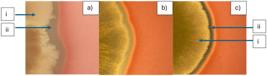
Hemolytic activity of bacteria on Blood Agar media: (a) isolate BT1.8; (b) isolate BT9.1; (c) isolate SM2.3. with (i) colony, (ii) clear zone.
The hemolytic activity observed in Bacillus spp. isolates indicates the production of lipopeptide biosurfactants capable of disrupting the integrity of lipid membranes. Given that fungal cell membranes are rich in ergosterol, the ability of biosurfactants to lyse erythrocytes through interaction with membrane lipid components may be mechanistically correlated with their ability to damage fungal cell membranes. Therefore, hemolysis activity can be used as an indirect indicator of membrane disruption-based antifungal potential, although specific confirmation of ergosterol interactions still requires further biochemical analysis.
The potential of the BT1.8 bacterial isolate was further analyzed to determine its species name through 16S rRNA gene detection. The 16S rRNA gene is an important component of the small ribosomal subunit in bacteria that plays a vital role in the translation and decoding of mRNA (Smith et al., 2018). This gene is known to be highly conservative and evolve slowly, making it an excellent marker for phylogenetic studies and bacterial taxonomic identification (Chappidi et al., 2019; Kim and Chun, 2014). The visualization results of the 16S rRNA gene of the BT1.8 bacterial isolate using 27F and 1492 R primers show the presence of this gene with a size of 1344 bp on a 1.5% agarose gel, as shown in Figure 6.

Electrophoresis result of DNA B. cereus BT1.8 marked with a band measuring 1344 bp. (M: Marker; S: Sample).
The results of the Basic Local Alignment Search Tools nucleotide (BLASTn) analysis and alignment with the database in GeneBank show that the BT1.8 bacterial isolate has an identical similarity to B. cereus of 99.11%, as shown in Table 3.
B. cereus is one of the species in the B. cereus group, which consists of B. cereus, B. anthracis, and B. thuringiensis. These three types of Bacillus are classified as pathogenic bacteria that can infect various types of living things. B. cereus plays a very important role in agriculture, as it can be used as a biological control agent and plant growth factor (PGPR). B. cereus can produce antimicrobial compounds that are very effective in fighting various plant pathogens, including bacteria and fungi, which help in controlling plant diseases (Azizoglu et al., 2025), such as Fusarium, which is effectively inhibited by the MH778713 strain, reducing the percentage of plant wilting from 96% to 12% (Ramírez et al., 2022). In addition, B. cereus can also be pathogenic to nematodes that attack plants, thereby reducing the damage caused by these parasites (Azizoglu et al., 2025).
Further analysis at the molecular level was also conducted to determine the presence of biosurfactant biosynthesis genes such as surfactin (srfAD) from the B. cereus BT1.8 isolate. The results showed that there was a DNA band from the srfADgene in B. cereus BT1.8, as shown in Figure 7. The srfAD gene produced by B. cereus BT1.8 is 729 bp in size.

Electrophoresis result of SrfAD gene of B. cereus BT1.8 marked with a band measuring 729 bp. (M: Marker; S: Sample).
Sequencing results and BLASTn analysis, as well as alignment with the database in GeneBank, show that there is a biosurfactant biosynthesis gene (srfAD) that has 98.22% similarity with B. cabrialesii strain WM_TN15 with access number CP189719.1. Based on BLASTp results, the protein in the srfAD gene of B. cereus BT1.8 has the highest similarity to the surfactin biosynthesis thioesterase srfAD from Bacillus halotolerans bacteria in GenBank, at 92.98% (GenBank access number WP_105955226.1). The similarity level of the srfAD gene possessed by B. cereus BT1.8 is more identical to B. halotolerans because the srfAD gene belongs to a conservative biosynthetic gene cluster, facilitating Horizontal Gene Transfer (HGT) and recombination (which allows similarity with B. halotolerans) (Danevčič et al., 2021). High genomic variability and lateral gene exchange result in one strain being closer to B. halotolerans in certain genes than to other B. cereus strains. The srf gene can be placed on elements that move between strains or between chromosomes. As in the B. cereus sensu lato group, whose biosynthetic genes include the srf cluster, HGT is known to occur frequently. Therefore, B. cereus can acquire genes from B. halotolerans or other strains through inter-strain transfer, while strains that do not possess these genes remain conservative (Böhm et al., 2015).
The presence of the srf gene in B. cereus BT1.8 is supported by (Ayangbenro and Babalola, 2020) research, which revealed that B. cereus NWUAB01 has a lipopeptide biosynthetic gene cluster related to metal complexing properties. B. cereus has a non-ribosomal peptide synthetase (NRPS) gene that is also involved in the biosynthesis of lipopeptides, which are a group of biosurfactant compounds.
The group of bacteria from the genus Bacillus is widely known as a producer of biosurfactant compounds, especially lipopeptides, namely surfactin. Surfactin is a protein encoded by the sfp gene group and the srf gene, one of which is the srfAD gene. The mechanism of surfactin formation involves several stages, such as initiation, elongation, and regulation. The initiation stage begins with the transfer of β-hydroxy fatty acids to the srfA enzyme, which then forms β-hydroxymyristoyl-glutamate. This process is supported by the thioesterase enzyme srfD, which increases the efficiency of initiation product formation (Steller et al., 2004). In the elongation process, surfactin peptides are synthesized by Non-Ribosomal Peptide Synthetase (NRPSs), which combine amino acids and fatty acids through a condensation reaction. NRPS enables the structural diversity of surfactin by combining various fatty acid isoforms (C12 to C17) (Théatre et al., 2021; Wang et al., 2024). Finally, at the regulatory stage, surfactin synthesis involves several genes and quorum sensing systems. The Spo0A gene plays an important role in regulating surfactin synthesis and cell differentiation. The quorum sensing system and the Sfp gene are also required for surfactin production (Rahman et al., 2021).
The combination of genetic evidence in the form of the presence of biosurfactant biosynthesis genes and phenotypic evidence in the form of hemolysis activity and increased inhibitory power in pellet and sonication treatments against plant pathogenic fungi shows that isolate BT1.8 has consistent molecular and functional capacity to produce active biosurfactant compounds, thus providing a coherent mechanistic basis as a candidate agent for antifungal products.
Advances in science and technology have made further research into the detection of active compounds produced by microbes as biocontrol agents easier with the use of molecular methods and analysis. Further analysis such as Whole Genome Sequencing (WGS), which is the analysis of the composition and interpretation of the entire microbial genome sequence. In the future, with this analysis, further research is needed to determine the various types of genes that encode the biosynthesis of active compounds/metabolites produced by B. cereus BT1.8.
4. Conclusions
Overall, B. cereus BT1.8 bacteria show strong potential as a biocontrol agent against the plant pathogen F. oxysporum, supported by phenotypic and molecular characteristics consistent with biosurfactant activity screening and pathogen inhibition mechanisms through membrane disruption. The integration of genetic evidence and observed biological activity provides a clear mechanistic basis for the antagonistic capacity of this bacterium, while reinforcing the relevance of local Bacillus isolates as promising biocontrol candidates. These findings underscore the importance of a mechanism-based approach in the selection and evaluation of biological agents, and open up opportunities for the development of more effective, sustainable, and applicable formulations and strategies for plant disease control at the field level.
Acknowledgements
Researchers would like to acknowledge the Dean of Science and Technology Faculty and the Chancellor of Universitas Airlangga, who have provided laboratory facilities. This research funded by internal funding, which is the Airlangga Research Fund Doctoral Dissertation Research scheme (ARF-PDDA) 2025, Research Institutions and Community Service (LPPM) of Universitas Airlangga research funding, Contract Number: 1815/B/UN3.LPPM/PT.01.03/2025. Researchers also thanks the research assistants and parties who participated in this research.
Data Availability Statement
The entire data set supporting the results of this study was published in the article itself.
References
-
AHMAD, T., XING, F., NIE, C., CAO, C., XIAO, Y., YU, X., MOOSA, A. and LIU, Y., 2023. Biocontrol potential of lipopeptides produced by the novel Bacillus subtilis strain Y17B against postharvest Alternaria fruit rot of cherry. Frontiers in Microbiology, vol. 14, pp. 1150217. https://doi.org/10.3389/fmicb.2023.1150217 PMid:37032895.
» https://doi.org/10.3389/fmicb.2023.1150217 -
AHMED, M.E., AHMED, Z.M. and THAMER, A., 2022 [viewed 3 November 2025]. The evolutionary effects of bacillin and S-pyocin bacteriocin and their effects on propionibacterium acnes and fungi. Biochemical and Cellular Archives [online], vol. 20, suppl. 2, pp. 3645-3649. Available from: https://connectjournals.com/03896.2020.20.3645
» https://connectjournals.com/03896.2020.20.3645 -
AJESH, K., SUDARSLAL, S., ARUNAN, C. and SREEJITH, K., 2013. Kannurin, a novel lipopeptide from Bacillus cereus strain AK1: isolation, structural evaluation and antifungal activities. Journal of Applied Microbiology, vol. 115, no. 6, pp. 1287-1296. https://doi.org/10.1111/jam.12324 PMid:23937170.
» https://doi.org/10.1111/jam.12324 - AUSUBEL, F.M., BRENT, R., KINGSTON, R.R., MOORE, D.D., SEIDMAN, J.G., SMITH, J.A. and STRUHL, K., 2003. Current protocols in molecular biology New Jersey: John Wiley & Sons.
-
AYANGBENRO, A.S. and BABALOLA, O.O., 2020. Genomic analysis of Bacillus cereus NWUAB01 and its heavy metal removal from polluted soil. Scientific Reports, vol. 10, no. 1, pp. 19660. https://doi.org/10.1038/s41598-020-75170-x PMid:33184305.
» https://doi.org/10.1038/s41598-020-75170-x -
AZIZOGLU, U., ARGENTEL‐MARTÍNEZ, L., PEÑUELAS‐RUBIO, O., HERRERA‐SEPÚLVEDA, A., IBAL, J.C., SHARAFI, R. and SANSINENEA, E., 2025. Natural products produced by the species of Bacillus cereus group: recent updates. Journal of Basic Microbiology, vol. 65, no. 3, e2400666. https://doi.org/10.1002/jobm.202400666 PMid:39569545.
» https://doi.org/10.1002/jobm.202400666 -
BAKKER, C.E., BARGHOUTH, Z., RAMLAWI, S. and AVIS, T.J., 2025. Biochemistry and differential mechanistic activity of antimicrobial lipopeptides from plant pathogen antagonists from the genus Bacillus. Canadian Journal of Plant Pathology, vol. 47, no. 1, pp. 98-110. https://doi.org/10.1080/07060661.2024.2425955
» https://doi.org/10.1080/07060661.2024.2425955 -
BAMBA, T., AOKI, R., HORI, Y., ISHIKAWA, S., YOSHIDA, K.I., TAOKA, N. and HASUNUMA, T., 2024. High-throughput evaluation of hemolytic activity through precise measurement of colony and hemolytic zone sizes of engineered Bacillus subtilis on blood agar. Biology Methods and Protocols, vol. 9, no. 1, pp. bpae044. https://doi.org/10.1093/biomethods/bpae044 PMid:38962661.
» https://doi.org/10.1093/biomethods/bpae044 -
BIDIMA, M.G.S., CHTAINA, N., EZZAHIRI, B., EL GUILLI, M., BARAKAT, I. and EL KAMLI, T. 2022. Antifungal activity of bioactive compounds produced by the endophyte Bacillus velezensis NC318 against the soil borne pathogen Sclerotium rolfsii Sacc.Journal of Plant Protection Research, 326-333. doi: 10.24425/jppr.2022.142139
» https://doi.org/10.24425/jppr.2022.142139 -
BÖHM, M.E., HUPTAS, C., KREY, V.M. and SCHERER, S., 2015. Massive horizontal gene transfer, strictly vertical inheritance and ancient duplications differentially shape the evolution of Bacillus cereus enterotoxin operons hbl, cytK and nhe. BMC Evolutionary Biology, vol. 10, no. 15, pp. 246. https://doi.org/10.1186/s12862-015-0529-4 PMid:26555390.
» https://doi.org/10.1186/s12862-015-0529-4 -
CHANG, W.T., CHEN, Y.C. and JAO, C.L., 2007. Antifungal activity and enhancement of plant growth by Bacillus cereus grown on shellfish chitin wastes. Bioresource Technology, vol. 98, no. 6, pp. 1224-1230. https://doi.org/10.1016/j.biortech.2006.05.005 PMid:16797180.
» https://doi.org/10.1016/j.biortech.2006.05.005 -
CHAPPIDI, S., VILLA, E.C. and CANTAREL, B.L., 2019. Using Mothur to determine bacterial community composition and structure in 16S ribosomal RNA datasets. Current Protocols in Bioinformatics, vol. 67, no. 1, e83. https://doi.org/10.1002/cpbi.83 PMid:31524992.
» https://doi.org/10.1002/cpbi.83 - CHEN, X. and DU, C., 2021. Advances in research on the phytohormone regulating interactions between plants and Fusarium oxysporum. Chinese Journal of Applied and Environmental Biology, vol. 27, no. 3, pp. 816-822.
-
CZYMMEK, K.J., FOGG, M., POWELL, D.H., SWEIGARD, J., PARK, S.-J. and KANG, S., 2007. In vivo time-lapse documentation using confocal and multi-photon microscopy reveals the mechanisms of invasion into the Arabidopsis root vascular system by Fusarium oxysporum. Fungal Genetics and Biology : FG & B, vol. 44, no. 10, pp. 1011-1023. https://doi.org/10.1016/j.fgb.2007.01.012 PMid:17379550.
» https://doi.org/10.1016/j.fgb.2007.01.012 -
DANEVČIČ, T., DRAGOŠ, A., SPACAPAN, M., STEFANIC, P., DOGSA, I. and MANDIC-MULEC, I., 2021. Surfacer inBacillus subtilis. Frontiers in Microbiology, vol. 14, no. 12, pp. 657407. https://doi.org/10.3389/fmicb.2021.657407 PMid:34054753.
» https://doi.org/10.3389/fmicb.2021.657407 -
DHARMA, K.S., SURYANTI, S. and WIDIASTUTI, A., 2024. Hormesis in pathogenic and biocontrol fungi: from inhibition to stimulation.Caraka Tani: Journal of Sustainable Agriculture, vol. 39, no. 2, pp. 281-296. https://doi.org/10.20961/carakatani.v39i2.83012
» https://doi.org/10.20961/carakatani.v39i2.83012 -
EDGAR, C.I., MCGRATH, K.C., DOMBRECHT, B., MANNERS, J.M., MACLEAN, D.C., SCHENK, P.M. and KAZAN, K., 2006. Salicylic acid mediates resistance to the vascular wilt pathogen Fusarium oxysporum in the model host Arabidopsis thaliana. Australasian Plant Pathology, vol. 35, no. 6, pp. 581-591. https://doi.org/10.1071/AP06060
» https://doi.org/10.1071/AP06060 -
GUO, Q., LI, S., DONG, L., SU, Z., WANG, P., LIU, X. and MA, P., 2023. Screening biocontrol agents for cash crop Fusarium wilt based on fusaric acid tolerance and antagonistic activity against Fusarium oxysporum. Toxins, vol. 15, no. 6, pp. 381. https://doi.org/10.3390/toxins15060381 PMid:37368682.
» https://doi.org/10.3390/toxins15060381 -
HAMMAD, M., ALI, H., HASSAN, N., TAWAB, A., SALMAN, M., JAWAD, I. and RASHID, M.H., 2023. Food safety and biological control; genomic insights and antimicrobial potential of Bacillus velezensis FB2 against agricultural fungal pathogens. PLoS One, vol. 18, no. 11, e0291975. https://doi.org/10.1371/journal.pone.0291975 PMid:37963161.
» https://doi.org/10.1371/journal.pone.0291975 -
IJAH, U.J.J. and OLARINOYE, R., 2012. Biosurfactant production by Bacillus strains RO7 AND R28 grown on diesel. Malaysian Journal of Science. Series B, Physical & Earth Sciences, vol. 31, no. 2, pp. 83-90. https://doi.org/10.22452/mjs.vol31no2.7
» https://doi.org/10.22452/mjs.vol31no2.7 -
ISLAM, T., DANISHUDDIN., TAMANNA, N.T., MATIN, M.N., BARAI, H.R. and HAQUE, M.A., 2024. Resistance mechanisms of plant pathogenic fungi to fungicide, environmental impacts of fungicides, and sustainable solutions. Plants, vol. 13, no. 19, pp. 2737. https://doi.org/10.3390/plants13192737 PMid:39409607.
» https://doi.org/10.3390/plants13192737 - JASIM, B., SREELAKSHMI, S., MATHEW, J. and RADHAKRISHNAN, E.K., 2016. Identification of endophytic Bacillus mojavensis with highly specialized broad spectrum antibacterial activity. Biotech, vol. 6, pp. 187.
-
JEONG, M.H., LEE, Y.S., CHO, J.Y., AHN, Y.S., MOON, J.H., HYUN, H.N. and KIM, K.Y., 2017. Isolation and characterization of metabolites from Bacillus licheniformis MH48 with antifungal activity against plant pathogens. Microbial Pathogenesis, vol. 110, pp. 645-653. https://doi.org/10.1016/j.micpath.2017.07.027 PMid:28733027.
» https://doi.org/10.1016/j.micpath.2017.07.027 -
KHADIRI, M., BOUBAKER, H., ASKARNE, L., EZRARI, S., RADOUANE, N., FARHAOUI, A. and LAHLALI, R., 2023. Bacillus cereus B8W8 an effective bacterial antagonist against major postharvest fungal pathogens of fruit. Postharvest Biology and Technology, vol. 200, pp. 112315. https://doi.org/10.1016/j.postharvbio.2023.112315
» https://doi.org/10.1016/j.postharvbio.2023.112315 -
KIM, M. and CHUN, J., 2014. 16S rRNA gene-based identification of bacteria and archaea using the EzTaxon server. London: Academic Press, pp. 61-74. Methods in microbiology, no. 41. https://doi.org/10.1016/bs.mim.2014.08.001
» https://doi.org/10.1016/bs.mim.2014.08.001 -
KUMAR, S.N., SREEKALA, S.R., CHANDRASEKARAN, D., NAMBISAN, B. and ANTO, R.J., 2014. Biocontrol of Aspergillus species on peanut kernels by antifungal diketopiperazine producing Bacillus cereus associated with entomopathogenic nematode. PLoS One, vol. 9, no. 8, e106041. https://doi.org/10.1371/journal.pone.0106041 PMid:25157831.
» https://doi.org/10.1371/journal.pone.0106041 -
LEE, H.H., PARK, J., LIM, J.Y., KIM, H., CHOI, G.J., KIM, J.C. and SEO, Y.S., 2015. Complete genome sequence of Bacillus velezensis G341, a strain with a broad inhibitory spectrum against plant pathogens. Journal of Biotechnology, vol. 211, pp. 97-98. https://doi.org/10.1016/j.jbiotec.2015.07.005 PMid:26187870.
» https://doi.org/10.1016/j.jbiotec.2015.07.005 -
LI, X.Y., MAO, Z.C., WU, Y.X., HO, H.H. and HE, Y.Q., 2015. Comprehensive volatile organic compounds profiling of Bacillus species with biocontrol properties by head space solid phase microextraction with gas chromatography-mass spectrometry. Biocontrol Science and Technology, vol. 25, no. 2, pp. 132-143. https://doi.org/10.1080/09583157.2014.960809
» https://doi.org/10.1080/09583157.2014.960809 -
LIU, W. and SUN, C., 2021. C17-fengycin B, produced by deep-sea-derived Bacillus subtilis, possessing a strong antifungal activity against Fusarium solani. Journal of Oceanology and Limnology, vol. 39, no. 5, pp. 1938-1947. https://doi.org/10.1007/s00343-020-0215-2
» https://doi.org/10.1007/s00343-020-0215-2 -
MILJAKOVIĆ, D., MARINKOVIĆ, J. and BALEŠEVIĆ-TUBIĆ, S., 2020. The significance of Bacillus spp. in disease suppression and growth promotion of field and vegetable crops. Microorganisms, vol. 8, no. 7, pp. 1037. https://doi.org/10.3390/microorganisms8071037 PMid:32668676.
» https://doi.org/10.3390/microorganisms8071037 -
MOHD ZAINUDIN, N.A.I., ABD MURAD, N.B. and SHAARI, F.N., 2024. Utilisation of plant-based product in post-harvest disease management of fruits. In: M. WONG, ed. Advances in tropical crop protection. Cham: Springer, pp. 121-155. https://doi.org/10.1007/978-3-031-59268-3_9
» https://doi.org/10.1007/978-3-031-59268-3_9 -
MYERS, P. and YOUSTEN, A.A., 1978. Toxic Activity of Bacillus sphaericus SSII-I for Mosquito Larvae. Infection and Immunity, vol. 19, no. 3, pp. 1047-1053. https://doi.org/10.1128/iai.19.3.1047-1053.1978 PMid:640722.
» https://doi.org/10.1128/iai.19.3.1047-1053.1978 -
MYO, E.M., LIU, B., MA, J., SHI, L., JIANG, M., ZHANG, K. and GE, B., 2019. Evaluation of Bacillus velezensis NKG-2 for bio-control activities against fungal diseases and potential plant growth promotion. Biological Control, vol. 134, pp. 23-31. https://doi.org/10.1016/j.biocontrol.2019.03.017
» https://doi.org/10.1016/j.biocontrol.2019.03.017 -
NAFIDIASTRI, F.A., SUSETYO, R.D., NURHARIYATI, T., SUPRIYANTO, A., GERALDI, A., NI’MATUZAHROH, N.M., FATIMAH, F. and SALAMUN, S., 2021. Biosurfactant activity of indigenous Bacillus sp. ES4.3 isolated from endemic breeding sites of dengue hemorrhagic fever vector in Surabaya, East Java, indonesia. Biodiversitas, vol. 22, no. 12, pp. 5375-5381. https://doi.org/10.13057/biodiv/d221219
» https://doi.org/10.13057/biodiv/d221219 -
NATIONAL CENTER FOR BIOTECHNOLOGY INFORMATION – NCBI, 2025 [viewed 3 November 2025]. GeneBank database [online]. Available from: https://www.ncbi.nlm.nih.gov/genbank/
» https://www.ncbi.nlm.nih.gov/genbank/ -
NURHARIYATI, T., SALAMUN, S., SUPRIYANTO, A., SINURAT, T.B.M., YULIANA, F., GERALDI, A. and IZZUDDIN, M., 2025. The potential of local Bacillus sp. BK7. 1, EG6. 4, and LSD4. 2 as biocontrol agents against the pathogenic fungus Fusarium oxysporum. Biodiversitas, vol. 26, no. 7. https://doi.org/10.13057/biodiv/d260719
» https://doi.org/10.13057/biodiv/d260719 -
PARK, G., NAM, J., KIM, J., SONG, J., KIM, P.I., MIN, H.J. and LEE, C.W., 2019. Structure and mechanism of surfactin peptide from Bacillus velezensis antagonistic to fungi plant pathogens. Bulletin of the Korean Chemical Society, vol. 40, no. 7, pp. 704-709. https://doi.org/10.1002/bkcs.11757
» https://doi.org/10.1002/bkcs.11757 - PEDRAZA, L.A., LÓPEZ, C.E. and URIBE-VÉLEZ, D., 2020. Mechanisms of action of Bacillus spp. (Bacillaceae) against phytopathogenic microorganisms during their interaction with plants. Acta Biologica Colombiana, vol. 25, no. 1, pp. 112-125.
-
PRASETYAWATI, E.T., SURTININGSIH, T., PURKAN, KHIFTIYAH, A.M. and SARI, S.K., 2023. Screening of biosurfactant production by Bacillus spp. potentially inhibiting the growth of Ralstonia solanacearum AIP Conference Proceedings, vol. 2554, no. 1, pp. 090008. https://doi.org/10.1063/5.0106613
» https://doi.org/10.1063/5.0106613 -
RAHMAN, F.B., SARKAR, B., MONI, R. and RAHMAN, M.S., 2021. Molecular genetics of surfactin and its effects on different sub-populations of Bacillus subtilis Biotechnology Reports, vol. 32, e00686. https://doi.org/10.1016/j.btre.2021.e00686 PMid:34786355.
» https://doi.org/10.1016/j.btre.2021.e00686 -
RAJAOFERA, M.J.N., WANG, Y., DAHAR, G.Y., JIN, P., FAN, L., XU, L. and MIAO, W., 2019. Volatile organic compounds of Bacillus atrophaeus HAB-5 inhibit the growth of Colletotrichum gloeosporioides. Pesticide Biochemistry and Physiology, vol. 156, pp. 170-176. https://doi.org/10.1016/j.pestbp.2019.02.019 PMid:31027577.
» https://doi.org/10.1016/j.pestbp.2019.02.019 -
RAMÍREZ, V., MARTÍNEZ, J., BUSTILLOS‐CRISTALES, M.D.R., CATAÑEDA‐ANTONIO, D., MUNIVE, J.A. and BAEZ, A., 2022. Bacillus cereus MH778713 elicits tomato plant protection against Fusarium oxysporum. Journal of Applied Microbiology, vol. 132, no. 1, pp. 470-482. https://doi.org/10.1111/jam.15179 PMid:34137137.
» https://doi.org/10.1111/jam.15179 -
RIU, F., RUDA, A., IBBA, R., SESTITO, S., LUPINU, I., PIRAS, S. and CARTA, A., 2022. Antibiotics and carbohydrate-containing drugs targeting bacterial cell envelopes: an overview. Pharmaceuticals, vol. 15, no. 8, pp. 942. https://doi.org/10.3390/ph15080942 PMid:36015090.
» https://doi.org/10.3390/ph15080942 -
RONCERO, M.I.G., HERA, C., RUIZ-RUBIO, M., GARCÍA MACEIRA, F.I., MADRID, M.P., CARACUEL, Z., CALERO, F., DELGADO-JARANA, J., ROLDÁN-RODRÍGUEZ, R., MARTÍNEZ-ROCHA, A.L., VELASCO, C., ROA, J., MARTÍN-URDIROZ, M., CÓRDOBA, D. and DI PIETRO, A., 2003. Fusarium as a model for studying virulence in soilborne plant pathogens. Physiological and Molecular Plant Pathology, vol. 62, no. 2, pp. 87-98. https://doi.org/10.1016/S0885-5765(03)00043-2
» https://doi.org/10.1016/S0885-5765(03)00043-2 - RUOFF, K.L., 2022. General approaches to identification of aerobic gram‐positive cocci. In: K.C. CARROLL, M.A. PFALLER, M.L. LANDRY, A.J. MCADAM, R. PATEL, S.S. RICHTER and D.W. WARNOCK, eds. Manual of clinical microbiology. Washington, D.C.: ASM Press, pp. 350-353.
- SALAMUN, FATIMAH, FINDAWATI, V., DANANGSUSETYO, R., AL-BATATI, N., NURHARIYATI, T., and SUPRIYANTO, A., 2020. Prospect of native entomopathogenic Bacilli from Baluran National Park as biological control of Dengue Fever Vector. Annals of Biology, vol. 36, no. 2, pp. 232-237.
-
SMITH, B.A., GUPTA, N., DENNY, K. and CULVER, G.M., 2018. Characterization of 16S rRNA processing with pre-30S subunit assembly intermediates from E. coli. Journal of Molecular Biology, vol. 430, no. 12, pp. 1745-1759. https://doi.org/10.1016/j.jmb.2018.04.009 PMid:29660326.
» https://doi.org/10.1016/j.jmb.2018.04.009 -
STELLER, S., SOKOLL, A., WILDE, C., BERNHARD, F., FRANKE, P. and VATER, J., 2004. Initiation of surfactin biosynthesis and the role of the SrfD-thioesterase protein. Biochemistry, vol. 43, no. 35, pp. 11331-11343. https://doi.org/10.1021/bi0493416
» https://doi.org/10.1021/bi0493416 -
THÉATRE, A., CANO-PRIETO, C., BARTOLINI, M., LAURIN, Y., DELEU, M., NIEHREN, J. and JACQUES, P., 2021. The surfactin-like lipopeptides from Bacillus spp.: natural biodiversity and synthetic biology for a broader application range. Frontiers in Bioengineering and Biotechnology, vol. 9, pp. 623701. https://doi.org/10.3389/fbioe.2021.623701 PMid:33738277.
» https://doi.org/10.3389/fbioe.2021.623701 -
TUYEN, D.T., TRUNG, N.T., THAO, N.T., LE THANH, N.S., DAI NGUYEN, N.P., TUYET, N.T.A., CUONG, N.T. and SHOW, P.L., 2023. Antifungal activity of secondary metabolites purified from Bacillus subtilis isolated in Vietnam and evaluated on in vitro and in vivo models. International Biodeterioration & Biodegradation, vol. 179, pp. 105558. https://doi.org/10.1016/j.ibiod.2022.105558
» https://doi.org/10.1016/j.ibiod.2022.105558 -
WANG, J.-F., HUANG, Q.-B., ZHANG, P.-D. and ZHANG, P.-P., 2024. Structure and biosynthesis of surfactin as well as its role in biological control. Shengwu Jishu Tongbao https://doi.org/10.13560/j.cnki.biotech.bull.1985.2023-0509
» https://doi.org/10.13560/j.cnki.biotech.bull.1985.2023-0509 -
WIJNTJES, C., WEBER, Y., HÖGER, S., NGUYEN, K.T., HOLLERT, H. and SCHÄFFER, A., 2022. Decelerated degradation of a sulfonylurea herbicide in four fungicide-treated soils. Environmental Science. Advances, vol. 1, no. 1, pp. 70-82. https://doi.org/10.1039/D1VA00021G
» https://doi.org/10.1039/D1VA00021G -
YUAN, J., RAZA, W., HUANG, Q. and SHEN, Q., 2012. The ultrasound‐assisted extraction and identification of antifungal substances from B. amyloliquefaciens strain NJN‐6 suppressing Fusarium oxysporum. Journal of Basic Microbiology, vol. 52, no. 6, pp. 721-730. https://doi.org/10.1002/jobm.201100560 PMid:22581589.
» https://doi.org/10.1002/jobm.201100560 -
YUE, Y., SUN, X., TIAN, S., YAN, S., SUN, W., MIAO, J. and ZHU, W., 2024. Multi-omics and gut microbiome: unveiling the pathogenic mechanisms of early-life pesticide exposure. Pesticide Biochemistry and Physiology, vol. 199, pp. 105770. https://doi.org/10.1016/j.pestbp.2024.105770 PMid:38458664.
» https://doi.org/10.1016/j.pestbp.2024.105770 -
ZERBIB, D., 2017. Bacterial cell envelopes: composition, architecture, and origin. In: D. MIKLAVČIČ, ed. Handbook of electroporation. Cham: Springer, pp. 417-436. https://doi.org/10.1007/978-3-319-32886-7_28
» https://doi.org/10.1007/978-3-319-32886-7_28 - ZHANG, Y., BI, Y., MU, X.-N., ZHENG, Z.-W., WANG, Z.-G. and XU, W.-H., 2025. Biocontrol characteristics of strain JB7 against Fusarium graminearum. Shengwu Jishu Tongbao
-
ZIHALIRWA KULIMUSHI, P., ARGÜELLES ARIAS, A., FRANZIL, L., STEELS, S. and ONGENA, M., 2017. Stimulation of fengycin-type antifungal lipopeptides in Bacillus amyloliquefaciens in the presence of the maize fungal pathogen Rhizomucor variabilis Frontiers in Microbiology, vol. 8, pp. 850. https://doi.org/10.3389/fmicb.2017.00850 PMid:28555132.
» https://doi.org/10.3389/fmicb.2017.00850
Edited by
-
Editor: Takako Matsumura Tundisi
Data citations
BIDIMA, M.G.S., CHTAINA, N., EZZAHIRI, B., EL GUILLI, M., BARAKAT, I. and EL KAMLI, T. 2022. Antifungal activity of bioactive compounds produced by the endophyte Bacillus velezensis NC318 against the soil borne pathogen Sclerotium rolfsii Sacc.Journal of Plant Protection Research, 326-333. doi: 10.24425/jppr.2022.142139


