Elicitation of Piper sarmentosum callus with heavy metals: enhanced secondary metabolites, antioxidant and antimicrobial activities
Abstract
Piper sarmentosum is a medicinal plant with rich bioactive secondary metabolites. This work investigated the effects of heavy metal triggers (cobalt, copper, zinc, and mercury) on callus growth, secondary metabolites, antioxidant, and antimicrobial activities in P. sarmentosum cell cultures. Callus was induced on MS solid medium with 2 mg/L 2,4-D and 0.5 mg/L BAP, then subcultured on MS liquid medium containing 1.5 mg/L 2,4-D and 1.5 mg/L BAP with the addition of elicitors at concentrations of 0.5; 1; and 2.5 mg/L. Elicitation did not contribute significantly to callus biomass (p>0.05). All extracts contained alkaloids and flavonoids, while terpenoids/steroids and saponins were not detected. GC-MS analysis identified 42 compounds. Cobalt and copper extracts accumulated fatty acid methyl esters (0.14-0.61%) and alkaloids, including pipataline (0.03%) in the copper extract. Zinc and mercury extracts were dominated by glycerides (0.23-1.79%). The highest antioxidant activity was shown by 1 mg/L cobalt (IC50 62.55 ± 2.08 µg/mL) and 2.5 mg/L copper (IC50 61.72 ± 1.31 µg/mL), while zinc and mercury showed very weak activity (IC50 >200 µg/mL). All extracts inhibited Staphylococcus aureus, Escherichia coli, and Candida albicans. Cobalt 2.5 mg/L at 750 mg/L concentration showed the strongest activity against S. aureus (40.37 ± 3.56 mm), zinc 2.5 mg/L at 750 mg/L concentration against E. coli (36.05 ± 3.86 mm), and cobalt 2.5 mg/L at 500 mg/L concentration against C. albicans (41.62 ± 4.83 mm). Cobalt and copper at optimal concentrations effectively enhanced antioxidant and antimicrobial activities. To our knowledge, this is the first comparative study of four heavy metal elicitors inP. sarmentosumcallus.
Keywords:
Piper sarmentosum; bioactivities; callus culture; heavy metals; secondary metabolites
Resumo
Piper sarmentosum é uma planta medicinal rica em metabólitos secundários bioativos. Este trabalho investigou os efeitos de metais pesados (cobalto, cobre, zinco e mercúrio) no crescimento de calos, nos metabólitos secundários e nas atividades antioxidantes e antimicrobianas em culturas celulares de P. sarmentosum. Os calos foram induzidos em meio sólido MS com 2 mg/L de 2,4-D e 0,5 mg/L de BAP, sendo depois subcultivado em meio líquido MS contendo 1,5 mg/L de 2,4-D e 1,5 mg/L de BAP, com a adição de elicitores nas concentrações de 0,5; 1; e 2,5 mg/L. A elicitação não contribuiu significativamente para a biomassa dos calos (p>0,05). Todos os extratos continham alcaloides e flavonoides, enquanto terpenoides/esteroides e saponinas não foram detectados. A análise por GC-MS identificou 42 compostos. Os extratos de cobalto e cobre acumularam ésteres metílicos de ácidos graxos (0,14-0,61%) e alcaloides, incluindo pipatalina (0,03%) no extrato de cobre. Os extratos de zinco e mercúrio foram dominados por glicerídeos (0,23-1,79%). A maior atividade antioxidante foi observada com 1 mg/L de cobalto (IC50 62,55 ± 2,08 µg/mL) e 2,5 mg/L de cobre (IC50 61,72 ± 1,31 µg/mL), enquanto o zinco e o mercúrio apresentaram atividade muito fraca (IC50 > 200 µg/mL). Todos os extratos inibiram Staphylococcus aureus, Escherichia coli e Candida albicans. O cobalto a 2,5 mg/L na concentração de 750 mg/L apresentou a atividade mais forte contra S. aureus (40,37 ± 3,56 mm), o zinco a 2,5 mg/L na concentração de 750 mg/L contra E. coli (36,05 ± 3,86 mm) e o cobalto a 2,5 mg/L na concentração de 500 mg/L contra C. albicans (41,62 ± 4,83 mm). O cobalto e o cobre, em concentrações ótimas, aumentaram efetivamente as atividades antioxidantes e antimicrobianas. Até onde sabemos, este é o primeiro estudo comparativo de quatro elicitores de metais pesados em calos de P. sarmentosum.
Palavras-chave:
Piper sarmentosum; bioatividades; cultura de calos; metais pesados; metabólitos secundários
1. Introduction
Piper sarmentosum Roxb. (Piperaceae) is a Southeast Asian medicinal plant traditionally used to treat headaches, toothaches, coughs, asthma, and skin infections (Sun et al., 2020; Salehi et al., 2019). Its pharmacological activities, including antioxidant and antimicrobial effects, are linked to its diverse secondary metabolites. However, its utilization is hindered by limited biomass and metabolite variation due to environmental factors (Efferth, 2019). This variability poses practical challenges for standardization and large-scale production of consistent-quality extracts for pharmaceutical or nutraceutical applications. While plant tissue culture offers a controlled production alternative, undifferentiated cultures like callus often produce lower metabolite levels than intact plants (Rao and Ravishankar, 2002). Therefore, strategies to enhance production are needed.
Elicitation, particularly with abiotic elicitors like heavy metals, is an effective approach to stimulate secondary metabolite biosynthesis by inducing stress responses (Zhao et al., 2005; Namdeo, 2007). Successful elicitation has been reported using various abiotic elicitors, including silver ions (Ag+) inDracocephalum ruyschiana(Weremczuk-Jeżyna et al., 2024), cadmium (Cd2+) inGymnema sylvestre(Bhuvaneswari et al., 2012), and copper (Cu2+) inCatharanthus roseus(Fouad and Hafez, 2020). Heavy metals were selected over other abiotic elicitors (e.g., UV radiation, osmotic stress, or phytohormones) because they can directly activate specific stress-signaling pathways (such as MAPK cascades and transcription factors like MYB and WRKY) that regulate the biosynthesis of alkaloids and phenolic compounds (Razzaq et al., 2025). Compared to physical elicitors, heavy metals provide a more controllable, dose-dependent, and long-lasting stress stimulus in submerged callus cultures (Açikgoz et al., 2023; Bhuvaneswari et al., 2012). Moreover, certain heavy metals (e.g., Cu and Zn) act as cofactors for key enzymes in the phenylpropanoid pathway, offering a dual role as both elicitor and nutrient. At controlled concentrations, heavy metals can trigger metabolic changes in plant cultures in vitro (Ramakrishna and Ravishankar, 2011; Maksymiec, 2007).
The four heavy metals were selected based on distinct rationales: (i) Cobalt (Co) is a component of vitamin B12 and has been reported to stimulate alkaloid accumulation in several plant species (Fouad and Hafez, 2020); (ii) Copper (Cu) and zinc (Zn) are essential micronutrients that serve as cofactors for antioxidant enzymes (e.g., superoxide dismutase) and can activate phenylpropanoid metabolism at sub-toxic concentrations (Shafi et al., 2019); (iii) Mercury (Hg), despite its high toxicity, was included as a positive abiotic stress control representing a non-essential, highly phytotoxic metal (Álvarez-Rivera et al., 2022). While Hg is unsuitable for large-scale production, its inclusion helps to understand whether extreme stress differentially affects secondary metabolite profiles. To our knowledge, no previous study has directly compared essential (Co, Cu, Zn) and non-essential (Hg) heavy metals in P. sarmentosum callus cultures.
Despite the known metabolite richness of thePipergenus, research on heavy metal elicitation inP. sarmentosumcallus cultures is limited. Comprehensive studies comparing different heavy metal elicitors, their impact on metabolite profiles (using GC-MS based metabolite profiling), and the resulting biological activities are lacking (Adams, 2017). Furthermore, the specific metabolites responsible for the bioactivity of elicited callus extracts have not been clearly identified (Caesar and Cech, 2019).
Accordingly, this study aimed to establishP. sarmentosumcallus cultures on Murashige and Skoog (MS) solid medium and evaluate the effects of four heavy metal elicitors—cobalt, copper, zinc, and mercury—at concentrations of 0.5, 1, and 2.5 mg/L on callus growth, secondary metabolite profiles, antioxidant activity, and antimicrobial activity againstStaphylococcus aureus,Escherichia coli, andCandida albicans.
2. Methods
2.1. Plant material and callus induction
Young leaves (2nd-4th from shoot tip) ofP. sarmentosumwere collected from the Universitas Airlangga botanical garden (Surabaya, Indonesia; coordinates 7°15’55”S 112°47’45”E). Voucher specimens were deposited at the Herbarium of the Department of Biology, Universitas Airlangga (specimen code PS-2023-01). The leaves were washed under running tap water with detergent for 5 min. Surface sterilization was performed in a laminar air flow cabinet by first immersing the leaves in 20% (v/v) sodium hypochlorite (containing approximately 1% active chlorine) for 5 min, followed by three rinses with sterile distilled water. The leaves were then immersed in 70% (v/v) ethanol for 1 min and rinsed three times with sterile distilled water. This reverse sequence (hypochlorite before ethanol) was adopted based on preliminary experiments comparing two sterilization protocols. The conventional sequence (70% ethanol for 1 min followed by 20% sodium hypochlorite for 5 min) resulted in a high contamination rate (63% of explants showed fungal or bacterial growth after 2 weeks). In contrast, the modified sequence (hypochlorite followed by ethanol) reduced contamination to only 22% and increased explant survival to 78% after 2 weeks. Therefore, the reverse sequence was used for all subsequent experiments. The reduced ethanol exposure time (1 min instead of 5 min) further minimized tissue damage while maintaining sterilization efficacy. The sterilized leaves were cut into 1 × 1 cm explants. A total of 30 explants were used per treatment (10 jars with 3 explants each). Explants were cultured on Murashige and Skoog (MS) solid medium supplemented with 30 g/L sucrose, 8 g/L agar, 2 mg/L 2,4-dichlorophenoxyacetic acid (2,4-D), and 0.5 mg/L 6-benzylaminopurine (BAP). The medium pH was adjusted to 5.8 prior to autoclaving (121 °C, 1.5 atm for 20 min). Cultures were incubated at 25 ± 2 °C under continuous light (cool white fluorescent lamps, 40 µmol m−2 s−1) for 8 weeks. The incubation temperature of 25 °C was selected as the standard growth temperature forPiperspecies in tissue culture, while continuous light was used to promote callus proliferation based on preliminary experiments. After 8 weeks, the induced callus was characterized by its friable, whitish-yellow appearance with a moist texture, and was harvested for subculture and elicitation experiments.
2.2. Elicitation treatments
Approximately 0.5 g (fresh weight) of 8-week-old callus was transferred to 50 mL of MS liquid medium supplemented with 1.5 mg/L 2,4-D and 1.5 mg/L BAP in 250 mL Erlenmeyer flasks. The culture was incubated on an orbital shaker at 100 rpm, 25 ± 2 °C, under continuous light (Namdeo, 2007). Four heavy metal elicitors—cobalt(II) chloride hexahydrate (CoCl2·6H2O, MW 237.93 g/mol), copper(II) sulphate pentahydrate (CuSO4·5H2O, MW 249.69 g/mol), zinc sulphate hexahydrate (ZnSO4·6H2O, MW 287.56 g/mol), and mercury(II) sulphate (HgSO4, MW 296.65 g/mol)—were added to the medium at final concentrations of 0.5, 1, and 2.5 mg/L (equivalent to approximately 2.1, 4.2, and 10.5 µM for Co; 2.0, 4.0, and 10.0 µM for Cu; 1.7, 3.5, and 8.7 µM for Zn; and 1.7, 3.4, and 8.4 µM for Hg). Elicitor concentrations refer to the final concentration of the metal salt in the culture medium. Control cultures received no heavy metal addition. After 4 weeks of incubation, callus was harvested to determine fresh weight, dry weight (after drying at 50 °C for 48 h), and for extract preparation. All treatments were performed in triplicate (three flasks per treatment), and each experiment was repeated twice.
2.3. Extraction
The dried callus was ground into powder using a mortar and pestle. The powdered sample was macerated in methanol at a ratio of 1:10 (w/v) in a sealed container on an orbital shaker at 100 rpm for 48 h at room temperature, then filtered through Whatman filter paper No. 1. The filtrate was then evaporated manually at room temperature (25 ± 2 °C) under a fume hood with continuous air circulation until a thick extract was obtained. The extract was then transferred to glass vials and further dried under a stream of air to remove residual solvent. The final dried extract was stored at -20 °C for further analysis (Wanti et al., 2025). Room temperature evaporation was chosen to prevent degradation of heat-labile compounds that might be damaged by rotary evaporation at elevated temperatures (40 °C). This method has been successfully used in previous studies onPiperspecies callus extracts (Junairiah et al., 2024).
2.4. Phytochemical screening
Phytochemical screening for alkaloids, flavonoids, terpenoids, steroids, and saponins was performed following Irfansyah et al. (2024). Alkaloids were detected using Wagner's, Mayer's, and Dragendorff's reagents, where precipitate formation indicated a positive result. Flavonoids were identified by the HCl-magnesium test (red, green, or yellow coloration). The Liebermann-Burchard test was used for terpenoids (reddish-brown ring) and steroids (blue-green ring). Saponins were detected using the foam test (stable foam for at least 1 min). All tests were performed at room temperature in triplicate.
2.5. Gas chromatography-mass spectrometry (GC-MS) analysis
GC-MS analysis was performed using a Thermo Scientific TRACE 1610 system coupled with mass spectrometry. Separation was carried out on a TG-5MS capillary column (30.0 m × 0.25 mm i.d. × 0.25 μm film thickness) with helium as the carrier gas at a flow rate of 0.80 mL/min. Injection (split mode 10:1) was performed at 280 °C. The oven temperature was programmed from 50 °C (held for 2 min) to 300 °C at 20 °C/min, with a total run time of 30 min. The mass spectrometer was operated in electron ionization (EI) mode at 70 eV, with the ion source and transfer line at 230 °C and 280 °C, respectively. Mass spectra were scanned over 20-500 amu. Compounds were identified by comparing mass spectra with the NIST library (Junairiah et al., 2024).
2.6. Antioxidant activity (DPPH assays)
Antioxidant activity was evaluated using the DPPH (2,2-diphenyl-1-picrylhydrazyl) radical scavenging method (Guchu et al., 2020) with slight modifications. A 50 mg/L DPPH working solution was prepared fresh daily from a 1000 mg/L stock solution in methanol. Extract and silymarin (positive control) stock solutions (1000 mg/L in methanol) were serially diluted in methanol to obtain final assay concentrations of 6.25, 10, 12.5, 15, 25, 30, 50, 75, 100, 125, 150, and 200 mg/L. In a 96-well microplate, 200 μL of each diluted sample was mixed with 100 μL of DPPH working solution (2:1 ratio). The DPPH solution was added last to each well to ensure consistent reaction initiation time across all samples. The negative control consisted of 100 μL methanol mixed with 100 μL DPPH solution. All concentrations refer to final concentrations in the reaction mixture (300 μL total volume). The plate was incubated in the dark for 1 h at room temperature (25 °C). Absorbance was measured at 517 nm using a microplate reader. The percentage of radical scavenging activity was calculated using the following Equation 1:
where Abs. control is the absorbance of the negative control (DPPH + methanol) and Abs. sample is the absorbance of the sample or standard solution.
The IC50 value (the concentration required to scavenge 50% of DPPH radicals) was determined by linear regression analysis (y = a + bx) using GraphPad Prism version 9.0 (GraphPad Software, San Diego, CA, USA), where the scavenging activity (%) was plotted against the log10-transformed sample concentration (mg/L). Only data points between 20% and 80% scavenging were included in the regression to ensure linearity. Antioxidant activity was categorized based on IC50 values as shown in Table 1 (Molyneux, 2004). All assays were performed in triplicate (three independent experiments, each with three technical replicates).
2.7. Antimicrobial activity (disc diffusion assays)
Antimicrobial activity against Staphylococcus aureus (ATCC 25923), Escherichia coli (ATCC 25922), and Candida albicans (ATCC 10231) was evaluated using the disc diffusion method following CLSI guidelines (CLSI, 2023). Microbial suspensions were prepared from overnight cultures and adjusted to an optical density of 0.1 at 600 nm (approximately 1.5 × 10⁸ CFU/mL for bacteria and 1.5 × 10⁶ CFU/mL for C. albicans using a McFarland standard). The suspension was swabbed evenly onto Mueller-Hinton agar (MHA) for bacteria or potato dextrose agar (PDA) for C. albicans using sterile cotton swabs. Sterile paper discs (6 mm diameter) were impregnated with 20 μL of extract solution, where the stated concentrations (250, 500, 750, and 1000 mg/L) refer to the concentration of the extract in the impregnating solution (i.e., the dose per disc was 5, 10, 15, or 20 μg of extract). Chloramphenicol (30 μg/disc) and nystatin (100 IU/disc) were used as positive controls for bacteria and fungi, respectively, while DMSO 10% served as the negative control. Plates were incubated at 37 °C for 24 h for bacteria or at 25 °C for 48 h for C. albicans. Inhibition zone diameters (ZOI) were measured using a digital caliper and included the 6 mm disc diameter. All assays were performed in duplicate with three technical replicates. Results are reported as mean ± SD.
2.8. Statistical analysis
Data are presented as mean ± SD. Two-way analysis of variance (ANOVA) followed by Tukey's Honestly Significant Difference (HSD) post-hoc test (p<0.05) was used to analyze the effects of elicitor type and concentration on callus biomass, antioxidant activity (IC50), and antimicrobial activity using SPSS version 27.0. Prior to ANOVA, normality of residuals was verified using the Shapiro-Wilk test (p>0.05), and homogeneity of variances was confirmed using Levene's test (p>0.05). For IC50 comparisons, each value was derived from three independent experiments, and the standard deviation of the IC50 was calculated using error propagation.
3. Results and Discussion
3.1. Effects of elicitation on callus biomass
Elicitation with heavy metals (0.5-2.5 mg/L) did not significantly affect callus biomass (Table 2), indicating that the tested concentrations were within the tolerance range ofP. sarmentosumcultures—an important prerequisite for enhancing metabolite production without sacrificing yield. Differential responses were observed among elicitors: zinc showed a non-significant slight increase in fresh weight, consistent with its role as an essential micronutrient (Sturikova et al., 2018); cobalt and copper exhibited optimal biomass at intermediate concentrations followed by decline at 2.5 mg/L, suggesting a hormetic response (Agathokleous and Calabrese, 2022); while mercury produced the most variable biomass due to its high toxicity through protein binding and oxidative stress induction (İşkil et al., 2022; Azevedo and Rodriguez, 2012), making it unsuitable for practical applications despite its potential for metabolite accumulation. The high variability in mercury treatment (SD 0.17) likely reflects differential toxicity tolerance among individual callus clumps. These findings align with previous studies onDracocephalum ruyschiana(Weremczuk-Jeżyna et al., 2024),Catharanthus roseus(Fouad and Hafez, 2020), andGymnema sylvestre(Bhuvaneswari et al., 2012), which similarly reported that optimal concentrations of heavy metals can enhance secondary metabolite production without inhibiting growth. Specifically, all these studies observed a response pattern where low to moderate metal concentrations (in the range of 0.5-2.5 mg/L or equivalent molar concentrations) did not significantly reduce biomass while enhancing metabolite accumulation, consistent with our observations.
3.2. Phytochemical screening
All extracts tested positive for alkaloids and flavonoids, while terpenoids/steroids and saponins were not detected (Table 3). The consistent presence of alkaloids and flavonoids across treatments indicates that these compound classes are regularly produced in P. sarmentosum callus under heavy metal stress. However, the term "conserved defense responses" may be too strong, as this conclusion is based on a single species and in vitro system. Further studies across multiple species and culture conditions are needed to confirm whether this pattern is universally conserved. The results align with previous studies identifying alkaloids and flavonoids as characteristic metabolites of Piper species (Sun et al., 2020; Salehi et al., 2019; Tuntiwachwuttikul et al., 2006).
Variation in alkaloid detection among reagents—cobalt extract (0.5 mg/L) was positive with all three reagents, while zinc and mercury extracts were negative with Dragendorff but positive with Wagner and Mayer—may reflect differential reagent sensitivities toward alkaloid structural classes. However, this interpretation should be treated cautiously, as qualitative phytochemical screening is not structure-specific and cannot definitively identify alkaloid subtypes. The apparent differences in reagent responses suggest, but do not prove, that cobalt may promote more diverse alkaloid profiles, consistent with GC-MS data where alkaloids were detected only in cobalt and copper extracts (Farnsworth, 1966). Flavonoids were detected in all extracts, indicating that flavonoid biosynthesis is a universal response to heavy metal stress, as flavonoids play crucial roles in protecting plants from abiotic stresses by acting as antioxidants and metal chelators through activation of the phenylpropanoid pathway (Sharma et al., 2019; Falcone-Ferreyra et al., 2012; Deng and Lu, 2017).
The absence of terpenoids/steroids and saponins in all extracts may be attributed to the undifferentiated state of callus cultures or limitations of GC-MS in detecting non-volatile glycosylated compounds (Tholl, 2015). Terpenoid biosynthesis is often developmentally regulated and may require tissue differentiation absent in undifferentiated callus (Ikeuchi et al., 2019), while saponins typically accumulate in specific tissues and may not be produced in undifferentiated cultures (Güçlü-Üstündağ and Mazza, 2007).
3.3. GC-MS profiling
GC-MS analysis revealed distinct metabolite profiles among elicitor treatments, with a total of 42 compounds identified across all extracts (Table 4). Cobalt and copper extracts were characterized by fatty acid methyl esters (FAMEs), including hexadecanoic acid methyl ester (0.22-0.61%) and methyl stearate (0.14-0.33%), which are known for antibacterial activity through membrane disruption (Chandrasekaran et al., 2008; Shaaban et al., 2021). Notably, alkaloids were detected only in cobalt and copper extracts: pipataline (0.03%), an alkamide marker of the Piper genus with documented antimicrobial activity (Salehi et al., 2019), was identified in the copper 0.5 mg/L extract, while 1,2,3,4-tetrahydroisoquinolin-6-ol-1-carboxylic acid, 7-methoxy-1-methyl- (0.19%), an isoquinoline alkaloid associated with anti-inflammatory activity (Yang et al., 2025), was detected in the cobalt 1 mg/L extract. The presence of these taxon-specific alkaloids suggests that alkamide and isoquinoline biosynthetic pathways remain active in in vitro cultures and may be influenced by specific elicitor treatments. However, this conclusion is based on metabolite profiling only; transcriptomic or enzymatic studies would be required to confirm selective pathway enhancement.
Major bioactive compounds identified by GC-MS in elicitedP. sarmentosumcallus extracts, with bioactivities assigned based on literature reports.
In contrast, zinc and mercury extracts were dominated by glycerides, particularly hexadecanoic acid, 2-hydroxy-1-(hydroxymethyl)ethyl ester (0.37-0.97% in zinc; 0.80-1.79% in mercury) and octadecanoic acid, 2,3-dihydroxypropyl ester (0.23-0.53% in zinc; 0.48-0.90% in mercury). These monoacylglycerol derivatives are involved in membrane lipid remodeling under stress and possess antimicrobial properties (Henschel et al., 2024; Yoon et al., 2018), with mercury treatments achieving the highest relative peak area (%) among all treatments for these glyceride compounds (up to 1.79% for hexadecanoic acid derivative). Additionally, 2-hydroxy-gamma-butyrolactone (0.20-0.35%), a lactone with documented antioxidant activity (Açikgoz et al., 2023; Nong et al., 2014), was detected in cobalt, zinc, and mercury extracts, likely contributing to the observed antioxidant activity. The bioactivities assigned to these compounds are based on previously published studies and include antibacterial, antimicrobial, antioxidant, and anti-inflammatory properties.
3.4. Antioxidant activity assays
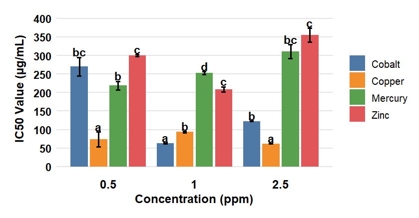
Antioxidant activity varied significantly among elicitor treatments (Table 5), with silymarin (IC50 19.55 ± 6.07 µg/mL) confirming assay reliability as a very strong positive control. Cobalt 1 mg/L and copper 2.5 mg/L extracts exhibited the strongest antioxidant activity among samples, with IC50 values of 62.55 ± 2.08 and 61.72 ± 1.31 µg/mL, respectively, categorized as strong according to Table 1. Copper 0.5 and 1 mg/L extracts also fell into the strong category (73.86 ± 5.42 and 93.31 ± 3.81 µg/mL), while cobalt 2.5 mg/L showed moderate activity (122.83 ± 1.31 µg/mL). All zinc and mercury extracts exhibited very weak activity (IC50 >200 µg/mL). Tukey's HSD test confirmed that cobalt 1 mg/L and copper 2.5 mg/L were significantly different from other treatments (p<0.05) but not from each other.
Figure 1 presents the antioxidant activity of selected extracts, showing representatives of the strong category (cobalt 1 mg/L and copper 2.5 mg/L), moderate activity (cobalt 2.5 mg/L), and a comparison within the same elicitor type (copper 0.5 mg/L). Although approximately three-fold less potent than silymarin, cobalt and copper extracts demonstrated promising antioxidant potential, with activity levels strongly associated with their distinct metabolite profiles—particularly the presence of alkaloids and fatty acid methyl esters in these extracts compared to the glyceride-dominated profiles of zinc and mercury treatments.
Antioxidant activity (IC50 values) ofP. sarmentosumcallus extracts from all elicitor treatments determined by DPPH radical scavenging assay. The bar chart shows IC50 values for cobalt, copper, zinc, and mercury at concentrations of 0.5, 1, and 2.5 mg/L.
The strong antioxidant activity of cobalt 1 mg/L and copper 2.5 mg/L extracts may be attributed to the presence of 2-hydroxy-gamma-butyrolactone, a lactone with documented radical-scavenging activity through hydrogen donation and metal chelation mechanisms (Açikgoz et al., 2023; Nong et al., 2014). Additionally, fatty acid methyl esters and alkaloids—such as the tetrahydroisoquinoline derivative detected in cobalt extracts—may contribute to the overall activity through synergistic effects, as reported for complex plant extracts (Granato et al., 2018; Caesar and Cech, 2019; Yang et al., 2025). The non-linear response of cobalt (optimal at 1 mg/L, lower at 2.5 mg/L) is consistent with a hormetic-like pattern, where moderate stress may activate defense mechanisms while excessive levels cause oxidative damage (Agathokleous and Calabrese, 2022; Jarin et al., 2025). However, because our study included only three concentrations (0.5, 1, and 2.5 mg/L), a full dose-response curve with more data points would be needed to definitively establish hormesis. The interpretation should therefore be considered preliminary. In contrast, copper showed consistent activity across concentrations, suggesting robust copper homeostasis mechanisms in P. sarmentosum callus, possibly involving phytochelatin synthesis and vacuolar sequestration (Mir et al., 2021; Sanusi et al., 2025).
The weak antioxidant activity of zinc and mercury extracts may be explained by their high glyceride content (>95% of total identified compounds), which could dilute minor antioxidant components such as the low levels of 2-hydroxy-gamma-butyrolactone detected in these extracts (Granato et al., 2018). Furthermore, matrix effects in complex mixtures can significantly impact apparent antioxidant activity, as non-antioxidant compounds may interfere with radical-probe interactions or alter the bioavailability of active constituents (Apak et al., 2016).
3.5. Antimicrobial activity
All extracts demonstrated antimicrobial activity against the test microorganisms, with significant effects of elicitor type, concentration, and their interaction (p < 0.005, two-way ANOVA; Table 6). The negative control (DMSO) produced no inhibition zone, while positive controls (chloramphenicol and nystatin) produced inhibition zones of 20.5-24.3 mm, validating the assay conditions. AgainstS. aureus, the cobalt 2.5 mg/L extract at 750 mg/L produced the largest inhibition zone (40.37 ± 3.56 mm), followed by cobalt 1 mg/L at 1000 mg/L (40.20 ± 4.69 mm). AgainstE. coli, the zinc 2.5 mg/L extract at 750 mg/L showed the largest inhibition zone among recommended elicitors (36.05 ± 3.86 mm), while mercury 2.5 mg/L at 500 mg/L showed similar activity (36.28 ± 2.71 mm) but is not recommended due to toxicity. AgainstC. albicans, the cobalt 2.5 mg/L extract at 500 mg/L exhibited the largest inhibition zone (41.62 ± 4.83 mm), followed by copper 1 mg/L at 500 mg/L (40.77 ± 3.98 mm). The very large inhibition zones (>40 mm) may reflect a combined effect of bioactive compounds and residual heavy metals, although metal concentrations in the final extract were not measured.
The observed antimicrobial activity is consistent with the metabolite profiles identified by GC-MS. Cobalt and copper extracts, which exhibited the strongest antibacterial activity againstS. aureus, contained alkaloids (pipataline and tetrahydroisoquinoline derivatives) and fatty acid methyl esters (methyl palmitate and methyl stearate), both compound classes with documented antibacterial properties (Chandrasekaran et al., 2008; Salehi et al., 2019; Yang et al., 2025). The activity of zinc extracts againstE. colimay be attributed to their high glyceride content, as monoacylglycerol derivatives have been reported to possess antimicrobial activity, particularly against Gram-negative bacteria (Henschel et al., 2024; Yoon et al., 2018). The potent anticandidal activity of cobalt and copper extracts correlates with the presence of alkaloids and 2-hydroxy-gamma-butyrolactone, which have documented antifungal properties (Nong et al., 2014; Açikgoz et al., 2023).
The antimicrobial activity of the extracts can be attributed to multiple bioactive compounds. Fatty acid methyl esters (e.g., methyl palmitate and methyl stearate) disrupt bacterial membranes by integrating into the lipid bilayer, causing permeability changes and cytoplasmic leakage, with greater efficacy against Gram-positive bacteria due to the absence of an outer membrane barrier (Casillas-Vargas et al., 2021; Shaaban et al., 2021; Breijyeh et al., 2020). Isoquinoline alkaloids, such as the tetrahydroisoquinoline derivative detected in cobalt extracts, intercalate with DNA and inhibit topoisomerase enzymes (Yang et al., 2025; Casciaro et al., 2020). Lactones like 2-hydroxy-gamma-butyrolactone influence quorum sensing and biofilm formation (Vadakkan et al., 2018), while pipataline (copper extracts) contributes to antimicrobial activity through membrane disruption and efflux pump inhibition (Salehi et al., 2019). Minor terpenoids (e.g., caryophyllene) also possess documented antifungal activity (Dahham et al., 2015; Fidyt et al., 2016).
The observation that 500 mg/L extract concentration frequently yielded optimal antimicrobial activity may reflect a non-linear, possibly hormetic-like response, where intermediate concentrations maximize efficacy while higher concentrations may reduce activity due to compound aggregation or antagonistic interactions (Klančnik et al., 2010; Caesar & Cech, 2019). As with the antioxidant data, confirmation of hormesis would require a more detailed concentration series. Differential activity against the three test microorganisms reflects their distinct cell wall architectures: S. aureus (Gram-positive) lacks an outer membrane, increasing susceptibility to hydrophobic compounds (Breijyeh et al., 2020); E. coli (Gram-negative) possesses an outer membrane restricting antimicrobial entry (Exner et al., 2017); and C. albicans (fungus) features ergosterol as its primary membrane sterol (Pristov and Ghannoum, 2019). The substantial antibacterial efficacy of cobalt and copper extracts, particularly against S. aureus and C. albicans, highlights their potential as natural antimicrobial agents in the context of rising multidrug-resistant infections (Tacconelli et al., 2018).
4. Future Prospects
The present findings open several avenues for further research. First, transcriptomic and proteomic analyses are needed to elucidate the molecular mechanisms by which cobalt and copper upregulate alkaloid and fatty acid methyl ester biosynthetic pathways in P. sarmentosum callus. Second, fed-batch or bioreactor cultivation using optimized Co (1 mg/L) and Cu (2.5 mg/L) concentrations should be explored to scale up bioactive compound production for industrial applications. Third, fractionation and purification of the active constituents (e.g., pipataline and tetrahydroisoquinoline derivatives) are required to confirm their individual and synergistic bioactivities. Fourth, in vivo toxicity and efficacy studies using animal models are necessary before these extracts can be considered for pharmaceutical or nutraceutical development. Finally, encapsulation or nanoformulation of the elicited callus extracts could improve their stability and targeted delivery for antimicrobial applications.
5. Conclusion
This study demonstrated that elicitation ofP. sarmentosumcallus with cobalt (1 mg/L) and copper (2.5 mg/L) produced extracts with relatively strong antioxidant activity (IC50 62.55 ± 2.08 and 61.72 ± 1.31 µg/mL, respectively) and very strong antimicrobial activity againstS. aureus(up to 40.37 mm),E. coli(up to 36.05 mm), andC. albicans(up to 41.62 mm). GC-MS analysis with high-confidence identification (SI ≥800, RSI 800-1000) revealed 42 compounds, with cobalt and copper extracts containing fatty acid methyl esters (0.14-0.61%) and taxon-specific alkaloids (e.g., pipataline), while zinc and mercury extracts accumulated glycerides (0.23-1.79%) and exhibited weak antioxidant activity. Two-way ANOVA confirmed significant effects of elicitor type and concentration on bioactivities (p<0.05) but not on callus biomass (p>0.05). These findings warrant further investigation of heavy metal elicitation, particularly using optimal concentrations of cobalt and copper, for producingP. sarmentosumcallus extracts with enhanced bioactive properties.
Acknowledgements
The authors would like to express their gratitude to the Directorate of Research, Technology, and Community Service, Directorate General of Higher Education, Ministry of Education, Culture, Research, and Technology of the Republic of Indonesia for funding this research through the research grant scheme with Contract Number 0419/C3/PT.05.00/2025 and Agreement/Contract Number 059/C3/DT.05.00/PL/2025; 2338/B/UN3.LPPM/PT.01.03/2025.
Data Availability Statement
All relevant data are fully included within the main text and tables of the manuscript.
References
- AÇIKGOZ, M.A., BATIAY, E., KARA, Ş.M., and AYGÜN, A., 2023. Influence of selected heavy metals on cell growth and camphor secretion inAchillea gypsicolaHub. Mor. in vitro cell cultures. Journal of Agricultural Faculty of Gaziosmanpaşa University, vol. 40, no. 1, pp. 11-18.
- ADAMS, R.P., 2017. Identification of essential oil components by gas chromatography/mass spectrometry. Carol Stream: Allured Business Media.
-
AGATHOKLEOUS, E. and CALABRESE, E.J., 2022. Hormesis: a general biological principle. Chemical Research in Toxicology, vol. 35, no. 4, pp. 547-549. https://doi.org/10.1021/acs.chemrestox.2c00032 PMid:35293720.
» https://doi.org/10.1021/acs.chemrestox.2c00032 -
ALVAREZ-RIVERA, G., SANZ, A., CIFUENTES, A., IBÁNEZ, E., PAAPE, T., LUCAS, M.M. and PUEYO, J.J., 2022. Flavonoid accumulation varies inMedicago truncatulain response to mercury stress. Frontiers in Plant Science, vol. 13, pp. 933209. https://doi.org/10.3389/fpls.2022.933209 PMid:35874019.
» https://doi.org/10.3389/fpls.2022.933209 -
APAK, R., ÖZYÜREK, M., GÜÇLÜ, K. and ÇAPANOĞLU, E., 2016. Antioxidant activity/capacity measurement. 1. Classification, physicochemical principles, mechanisms, and electron transfer (ET)-based assays. Journal of Agricultural and Food Chemistry, vol. 64, no. 5, pp. 997-1027. https://doi.org/10.1021/acs.jafc.5b04739 PMid:26728425.
» https://doi.org/10.1021/acs.jafc.5b04739 -
APARNA, V., DILEEP, K.V., MANDAL, P.K., KARTHE, P., SADASIVAN, C. and HARIDAS, M., 2012. Anti-inflammatory property of n-hexadecanoic acid: structural evidence and kinetic assessment. Chemical Biology & Drug Design, vol. 80, no. 3, pp. 434-439. https://doi.org/10.1111/j.1747-0285.2012.01418.x PMid:22642495.
» https://doi.org/10.1111/j.1747-0285.2012.01418.x -
AZEVEDO, R. and RODRIGUEZ, E., 2012. Phytotoxicity of mercury in plants: a review. Journal of Botany, vol. 2012, pp. 848614. https://doi.org/10.1155/2012/848614
» https://doi.org/10.1155/2012/848614 -
BHUVANESWARI, C., RAO, K., GANDI, S. and GIRI, A., 2012. Abiotic elicitation of gymnemic acid in the suspension cultures of Gymnema sylvestre. World Journal of Microbiology & Biotechnology, vol. 28, no. 2, pp. 741-747. https://doi.org/10.1007/s11274-011-0870-8 PMid:22806870.
» https://doi.org/10.1007/s11274-011-0870-8 -
BREIJYEH, Z., JUBEH, B. and KARAMAN, R., 2020. Resistance of gram-negative bacteria to current antibacterial agents and approaches to resolve it. Molecules, vol. 25, no. 6, pp. 1340. https://doi.org/10.3390/molecules25061340 PMid:32187986.
» https://doi.org/10.3390/molecules25061340 -
CAESAR, L.K. and CECH, N.B., 2019. Synergy and antagonism in natural product extracts: when 1 + 1 does not equal 2. Natural Product Reports, vol. 36, no. 6, pp. 869-888. https://doi.org/10.1039/C9NP00011A PMid:31187844.
» https://doi.org/10.1039/C9NP00011A -
CASCIARO, B., MANGIARDI, L., CAPPIELLO, F., ROMEO, I., LOFFREDO, M.R., IAZZETTI, A., CALCATERRA, A., GOGGIAMANI, A., GHIRGA, F., MANGONI, M.L., BOTTA, B. and QUAGLIO, D., 2020. Naturally-occurring alkaloids of plant origin as potential antimicrobials against antibiotic-resistant infections. Molecules, vol. 25, no. 16, pp. 3619. https://doi.org/10.3390/molecules25163619 PMid:32784887.
» https://doi.org/10.3390/molecules25163619 -
CASILLAS-VARGAS, G., OCASIO-MALAVÉ, C., MEDINA, S., MORALES-GUZMÁN, C., DEL VALLE, R.G., CARBALLEIRA, N.M. and SANABRIA-RÍOS, D.J., 2021. Antibacterial fatty acids: an update of possible mechanisms of action and implications in the development of the next-generation of antibacterial agents. Progress in Lipid Research, vol. 82, pp. 101093. https://doi.org/10.1016/j.plipres.2021.101093 PMid:33577909.
» https://doi.org/10.1016/j.plipres.2021.101093 -
CHANDRASEKARAN, M., KANNATHASAN, K. and VENKATESALU, V., 2008. Antimicrobial activity of fatty acid methyl esters of some members of Chenopodiaceae. Zeitschrift für Naturforschung. C, A Journal of Biosciences, vol. 63, no. 5-6, pp. 331-336. https://doi.org/10.1515/znc-2008-5-604 PMid:18669016.
» https://doi.org/10.1515/znc-2008-5-604 - CLINICAL AND LABORATORY STANDARDS INSTITUTE – CLSI, 2023. Performance standards for antimicrobial susceptibility testing. 33rd ed. Wayne: CLSI. CLSI supplement M100.
-
DAHHAM, S.S., TABANA, Y.M., IQBAL, M.A., AHAMED, M.B., EZZAT, M.O., MAJID, A.S. and MAJID, A.M., 2015. The anticancer, antioxidant and antimicrobial properties of the sesquiterpene β-caryophyllene from the essential oil of Aquilaria crassna. Molecules, vol. 20, no. 7, pp. 11808-11829. https://doi.org/10.3390/molecules200711808 PMid:26132906.
» https://doi.org/10.3390/molecules200711808 -
DENG, Y. and LU, S., 2017. Biosynthesis and regulation of phenylpropanoids in plants. Critical Reviews in Plant Sciences, vol. 36, no. 4, pp. 257-290. https://doi.org/10.1080/07352689.2017.1402852
» https://doi.org/10.1080/07352689.2017.1402852 -
EFFERTH, T., 2019. Biotechnology applications of plant callus cultures. Engineering, vol. 5, no. 1, pp. 50-59. https://doi.org/10.1016/j.eng.2018.11.006
» https://doi.org/10.1016/j.eng.2018.11.006 -
EXNER, M., BHATTACHARYA, S., CHRISTIANSEN, B., GEBEL, J., GORONCY-BERMES, P., HARTEMANN, P., 2017. Antibiotic resistance: what is so special about multidrug-resistant Gram-negative bacteria? GMS Hygiene and Infection Control, vol. 12, pp. Doc05. https://doi.org/10.3205/dgkh000290
» https://doi.org/10.3205/dgkh000290 -
FALCONE-FERREYRA, M.L., RIUS, S.P. and CASATI, P., 2012. Flavonoids: Biosynthesis, biological functions, and biotechnological applications. Frontiers in Plant Science, vol. 3, pp. 222. https://doi.org/10.3389/fpls.2012.00222 PMid:23060891.
» https://doi.org/10.3389/fpls.2012.00222 -
FARNSWORTH, N.R., 1966. Biological and phytochemical screening of plants. Journal of Pharmaceutical Sciences, vol. 55, no. 3, pp. 225-276. https://doi.org/10.1002/jps.2600550302 PMid:5335471.
» https://doi.org/10.1002/jps.2600550302 -
FIDYT, K., FIEDOROWICZ, A., STRZĄDAŁA, L. and SZUMNY, A., 2016. β-caryophyllene and β-caryophyllene oxide: natural compounds of anticancer and analgesic properties. Cancer Medicine, vol. 5, no. 10, pp. 3007-3017. https://doi.org/10.1002/cam4.816 PMid:27696789.
» https://doi.org/10.1002/cam4.816 -
FOUAD, A.S. and HAFEZ, R.M., 2020. Effects of cobalt ions and cobalt nanoparticles on transient expression of gus gene in Catharanthus roseus suspension cultures. Journal of Radiation Research and Applied Sciences, vol. 13, no. 1, pp. 765-775. https://doi.org/10.1080/16878507.2020.1847386
» https://doi.org/10.1080/16878507.2020.1847386 -
GRANATO, D., SHAHIDI, F., WROLSTAD, R., KILMARTIN, P., MELTON, L.D., HIDALGO, F.J., MIYASHITA, K., CAMP, J.V., ALASALVAR, C., ISMAIL, A.B., ELMORE, S., BIRCH, G.G., CHARALAMPOPOULOS, D., ASTLEY, S.B., PEGG, R., ZHOU, P. and FINGLAS, P., 2018. Antioxidant activity, total phenolics and flavonoids contents: should we ban in vitro screening methods? Food Chemistry, vol. 264, pp. 471-475. https://doi.org/10.1016/j.foodchem.2018.04.012 PMid:29853403.
» https://doi.org/10.1016/j.foodchem.2018.04.012 -
GUCHU, B.M., MACHOCHO, A.K.O., MWIHIA, S.K. and NGUGI, M.P., 2020. In vitro antioxidant activities of methanolic extracts of Caesalpinia volkensii Harms., Vernonia lasiopus O. Hoffm., and Acacia hockii de Wild. Evidence-Based Complementary and Alternative Medicine : eCAM, vol. 2020, no. 1, pp. 3586268. https://doi.org/10.1155/2020/3586268 PMid:33062006.
» https://doi.org/10.1155/2020/3586268 -
GÜÇLÜ-ÜSTÜNDAĞ, Ö. and MAZZA, G., 2007. Saponins: properties, applications and processing. Critical Reviews in Food Science and Nutrition, vol. 47, no. 3, pp. 231-258. https://doi.org/10.1080/10408390600698197 PMid:17453922.
» https://doi.org/10.1080/10408390600698197 -
HENSCHEL, J.M., ANDRADE, A.N., SANTOS, J.B.L., SILVA, R.R., MATA, D.A., SOUZA, T. and BATISTA, D.S., 2024. Lipidomics in plants under abiotic stress conditions: an overview. Agronomy, vol. 14, no. 8, pp. 1670. https://doi.org/10.3390/agronomy14081670
» https://doi.org/10.3390/agronomy14081670 -
IKEUCHI, M., FAVERO, D.S., SAKAMOTO, Y., IWASE, A., COLEMAN, D., RYMEN, B. and SUGIMOTO, K., 2019. Molecular mechanisms of plant regeneration. Annual Review of Plant Biology, vol. 70, no. 1, pp. 377-406. https://doi.org/10.1146/annurev-arplant-050718-100434 PMid:30786238.
» https://doi.org/10.1146/annurev-arplant-050718-100434 -
IRFANSYAH, F.D., FATIMAH and JUNAIRIAH, J., 2024. Phytochemical screening and antioxidant activity of three tabebuya species (Tabebuia spp.). Berita Biologi, vol. 23, no. 1, pp. 49-59. https://doi.org/10.55981/beritabiologi.2024.1668
» https://doi.org/10.55981/beritabiologi.2024.1668 -
İŞKIL, R., SURGUN-ACAR, Y., ÇATAV, Ş.S., ZEMHERI-NAVRUZ, F. and ERDEN, Y., 2022. Mercury toxicity affects oxidative metabolism and induces stress responsive mechanisms in wheat (Triticum aestivum L.). Physiology and Molecular Biology of Plants : an International Journal of Functional Plant Biology, vol. 28, no. 4, pp. 911-920. https://doi.org/10.1007/s12298-022-01171-x PMid:35592475.
» https://doi.org/10.1007/s12298-022-01171-x -
JARIN, A.S., KHAN, M.A.R., APON, T.A., ISLAM, M.A., RAHAT, A., AKTER, M., ANIK, T.R., NGUYEN, H.M., NGUYEN, T.T., HA, C.V. and TRAN, L.P., 2025. Plant responses to heavy metal stresses: Mechanisms, defense strategies, and nanoparticle-assisted remediation. Plants, vol. 14, no. 24, pp. 3834. https://doi.org/10.3390/plants14243834 PMid:41470716.
» https://doi.org/10.3390/plants14243834 -
JUNAIRIAH, J., SUHARGO, L., NURHARIYATI, T. and ZURAIDASSANAAZ, N.I., 2024. Secondary metabolite profiles, antimicrobial and antioxidant activities of callus, and leaves extract of Piper sarmentosum Roxb. Journal of Applied Biology and Biotechnology, vol. 12, no. 5, pp. 81-88. https://doi.org/10.7324/JABB.2024.190553
» https://doi.org/10.7324/JABB.2024.190553 -
KLANČNIK, A., PISKERNIK, S., JERŠEK, B. and MOŽINA, S.S., 2010. Evaluation of diffusion and dilution methods to determine the antibacterial activity of plant extracts. Journal of Microbiological Methods, vol. 81, no. 2, pp. 121-126. https://doi.org/10.1016/j.mimet.2010.02.004 PMid:20171250.
» https://doi.org/10.1016/j.mimet.2010.02.004 -
MAKSYMIEC, W., 2007. Signaling responses in plants to heavy metal stress. Acta Physiologiae Plantarum, vol. 29, no. 3, pp. 177-187. https://doi.org/10.1007/s11738-007-0036-3
» https://doi.org/10.1007/s11738-007-0036-3 -
MIR, A.R., PICHTEL, J. and HAYAT, S., 2021. Copper: uptake, toxicity and tolerance in plants and management of Cu-contaminated soil. Biometals : An International Journal on the Role of Metal Ions in Biology, Biochemistry, and Medicine, vol. 34, no. 4, pp. 737-759. https://doi.org/10.1007/s10534-021-00306-z PMid:33909216.
» https://doi.org/10.1007/s10534-021-00306-z - MOLYNEUX, P., 2004. The use of the stable free radical diphenylpicryl-hydrazyl (DPPH) for estimating antioxidant activity. Songklanakarin Journal of Science and Technology, vol. 26, no. 2, pp. 211-219.
- NAMDEO, A.G., 2007. Plant cell elicitation for production of secondary metabolites: a review. Pharmacognosy Reviews, vol. 1, no. 1, pp. 69-79.
-
NONG, X.H., WANG, Y.F., ZHANG, X.Y., ZHOU, M.P., XU, X.Y. and QI, S.H., 2014. Territrem and butyrolactone derivatives from a marine-derived fungus Aspergillus terreus. Marine Drugs, vol. 12, no. 12, pp. 6113-6124. https://doi.org/10.3390/md12126113 PMid:25522319.
» https://doi.org/10.3390/md12126113 - PEREZ-GUTIERREZ, R.M., NEIRA GONZALEZ, A.M. and HOYO-VADILLO, C., 2013. Alkaloids from Piper: A review of its phytochemistry and pharmacology. Mini Reviews in Medicinal Chemistry, vol. 13, no. 2, pp. 163-193. PMid:23279257.
-
PRISTOV, K.E. and GHANNOUM, M.A., 2019. Resistance of Candida to azoles and echinocandins worldwide. Clinical Microbiology and Infection: The Official Publication of the European Society of Clinical Microbiology and Infectious Diseases, vol. 25, no. 7, pp. 792-798. https://doi.org/10.1016/j.cmi.2019.03.028 PMid:30965100.
» https://doi.org/10.1016/j.cmi.2019.03.028 -
RAMAKRISHNA, A. and RAVISHANKAR, G.A., 2011. Influence of abiotic stress signals on secondary metabolites in plants. Plant Signaling & Behavior, vol. 6, no. 11, pp. 1720-1731. https://doi.org/10.4161/psb.6.11.17613 PMid:22041989.
» https://doi.org/10.4161/psb.6.11.17613 -
RAO, R.S. and RAVISHANKAR, G.A., 2002. Plant cell cultures: chemical factories of secondary metabolites. Biotechnology Advances, vol. 20, no. 2, pp. 101-153. https://doi.org/10.1016/S0734-9750(02)00007-1 PMid:14538059.
» https://doi.org/10.1016/S0734-9750(02)00007-1 -
RAZZAQ, A., ZAFAR, M.M., ALI, A., IHSAN, L., QADIR, F., KHAN, M.N., ZHANG, Y., GAO, L., CONG, H., IQBAL, R., JIANG, X. and QIAO, F., 2025. Elicitor-mediated enhancement of secondary metabolites in plant species: a review. Frontiers in Plant Science, vol. 16, pp. 1706600. https://doi.org/10.3389/fpls.2025.1706600 PMid:41200498.
» https://doi.org/10.3389/fpls.2025.1706600 -
SALEHI, B., ZAKARIA, Z.A., GYAWALI, R., IBRAHIM, S.A., RAJKOVIC, J., SHINWARI, Z.K., KHAN, T., SHARIFI-RAD, J., OZLEYEN, A., TURKDONMEZ, E., VALUSSI, M., TUMER, T.B., MONZOTE FIDALGO, L., MARTORELL, M. and SETZER, W.N., 2019. Piper species: a comprehensive review on their phytochemistry, biological activities and applications. Molecules, vol. 24, no. 7, pp. 1364. https://doi.org/10.3390/molecules24071364 PMid:30959974.
» https://doi.org/10.3390/molecules24071364 -
SANUSI, I.O., ADEPOJU, A.A. and ABDULRAHMAN, B.D., 2025. Heavy metal contamination, human health impact, and remediation techniques in water bodies: a review. Discover Environment, vol. 3, no. 1, pp. 268. https://doi.org/10.1007/s44274-025-00475-5
» https://doi.org/10.1007/s44274-025-00475-5 -
SHAABAN, M.T., GHALY, M.F. and FAHMI, S.M., 2021. Antibacterial activities of hexadecanoic acid methyl ester and green-synthesized silver nanoparticles against multidrug-resistant bacteria. Journal of Basic Microbiology, vol. 61, no. 6, pp. 557-568. https://doi.org/10.1002/jobm.202100061 PMid:33871873.
» https://doi.org/10.1002/jobm.202100061 -
SHAFI, A., ZAHOOR, I., GILL, T., AHUJA, P.S., KUMAR, S. and SINGH, A.K., 2019. Ectopic co-expression of the SOD and APX genes enhanced callus growth and in vitro regeneration in Arabidopsis. Plant Biotechnology Reports, vol. 13, no. 3, pp. 273-283. https://doi.org/10.1007/s11816-019-00535-2
» https://doi.org/10.1007/s11816-019-00535-2 -
SHARMA, A., SHAHZAD, B., REHMAN, A., BHARDWAJ, R., LANDI, M. and ZHENG, B., 2019. Response of phenylpropanoid pathway and the role of polyphenols in plants under abiotic stress. Molecules, vol. 24, no. 13, pp. 2452. https://doi.org/10.3390/molecules24132452 PMid:31277395.
» https://doi.org/10.3390/molecules24132452 -
STURIKOVA, H., KRYSTOFOVA, O., HUSKA, D. and ADAM, V., 2018. Zinc, zinc nanoparticles and plants. Journal of Hazardous Materials, vol. 349, pp. 101-110. https://doi.org/10.1016/j.jhazmat.2018.01.040 PMid:29414741.
» https://doi.org/10.1016/j.jhazmat.2018.01.040 -
SUN, X., CHEN, W., DAI, W., XIN, H., RAHMAND, K., WANG, Y., ZHANG, J., ZHANG, S., XU, L. and HAN, T., 2020. Piper sarmentosum Roxb.: a review on its botany, traditional uses, phytochemistry, and pharmacological activities. Journal of Ethnopharmacology, vol. 263, pp. 112897. https://doi.org/10.1016/j.jep.2020.112897 PMid:32620264.
» https://doi.org/10.1016/j.jep.2020.112897 -
TACCONELLI, E., CARRARA, E., SAVOLDI, A., HARBARTH, S., MENDELSON, M., MONNET, D.L., PULCINI, C., KAHLMETER, G., KLUYTMANS, J., CARMELI, Y., OUELLETTE, M., OUTTERSON, K., PATEL, J., CAVALERI, M., COX, E.M., HOUCHENS, C.R., GRAYSON, M.L., HANSEN, P., SINGH, N., THEURETZBACHER, U., MAGRINI, N., ABODERIN, A.O., AL-ABRI, S.S., AWANG JALIL, N., BENZONANA, N., BHATTACHARYA, S., BRINK, A.J., BURKERT, F.R., CARS, O., CORNAGLIA, G., DYAR, O.J., FRIEDRICH, A.W., GALES, A.C., GANDRA, S., GISKE, C.G., GOFF, D.A., GOOSSENS, H., GOTTLIEB, T., GUZMAN BLANCO, M., HRYNIEWICZ, W., KATTULA, D., JINKS, T., KANJ, S.S., KERR, L., KIENY, M.-P., KIM, Y.S., KOZLOV, R.S., LABARCA, J., LAXMINARAYAN, R., LEDER, K., LEIBOVICI, L., LEVY-HARA, G., LITTMAN, J., MALHOTRA-KUMAR, S., MANCHANDA, V., MOJA, L., NDOYE, B., PAN, A., PATERSON, D.L., PAUL, M., QIU, H., RAMON-PARDO, P., RODRÍGUEZ-BAÑO, J., SANGUINETTI, M., SENGUPTA, S., SHARLAND, M., SI-MEHAND, M., SILVER, L.L., SONG, W., STEINBAKK, M., THOMSEN, J., THWAITES, G.E., VAN DER MEER, J.W.M., VAN KINH, N., VEGA, S., VILLEGAS, M.V., WECHSLER-FÖRDÖS, A., WERTHEIM, H.F.L., WESANGULA, E., WOODFORD, N., YILMAZ, F.O. and ZORZET, A., 2018. Discovery, research, and development of new antibiotics: the WHO priority list of antibiotic-resistant bacteria and tuberculosis. The Lancet. Infectious Diseases, vol. 18, no. 3, pp. 318-327. https://doi.org/10.1016/S1473-3099(17)30753-3 PMid:29276051.
» https://doi.org/10.1016/S1473-3099(17)30753-3 -
THOLL, D., 2015. Biosynthesis and biological functions of terpenoids in plants. Advances in Biochemical Engineering/Biotechnology, vol. 148, pp. 63-106. https://doi.org/10.1007/10_2014_295 PMid:25583224.
» https://doi.org/10.1007/10_2014_295 -
TUNTIWACHWUTTIKUL, P., PHANSA, P., POOTAENG-ON, Y. and CHARLES, W., 2006. Chemical constituents of the roots of Piper sarmentosum. Chemical & Pharmaceutical Bulletin, vol. 54, no. 2, pp. 149-151. https://doi.org/10.1248/cpb.54.149 PMid:16462055.
» https://doi.org/10.1248/cpb.54.149 -
VADAKKAN, K., CHOUDHURY, A.A., GUNASEKARAN, R., HEMAPRIYA, J. and VIJAYANAND, S., 2018. Quorum sensing intervened bacterial signaling: pursuit of its cognizance and repression. Journal of Genetic Engineering and Biotechnology, vol. 16, no. 2, pp. 239-252. https://doi.org/10.1016/j.jgeb.2018.07.001 PMid:30733731.
» https://doi.org/10.1016/j.jgeb.2018.07.001 - WANTI, Y.T.N.I., UTAMI, E.S.W., and JUNAIRIAH, 2025. Utilization of chitosan elicitation on biomass, total flavonoids, and antioxidant activity of black betel callus (Piper betle L. var. Nigra). Journal of Tropical Life Science, vol. 15, no. 2, pp. 343-350.
-
WEREMCZUK-JEŻYNA, I., GOMULSKI, J., KISS, A.K. and GRZEGORCZYK-KAROLAK, I., 2024. Effect of Ag+ and Cd2+ elicitation on polyphenol production in shoot culture of Dracocephalum ruyschiana L. Molecules, vol. 29, no. 22, pp. 5263. https://doi.org/10.3390/molecules29225263
» https://doi.org/10.3390/molecules29225263 -
YANG, X., BU, T., MA, Y., YU, X., GONG, Z., WANG, J., LIU, X., JENIS, J., HU, H., MIAO, X. and SHANG, X., 2025. An updated review of isoquinoline alkaloids: biological activity and mode of action, structural modifications, and agricultural applications. Industrial Crops and Products, vol. 234, pp. 121591. https://doi.org/10.1016/j.indcrop.2025.121591
» https://doi.org/10.1016/j.indcrop.2025.121591 -
YOON, B.K., JACKMAN, J.A., VALLE-GONZÁLEZ, E.R. and CHO, N.J., 2018. Antibacterial free fatty acids and monoglycerides: biological activities, experimental testing, and therapeutic applications. International Journal of Molecular Sciences, vol. 19, no. 4, pp. 1114. https://doi.org/10.3390/ijms19041114 PMid:29642500.
» https://doi.org/10.3390/ijms19041114 -
ZHAO, J., DAVIS, L.C. and VERPOORTE, R., 2005. Elicitor signal transduction leading to production of plant secondary metabolites. Biotechnology Advances, vol. 23, no. 4, pp. 283-333. https://doi.org/10.1016/j.biotechadv.2005.01.003 PMid:15848039.
» https://doi.org/10.1016/j.biotechadv.2005.01.003
Edited by
-
Editor:
Takako Matsumura Tundisi