
 Antimicrobial susceptibility of Escherichia coli to extracts of plant species Zingiber officinale and Hedychium coronarium
Abstract
Antimicrobial resistance is a growing global concern, intensifying the demand for effective and sustainable treatment alternatives. This study adopts a novel approach by assessing the antimicrobial activity of Zingiber officinale and Hedychium coronarium extracts against both standard (Escherichia coli ATCC 25922) and environmental isolates. Plant materials were sequentially fractionated with five solvents of increasing polarity, and the resulting extracts were characterized by GC–MS. Non-polar fractions of Zingiber officinale displayed the strongest activity, particularly against the reference strain, while environmental isolates exhibited greater resistance. This integrative approach offers a realistic evaluation of antimicrobial potential under environmental conditions. Findings highlight the role of solvent selection in maximizing the recovery of bioactive compounds and position Zingiber officinale as a promising candidate for phytotherapeutic development. These results strengthen the case for using phytochemicals as alternative antimicrobials and provide a basis for future veterinary and translational research.
Keywords:
antimicrobial resistance; Escherichia coli; medicinal plants; phytochemical compounds
Resumo
A resistência antimicrobiana é uma preocupação global crescente, intensificando a demanda por alternativas de tratamentos eficazes e sustentáveis. Este estudo adota uma abordagem inovadora ao avaliar a atividade antimicrobiana dos extratos de Zingiber officinale e Hedychium coronarium frente à cepa padrão (Escherichia coli ATCC 25922) e a isolados ambientais. Os materiais vegetais foram fracionados utilizando cinco solventes de polaridade crescente e a composição dos extratos foi caracterizada por GC-MS. As frações apolares de Zingiber officinale apresentaram a atividade mais intensa, especialmente contra a cepa de referência, enquanto os isolados ambientais exibiram maior resistência. Esta abordagem integrativa oferece uma avaliação realista do potencial antimicrobiano em condições ambientais. Os resultados destacam o papel da escolha do solvente na maximização da recuperação de compostos bioativos e posicionam Zingiber officinale como um candidato promissor ao desenvolvimento de fitoterápicos. Estes achados reforçam o uso de fitoquímicos como antimicrobianos alternativos e fornecem uma base para futuras pesquisas veterinárias e translacionais.
Palavras-chave:
resistência antimicrobiana; Escherichia coli; plantas medicinais; compostos fitoquímicos
1. Introduction
Throughout history, plants have served numerous purposes, particularly in traditional medicine. The earliest recorded evidence of their medicinal use dates back approximately 5,000 years to a Sumerian clay tablet from Nagpur, which references around 1,000 plant species used for therapeutic purposes (Süntar 2020; Andrade et al., 2021). By the early 19th century, over 80% of medicinal treatments relied on plants, significantly influencing the pharmaceutical industry’s development (Marinho et al., 2022; Abdallah et al., 2023; Ahmed et al., 2024). The improper use of antibiotics in agriculture and healthcare, poor sanitation, and ineffective infection control measures have fueled the emergence and spread of antimicrobial-resistant bacteria (Boonyasiri et al., 2014; WHO, 2019; Moretto et al., 2022). As a result, antibiotic resistance in pathogens such as Klebsiella pneumoniae, Salmonella typhimurium, Listeria monocytogenes, Candida albicans, Proteus vulgaris, Escherichia coli (E. coli) is a prominent concern in modern veterinary and human medicine (Caneschi et al., 2023).
The Zingiberaceae family, comprising around 1,300 species of aromatic, perennial herbs with creeping or tuberous rhizomes, is widely distributed across the Americas, Africa, and Asia (Taylor et al., 2009). These species are known for their medicinal properties, including anti-inflammatory, antimicrobial, antioxidant, and neuroprotective effects, and are used across various cultures (Mao et al., 2019; Deng et al., 2022; Asghar et al., 2023). Although the biological potential of these plants has been investigated, scientific evidence is still limited regarding the use of solvent-partitioned fractions in antimicrobial assays to evaluate the efficacy of distinct phytochemical groups against resistant E. coli strains isolated from environmental compartments (Wang et al., 2020; Yit and Zainal-Abidin, 2024).
This research gap warrants attention, as it allows a more realistic assessment of the therapeutic potential of plant extracts against resistant bacteria under environmentally relevant conditions, unlike laboratory strains that have not experienced selective pressure in situ. Environmental E. coli isolates often exhibit greater resistance to commercial antimicrobials due to adaptation to hostile and dynamic ecosystems (Anjum et al., 2021; Jang et al., 2017). Considering this expected resistance, the study was guided by the following question: Which solvent-based fractions and phytochemical compounds from Zingiber officinale and Hedychium coronarium show the highest antimicrobial activity against environmental E. coli strains? We tested rhizome extracts of Z. officinale (ginger) and H. coronarium (white garland lily) against both E. coli ATCC 25922 and environmental isolates. This comparative analysis offers broader insights into extract efficacy across laboratory and real-world contexts, supporting the development of plant-based strategies to address antimicrobial resistance.
2. Material and Methods
2.1. Description of plants and sample collection
In June 2022, Z. officinale rhizomes were sourced from a local store, while H. coronarium rhizomes were collected near the Federal University of Santa Maria, Frederico Westphalen campus (Rio Grande do Sul, Brazil; -27.39385, -53.42826). Table 1 outlines the general characteristics of the plants used.
2.2. Preparation of plant extracts
The plant rhizomes were dried in an air circulation oven at 50 °C for five days, and subsequently ground into a fine powder. The resulting powder was weighed into cellulose cartridges and subjected to methanol (99.9%; Sigma-Aldrich) extraction using Soxhlet equipment (in duplicate). Afterward, residual methanol was evaporated in a fume hood. The extract was fractionated with solvents in increasing polarity (hexane, ethyl ether, chloroform, ethyl acetate, and water), each fraction was separated, and the solvents were removed by rotary evaporation.
2.3. Analysis of the phytocompounds of the plants
A CG-3900 gas chromatograph coupled to a Varian Saturn 2100T mass spectrometer (GC/MS-SIS) with an automatic CP-8400 autosampler was used to analyze the extracts. The system featured a VF-5MS capillary column (30 m × 0.25 mm ID, 0.25 µm DF) with 5% phenyl and 95% dimethyl polysiloxane. Compound identification was based on the NIST spectral library, and relative component distribution was determined by normalizing peak areas. Operating conditions included a max temperature of 325 °C, helium flow at 2 mL/min, injector temperature of 250 °C, a 1 µL injection via a 10 µL syringe, an initial furnace temperature of 70 °C for 1 minute, followed by a 20 °C/min ramp to 280 °C. Compounds were fragmented via electron impact at 70V.
2.4. Microbial culture
Water samples were collected from the Lajeado Pardo River, located in Frederico Westphalen (Rio Grande do Sul, Brazil), for the isolation of Gram-negative bacteria. Raw water (100 mL) was filtered through cellulose acetate filters (0.45 µm) using vacuum to retain microorganisms. The filters were aseptically transferred to McConkey Agar and incubated at 35-37 °C for 18-24h to colony growth. A pure colony was inoculated in EC medium broth and grown on Muller-Hinton agar. Afterward, a pure colony was stored in brain and heart infusion broth (BHI) stock culture to continue subsequent testing. Environmental isolates colonies (S1, S2, S3, and S4) were compared with E. coli ATCC 25922 TM concerning its morphology. Stock cultures were stored at -20 °C in 80% BHI and 20% glycerol (Kouadio-Ngbesso et al., 2019).
2.5. PCR and amplification of isolated bacterial strains
The oligonucleotide primers used in these amplifications were selected based on previously published sequences (Smith et al., 1992), available in GenBank under accession numbers M84024 (gadA) and M84025 (gadB). Primer optimization was performed using the automated search tool of the Oligo 4.1 software (National Biosciences, Plymouth, Minnesota, USA). The forward primer targeting the gadA/B gene, corresponding to base positions 307-324, was 5′-ACCTGCGTTGCGTAAATA-3′, and the reverse primer, corresponding to positions 959-976, was 5′-GGGCGGGAGAAGTTGATG-3′. DNA extraction was performed using the boiling method, for its simplicity, low cost, speed, and reliability (Yamagishi et al., 2016). The Master Mix PCR Kit was used to amplify nucleic acids via the polymerase chain reaction (PCR). The formulation includes recombinant Taq DNA polymerase, MgCl2, and dNTPs, provided at concentrations suitable for preparing 100 reactions of 25 µL each. The reaction mix was supplied as a 2× concentrated Pre-Mix, which, upon dilution to 1× working concentration, contained all essential components required for PCR: a reaction buffer with Tris-HCl/KCl (pH 8.4), 2.0 mM MgCl2, 0.2 mM of each dNTP, and 2.5 U of recombinant Taq DNA polymerase.
Primers were prepared by diluting the stock solution tenfold. For the working solution, 10 μL of stock was mixed with 100 μL of ultrapure water, yielding a 1 pmol/μL concentration. Each reaction, in a 200 μL DNase- and RNase-free microtube, contained 12.5 μL of Pre-Mix, 2.5 μL of each primer (forward and reverse), 6.5 μL of ultrapure water, and 1 μL of template DNA (~5 ng, as measured by UV spectrophotometry at λ=260 nm). All steps followed the guidelines provided by the Ludwig Manufacturing Company (Walker et al., 2017). The thermal cycling protocol consisted of an initial denaturation at 95 °C for 5 minutes, followed by 40 cycles of 94 °C for 30 seconds, 55 °C for 30 seconds, and 72 °C for 2.5 minutes, with a final extension step at 72 °C for 5 minutes.
2.6. Bacterial susceptibility assay of plant extracts
The broth microdilution method was employed for in vitro tests against E. coli strains. Analyses were performed in duplicate using sterile 96-well acrylic plates (300 μL/well), with a negative control (a standard Salmonella strain from Fundação Oswaldo Cruz), a positive control (E. coli ATCC 25922TM), and five extract fractions tested at eight concentrations ranging from 1 to 320 μg/mL. Extracts were diluted in Mueller-Hinton broth with 2.5% DMSO. Each well received 100 μL of broth containing the extract at the desired concentration and 100 μL of bacterial suspension (0.85% saline solution) at a concentration equivalent to 10 on the McFarland scale. The plates were incubated in a B.O.D. incubator at 35 ± 2 °C for 18 hours. After incubation, 20 µL of a 3% 2,3,5-triphenyl tetrazolium chloride solution, prepared in sterile water, was added to each well to confirm bacterial growth. The plates were further incubated for 1 hour. Wells exhibiting red turbidity indicated viable bacterial cells, confirming the absence of an inhibitory effect (Santos et al., 2022).
2.7. Data analysis
Minimum Inhibitory Concentration (MIC) and Minimum Lethal Concentration (MLC) were determined for all extracts of solvents and water (CLSI, 2020). Heat maps were constructed to compare the effectiveness of the tested solvents. Correlation analyses between the MIC values of the different extracts were performed using Statgraphics 19 and SankeyMATIC diagrams to relate the compounds with the extracts.
3. Results
3.1. Yields of extracts of plants
The methanol extract yield exceeded 20% for both species (25.22% for Z. officinale and 34.79% for H. coronarium), while solvent fractions ranged from 0.61% to 65.21% (Figure 1). Both species showed extraction percentages below 3% for chloroform and ethyl acetate; however, H. coronarium had a higher recovery with chloroform and Z. officinale with ethyl acetate. Notably, the hexane fraction yielded higher percentages, at 21.73% for Z. officinale and 26.22% for H. coronarium, whereas the aqueous extract had the highest yield, exceeding 60%.

Extraction yield of Zingiber officinale and Hedychium coronarium by solvent. Pie charts indicate the percentage yield of the crude methanolic extracts for Z. officinale and H. coronarium. Bar plots show the yields of individual fractions (in mg/g of plant material), obtained using five solvents of increasing polarity: hexane, ethyl ether, chloroform, ethyl acetate, and water. Error bars show the standard deviation from duplicate analyses.
3.2. Confirmation of genes (PCR)
Comparison of E. coli species through molecular weight patterns following amplification showed positive results for all environmental isolates in the selective bacterial growth medium, confirmed by colilert and PCR tests.
3.3. Secondary metabolites and antimicrobial activity of plant extracts
The GC/MS-SIS chromatograms identified compounds based on retention times that matched the NIST library. Seven compounds were identified, with retention times between 18.48 and 28.47 minutes, mainly terpenes, including linalool, 4,4-dimethyl-2-pentanal, neral, nerol, geranial, 2-undecanone, and farnesol. Z. officinale extracts showed a higher variety of compounds compared with H. coronarium (Figure 2).
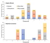
Distribution of identified compounds in percentage of total area in solvent fractions of Z. officinale and H. coronarium. Compounds are presented according to their retention times (RT): [1] Linalol (terpene, RT 18.48 min.), [2] 4.4-Dimetil-2-pentinal (aldehyde, RT 21.59 min), [3] Neral (terpene, RT 23.65 min), [4] Nerol (monoterpene, RT 24.13 min), [5] Geranial (acyclic monoterpene aldehyde, RT 24.70 min), [6] 2-Undecanona (ketone, 25.48 min), [7] Farnesol (sesquiterpene alcohol, RT 28.47 min), [8] Unid (unidentified, RT 30.05 min).
For example, the chloroform and ethyl ether extracts of Z. officinale contained five constituents, and six components were detected in the ethyl acetate extract. In contrast, H. coronarium extracts showed a maximum of two compounds per fraction (hexane and ethyl ether).
The MIC results (Figure 3A) show positive antimicrobial activity for E. coli ATCC and all environmental isolates from both plants, ranging from 5 to 320 µg/mL. All solvents demonstrated strong antimicrobial effects against E. coli ATCC, with MIC values between 5 and 10 µg/mL. For environmental isolates (S1 to S4), the MIC values varied: hexane ranged from 80 to 320 µg/mL for Z. officinale and 320 µg/mL for H. coronarium; ethyl ether ranged from 20 to 80 µg/mL for Z. officinale and 80 to 320 µg/mL for H. coronarium; chloroform ranged from 20 to 160 µg/mL; and ethyl acetate ranged from 20 to 320 µg/mL. The aqueous extract showed MIC values exceeding 320 µg/mL for all isolates.

(A) Heat maps of Minimum Inhibitory Concentration (MIC) and Minimum Lethal Concentration (MLC) for Z. officinale and H. coronarium extracts (µg/mL); (B) Correlation of MIC values between extract fractions and Sankey diagram linking compounds and solvent fractions in Z. officinale.
The MLC results were less conclusive, with most values exceeding 320 µg/mL. However, positive antimicrobial activity was observed at 320 µg/mL for the ethyl ether and chloroform extracts of Z. officinale; and at 160 µg/mL (ethyl ether) and 320 µg/mL (ethyl acetate) for H. coronarium against E. coli ATCC. Notably, all E. coli isolates displayed an MLC of 320 µg/mL for the ethyl acetate extracts of H. coronarium. Figure 3B shows the correlation matrix of MIC values across different extract fractions of Z. officinale. Moderate to strong positive correlations were observed between several solvents, particularly between hexane and ethyl ether (r = 0.71), chloroform and hexane (r = 0.82), and hexane and ethyl acetate (r = 0.82). The Sankey diagram shows the relationship between major phytochemical compounds identified and their distribution across solvent fractions, highlighting ethyl acetate and ethyl ether as the fractions containing the greatest diversity of compounds.
4. Discussion
4.1. Yields of extracts of plants
The extraction yields using non-polar solvents like hexane and ethyl ether highlight the significant presence of non-polar compounds. The aqueous extract, with yields exceeding 60%, along with chloroform and acetate fractions, suggests a notable presence of polar compounds in both plants, surpassing the aqueous yields reported by Yeh et al. (2014) for two Z. officinale varieties (15.84% and 10.86%). Hexane had the highest recovery, 26.22% for H. coronarium and 22% for Z. officinale (Figure 1), efficiently extracting non-polar compounds such as monoterpenes and essential oils (Syafitri et al., 2018). Lower yields for chloroform and ethyl acetate fractions, as reported by Cabana et al. (2013), may be attributed to factors such as extraction time, temperature, and solvent-specific characteristics (e.g., solubility and molecular structure). The selection of solvents, influenced by polarity, plays a decisive role in determining the phytochemical profile of extracts, thereby influencing both biological activity and commercial value.
4.2. Analysis of the phytochemical composition of solvent extracts
The identification of phytochemical compounds of plant species is key for understanding their secondary metabolites. Fractionating the extracts into different solvents allows for a more efficient and targeted evaluation of these bioactive compounds (Cunha and Graça, 2009). In a study by Gomes et al. (2016), 18 organic compounds were identified in the essential oil fraction of Z. officinale rhizomes, including neral (9.64%), farnesol (1.27%), nerol (1.07%), 2-undecanone (0.63%), and 1,1-dicyclopropylene (0.55%). Similarly, this research reports many of the same compounds, though in varying proportions due to differences in solvent extractions. This reinforces the shared chemical composition of plants within the Zingiberaceae family.
The extraction efficiency of compounds from Z. officinale and H. coronarium varies based on their physicochemical properties and the solvent's affinity for them. For instance, the high recovery of geranial in hexane (58.00%) from Z. officinale indicates a strong solvent-compound interaction, which aligns with the known solubility of terpenoids in non-polar solvents (Smith et al., 2020). This highlights the importance of selecting appropriate solvents to optimize the extraction of specific bioactive compounds.
For H. coronarium, the high extraction efficiency of nerol in hexane (85.13%) and farnesol in ethyl ether and chloroform (42.33% and 39.50%, respectively) demonstrates the selectivity of these solvents for specific compounds. This corroborates previous findings on the selective capacity of solvents in extracting terpenes and sesquiterpenes (Johnson et al., 2019; Lee et al., 2021). Similarly, the extraction of nerol from Z. officinale using hexane (12.98%) and ethyl acetate (49.50%) indicates its strong affinity for solvents with varying polarities. Certain studies confirm that non-polar and semi-polar solvents are highly effective in extracting monoterpenes, highlighting the versatility of hexane and ethyl acetate for extracting compounds from various chemical classes (Gomes et al., 2019). The substantial presence of 2-undecanone in ethyl ether (49.85%) and ethyl acetate (9.66%) further underscores the efficiency of these solvents in extracting ketones.
Nerol and geraniol are well-documented in ginger essential oils for their antimicrobial, antioxidant, and anti-inflammatory properties (Pinheiro Silva and Martins de Paiva, 2021). Notably, significant variations in secondary metabolite content between fresh and dried Z. officinale oils have been reported (Munda et al., 2018), with lower levels of acyclic monoterpenes found in dried ginger, suggesting that drying leads to compound loss. The concentrations of nerol (1.07%) and farnesol (1.27%) found in this study exceed those previously reported in ginger rhizome and leaf extracts (Cutrim et al., 2019).
Andrade et al. (2012) reported similar findings, where ginger essential oil extracted with various solvents revealed compounds such as linalool (1.80%), neral (16.47%), geranial (25.06%), and 2-undecanone (0.60%). Liu et al. (2019) identified 194 volatile components, including nerol, linalool, farnesol, 2-undecanone, neral, and geranial, in Z. officinale from different regions of China, highlighting the influence of the plant’s growth environment on its chemical composition.
Lastly, the high extraction yield of unidentified compounds in chloroform (60.50%) and ethyl acetate (73.32%) for H. coronarium can be attributed to the solvent's ability to solubilize a broad range of compounds with varying chemical properties. Chloroform and ethyl acetate are widely recognized for their effectiveness in extracting volatile and semi-volatile compounds from medicinal plants, potentially capturing not yet identified bioactive components (Oliveira et al., 2019; Martinez et al., 2021). Additionally, the high extraction of neral in ethyl ether (57.68%) confirms its selective affinity for terpenes, as reported in other plant matrices (Hernandez et al., 2020).
4.3. Antimicrobial potentiality of extracts of Zingiber officinale and Hedychium coronarium
MIC and MLC values were used to evaluate the inhibitory and bactericidal activity of the extracts against both the standard strain and environmental E. coli isolates. According to Aligiannis et al. (2001), MIC values are classified as follows: up to 500 µg/mL indicates strong inhibition, 600-1,000 µg/mL denotes moderate inhibition, and values above 1,000 µg/mL reflect weak inhibition. Based on this scale, the results obtained indicate strong inhibitory activity. The E. coli ATCC 25922 exhibited greater sensitivity to the extracts, with inhibition observed at the lowest concentrations tested.
The highest inhibition results against strains of environmental isolates were obtained from the ethyl ether and ethyl acetate fractions of Z. officinale (average MIC of 35 and 65 µg/mL, respectively), and in the chloroform extract (average of 100 µg/mL) for H. coronarium extracts, suggesting: (1) differences in the extracted compounds, (2) the compounds extracted from H. coronarium are less effective against these specific isolates, and (3) the compounds extracted by ethyl ether from Z. officinale are more effective against the E. coli isolates.
The high extraction of geranial (58.00%) from Z. officinale hexane extract is consistent with its antimicrobial activity (5 µg/mL for E. coli-ATCC; Smith et al., 2020). Furthermore, 2-undecanone and farnesol can contribute to antimicrobial activity (Gomes et al., 2019). In the hexane extract of H. coronarium, the MIC for E. coli isolates was 320 µg/mL, despite the high concentration of nerol (85.13%) in the fraction. Although nerol is known for its antimicrobial activity, its effectiveness may be influenced by potential interactions with co-occurring compounds, such as 2-undecanone (Hernandez et al., 2020).
Z. officinale ethyl ether extract contains linalool, 4,4-dimethyl-2-pentinal, geranial, farnesol, and 2-undecanone (most abundant, 49.85%). This mixture of compounds contributes to the low MIC of 20 µg/mL for E. coli S1 to S3, and 80 µg/mL for S4 isolates, reflecting the combined efficacy of these compounds. Several studies confirm that compounds such as linalool and geranial have potent antimicrobial properties, especially against gram-negative bacteria (Smith et al., 2020; Lee et al., 2021).
For H. coronarium, only neral (57.68%) and farnesol (42.33%) were reported in this extract, with MIC values of 320 µg/mL for E. coli isolates S1 to S3, and greater than 320 µg/mL for S4 isolates. Although neral is known for its antimicrobial activity, its efficacy may be reduced when not complemented by synergistic compounds (Hernandez et al., 2020). This may explain the higher MIC values observed for the H. coronarium extract, particularly against environmental isolate S4, which exhibited resistance to all tested fractions. This strain, isolated from the Lajeado Pardo River, likely acquired antimicrobial resistance genes (both synthetic and naturally occurring) through adaptive processes. One of the predominant mechanisms of resistance in E. coli is horizontal gene transfer, often mediated by plasmids and transposons, which facilitates the rapid spread of β-lactamase and carbapenemase genes. Additionally, E. coli can develop resistance by altering membrane permeability (via porin mutations), activating efflux pumps to expel antimicrobial agents, or forming biofilms and persister cells that increase survival under antibiotic pressure. These mechanisms are tightly regulated by genetic networks responsive to environmental stress (Poirel et al., 2018; Zhang et al., 2024; Nasrollahian et al., 2024).
In contrast, chloroform extracts of Z. officinale, composed primarily of geranial (54.13%), 4,4-dimethyl-2-pentanal (25.41%), and nerol (9.81%), exhibited a MIC of 5 µg/mL against the E. coli ATCC strain. The combination of geranial and nerol is highly effective against Gram-negative bacteria (Smith et al., 2020). Conversely, H. coronarium extracts rich in unidentified compounds (60.50%) and farnesol (39.50%) showed lower activity (MIC= 80 µg/mL), suggesting that these unidentified constituents may not significantly contribute to the antimicrobial effect (Johnson et al., 2019). The solvent ethyl acetate primarily extracted nerol (49.50%) and neral (11.30%) from Z. officinale, and neral (26.68%) from H. coronarium, resulting in a MIC ranging from 20 to 320 µg/mL for E. coli isolates (except E. coli-S4, which exceeded 320 µg/mL for H. coronarium). Nerol and neral are known for their antimicrobial properties, and their high concentration contributes to the extract's efficacy (Smith et al., 2020). Agrawal et al. (2018) obtained a value of 625 µg/mL for MIC (moderate inhibitory factor) for essential oil from the ginger rhizome against E. coli, a higher concentration than the maximum tested in this study (320 µg/mL). E. coli stood out as the most resistant compared to the others tested by Agrawal.
The aqueous extract of Z. officinale showed no inhibitory action against any of the strains of E. coli tested, even at the maximum concentration, indicating that the more polar fraction of Z. officinale has higher antimicrobial activity in Gram-positive bacteria (Agrawal et al., 2018). A similar result was evidenced by Yousfi et al. (2021), with no antimicrobial effect observed in the aqueous extract of the same plant species against the standard strain E. coli ATCC (25922). The limited action of the aqueous extract is due to the insolubility of the bioactive compounds present in Z. officinale. The unidentified compounds (73.32%) in the water extract of H. coronarium suggest that these unknown compounds are not highly effective against E. coli (Martinez et al., 2021).
The high proportion of unidentified compounds in some extracts may partially explain the variability in antimicrobial activity. Medicinal plant extracts typically contain hundreds of bioactive constituents at varying concentrations, making it difficult to determine which compounds are primarily responsible for the observed effects. Additionally, the analytical limitations of GC-MS, such as its bias toward detecting non-polar and high-molecular-weight compounds, may result in the underrepresentation of polar or low-abundance constituents. Broader chromatographic strategies or complementary analytical techniques are needed to fully characterize these complex mixtures and clarify the contribution of unidentified compounds to antimicrobial activity (van Vuuren and Viljoen, 2011; Rohloff, 2015; Chew et al., 2021).
Many studies have reported that the solubility and affinity of compounds for different solvents directly influence the antimicrobial efficacy of the extracts, highlighting the importance of selecting suitable solvents for the effective extraction of bioactive compounds (Gomes et al., 2019; Lee et al., 2021). The correlation analysis (Figure 3) reveals significant relationships between the various extractive fractions and a node graph for Z. officinale (a plant with lower mic values in the extracts). The analysis emphasizes that solvent selection directly affects antimicrobial efficacy due to the varying ability of each solvent to extract bioactive compounds based on their polarity. Correlation coefficients greater than 0.70 indicate consistent bacterial inhibition patterns across the extracts from both plants, suggesting they share bioactive compounds with similar mechanisms of action. Strong correlations (R > 0.70) were observed between (a) hexane and ethyl ether (R=0.82): Both extracts contained farnesol, 2-undecanone, geranial, and nerol, demonstrating similar chemical profiles, (b) hexane and chloroform (R=0.82): Both solvents efficiently extracted terpenes, particularly for Z. officinale nerol (9-13%) and geranial (54-58%), which were present in comparable proportions, contributing to the extracts’ antimicrobial properties, and (c) ethyl ether and ethyl acetate (R=0.82): These richer fractions extracted both polar and non-polar compounds, broadening the bioactive spectrum.
The node graph or Sankey diagram for Z. officinale (Figure 3) further illustrates the distribution of key bioactive compounds, underscoring the plant's superior performance across all solvents, which yielded the lowest MIC values. The largest nodes correspond to potent bioactive compounds such as geranial (a monoterpenoid alcohol), farnesol (a sesquiterpenoid alcohol), 2-undecanone (a linear aliphatic ketone), and nerol (a monoterpene) (Gomes et al., 2019). These compounds were identified across multiple fractions, emphasizing their contribution to antimicrobial activity and highlighting the crucial role of non-polar fractions in inhibiting E. coli strains, with MICs ranging from 20-80 µg/mL for environmental isolates and 5-10 µg/mL for the ATCC reference strain. The graph also reveals that fractions containing geranial and farnesol tend to form clusters associated with lower MIC values. This suggests potential synergistic effects, which could be further exploited to develop standardized extracts with enhanced potency (Jadimurthy et al., 2023). The superior antimicrobial performance of these non-polar fractions may be attributed to synergistic interactions among multiple phytochemicals that can involve pharmacodynamic synergy targeting different cellular pathways or pharmacokinetic enhancement, such as increased solubility or bioavailability. Some compounds may inhibit resistance mechanisms like efflux pumps or β-lactamases, while others may destabilize bacterial membranes or biofilms, making bacteria more vulnerable. These combined effects contribute to the heightened efficacy observed in complex mixtures compared to isolated compounds (Vaou et al., 2022; Shahin et al., 2025).
These reports support the growing interest in medicinal plants as alternative antimicrobial agents, particularly in addressing multidrug-resistant strains such as those observed in environmental isolates. E. coli is the primary facultative microorganism in the gastrointestinal systems of humans and animals, serving as a key indicator of fecal contamination (Peng et al., 2024). However, certain strains have acquired the ability to cause infections in the gastrointestinal, urinary, and central nervous systems, and prolonged exposure to antibiotics can lead to antibiotic resistance (Azimi et al., 2018; Pormohammad et al., 2019). For instance, Ozaki et al. (2011) identified E. coli strains in broiler farms that exhibited antimicrobial resistance, even without antibiotic use. Similarly, Wasyl et al. (2013) found E. coli resistance to 14 antimicrobials in poultry and livestock, with tetracycline, ampicillin, and ciprofloxacin being the most prevalent. Dubale et al. (2023) recently evaluated the antimicrobial effects of medicinal plant extracts against Pseudomonas aeruginosa, Staphylococcus aureus, E. coli, and Candida albicans, identifying key phytochemicals such as flavonoids, alkaloids, glycosides, phenols, saponins, steroids, and terpenoids. Flavonoids, alkaloids, and phenols were the most prominent, with crude and chloroform fractions exhibiting significant antimicrobial activity. This underscores the potential of secondary metabolites in traditionally used medicinal plants to combat microbial pathogens. The diverse phytochemical composition of medicinal plants offers promising antimicrobial properties, providing valuable alternatives for addressing the growing issue of antibiotic-resistant microorganisms (Yang et al., 2018; Jadimurthy et al., 2023).
5. Conclusions
Chromatographic analysis revealed that both Z. officinale and H. coronarium share several phytochemical compounds; however, their concentrations and efficacy were influenced by internal factors (genetic makeup) and external conditions (environment and harvesting). Z. officinale exhibited higher concentrations of phytochemicals, likely contributing to its stronger antimicrobial performance.
Antimicrobial assays using MIC and MLC methods confirmed inhibitory effects at the lowest concentrations tested in the hexane, ethyl ether, chloroform, and ethyl acetate fractions against the standard E. coli ATCC strain in both extracts. Nevertheless, environmental isolates displayed varying sensitivity, with Z. officinale showing greater efficacy across most fractions, except the aqueous one, which showed no notable activity in either plant extract.
The high extraction yields of non-polar solvents underscore the importance of using these solvents to extract bioactive compounds efficiently. However, the higher MLC values (>320 µg/mL) observed for several environmental isolates reflect the complexity of addressing resistant bacterial strains. This aligns with the growing global concern about antimicrobial resistance, highlighting the need to explore medicinal plants as complementary agents to conventional treatments.
Future research should prioritize toxicological evaluation of active fractions, formulation development, and in vivo validation to ensure safety and therapeutic efficacy. Moreover, integrating chemical profiling with molecular analyses of resistance genes may support the development of standardized phytotherapeutic products. These advances would contribute meaningfully to translational research aimed at combating antimicrobial-resistant pathogens in both human and animal health contexts.
One limitation of this study is the absence of genetic analysis of resistance determinants. Although phenotypic resistance was observed in environmental E. coli isolates, future work should include PCR-based amplification of specific resistance genes to better elucidate the underlying mechanisms. Additionally, while GC-MS was effective in identifying several major compounds, the use of complementary analytical methods, such as high-performance liquid chromatography, could enhance the detection of polar or low-abundance phytochemicals not captured in the present analysis.
The study’s findings have promising implications for applied use. The strong antimicrobial performance of Z. officinale extracts, particularly in non-polar fractions, suggests potential for development as plant-based antibacterial agents. In veterinary medicine, these extracts or their bioactive constituents could serve as natural alternatives to conventional antibiotics, especially in livestock production. This may contribute to reducing the overuse of antibiotics in agriculture, a known driver of antimicrobial resistance worldwide.
Data Availability Statement
The entire data set that supports the results of this study was published in the article itself.
References
-
ABDALLAH, E.M., ALHATLANI, B.Y., DE PAULA MENEZES, R. and MARTINS, C.H.G., 2023. Back to Nature Medicinal plants as promising sources for antibacterial drugs in the post-antibiotic era. Plants, vol. 12, no. 17, pp. 3077. http://doi.org/10.3390/plants12173077 PMid:37687324.
» http://doi.org/10.3390/plants12173077 - AGRAWAL, P., KOTAGIRI, D. and KOLLURU, V.C., 2018. Comparative analysis of antimicrobial activity of herbal extracts against pathogenic microbes. Advances in Biochemistry and Biotechnology, vol. 10, pp. 2574-2578.
-
AHMED, M.H., KARKUSH, S.I., ALI, S.A. and MOHAMMED, A.A., 2024. Phytochemicals: a new arsenal in drug discovery. International Journal of Medical Science and Dental Health, vol. 10, no. 1, pp. 29-44. http://doi.org/10.55640/ijmsdh-10-01-03
» http://doi.org/10.55640/ijmsdh-10-01-03 -
ALIGIANNIS, N., KALPOUTZAKIS, E., MITAKU, S. and CHINOU, I.B., 2001. Composition and antimicrobial activity of the essential oils of two Origanum species. Journal of Agricultural and Food Chemistry, vol. 49, no. 9, pp. 4168-4170. http://doi.org/10.1021/jf001494m PMid:11559104.
» http://doi.org/10.1021/jf001494m -
ANDRADE, M.A., CARDOSO, M.G., BATISTA, L.R., MALLET, A.C. and MACHADO, S.M., 2012. Essential oils of Cinnamomum zeylanicum, Cymbopogon nardus and Zingiber officinale: composition, antioxidant and antibacterial activities. Revista Ciência Agronômica, vol. 43, no. 2, pp. 399. http://doi.org/10.1590/S1806-66902012000200025
» http://doi.org/10.1590/S1806-66902012000200025 -
ANDRADE, N.D., ALMEIDA, B.M., SOUSA, R.M.S. and SANTOS ARAÚJO, M., 2021. Uso das plantas medicinais para fins terapêuticos por estudantes do Ensino Médio. Research. Social Development, vol. 10, no. 4, e59510414484. http://doi.org/10.33448/rsd-v10i4.14484
» http://doi.org/10.33448/rsd-v10i4.14484 -
ANJUM, M.F., SCHMITT, H., BÖRJESSON, S., BERENDONK, T.U., DONNER, E., STEHLING, E.G., BOERLIN, P., TOPP, E., JARDINE, C., LI, X., LI, B., DOLEJSKA, M., MADEC, J.-Y., DAGOT, C., GUENTHER, S., WALSH, F., VILLA, L., VELDMAN, K., SUNDE, M., KRZEMINSKI, P., WASYL, D., POPOWSKA, M., JÄRHULT, J., ÖRN, S., MAHJOUB, O., MANSOUR, W., THÁI, Đ.N., ELVING, J. and PEDERSEN, K., 2021. The potential of using E. coli as an indicator for the surveillance of antimicrobial resistance (AMR) in the environment. Current Opinion in Microbiology, vol. 64, pp. 152-158. http://doi.org/10.1016/j.mib.2021.09.011 PMid:34739920.
» http://doi.org/10.1016/j.mib.2021.09.011 -
ASGHAR, M.U., RAHMAN, A., HAYAT, Z., RAFIQUE, M.K., BADAR, I.H., YAR, M.K. and IJAZ, M., 2023. Exploration of Zingiber officinale effects on growth performance, immunity and gut morphology in broilers. Brazilian Journal of Biology = Revista Brasileira de Biologia, vol. 83, e250296. http://doi.org/10.1590/1519-6984.250296 PMid:34669804.
» http://doi.org/10.1590/1519-6984.250296 -
AZIMI, T., NASIRI, M.J., CHIRANI, A.S., POURIRAN, R. and DABIRI, H., 2018. The role of bacteria in the inflammatory bowel disease development: a narrative review. APMIS, vol. 126, no. 4, pp. 275-283. http://doi.org/10.1111/apm.12814 PMid:29508438.
» http://doi.org/10.1111/apm.12814 -
BOONYASIRI, A., TANGKOSKUL, T., SEENAMA, C., SAIYARIN, J., TIENGRIM, S. and THAMLIKITKUL, V., 2014. Prevalence of antibiotic resistant bacteria in healthy adults, foods, food animals, and the environment in selected areas in Thailand. Pathogens and Global Health, vol. 108, no. 5, pp. 235-245. http://doi.org/10.1179/2047773214Y.0000000148 PMid:25146935.
» http://doi.org/10.1179/2047773214Y.0000000148 -
CABANA, R., SILVA, L.R., VALENTÃO, P., VITURRO, C.I. and ANDRADE, P.B., 2013. Effect of different extraction methodologies on the recovery of bioactive metabolites from Satureja parvifolia (Phil.) Epling (Lamiaceae). Industrial Crops and Products, vol. 48, pp. 49-56. http://doi.org/10.1016/j.indcrop.2013.04.003
» http://doi.org/10.1016/j.indcrop.2013.04.003 -
CANESCHI, A., BARDHI, A., BARBAROSSA, A. and ZAGHINI, A., 2023. The use of antibiotics and antimicrobial resistance in veterinary medicine, a complex phenomenon: a narrative review. Antibiotics, vol. 12, no. 3, pp. 487. http://doi.org/10.3390/antibiotics12030487 PMid:36978354.
» http://doi.org/10.3390/antibiotics12030487 -
CHEW, Y.L., KHOR, M.A. and LIM, Y.Y., 2021. Choices of chromatographic methods as stability-indicating assays for pharmaceutical products: a review. Heliyon, vol. 7, no. 3, e06553. http://doi.org/10.1016/j.heliyon.2021.e06553 PMid:33855234.
» http://doi.org/10.1016/j.heliyon.2021.e06553 - CLINICAL AND LABORATORY STANDARDS INSTITUTE – CLSI, 2020. Performance standards for antimicrobial susceptibility testing. 30th ed. Wayne, PA: CLSI. CLSI supplement M100.
- CUNHA, A. P. and GRAÇA, J. A. B., 2009. Farmacognosia e fitoquímica. Lisboa: Fundação Calouste Gulbenkian.
-
CUTRIM, E.S.M., TELES, A.M., MOUCHREK, A.N., MOUCHREK FILHO, V.E. and EVERTON, G.O., 2019. Avaliação da atividade antimicrobiana e antioxidante dos óleos essenciais e extratos hidroalcoólicos de Zingiber officinale (Gengibre) e Rosmarinus officinalis (Alecrim). Revista Virtual de Química, vol. 11, no. 1, pp. 60-81. http://doi.org/10.21577/1984-6835.20190006
» http://doi.org/10.21577/1984-6835.20190006 -
DENG, M., YUN, X., REN, S., QING, Z. and LUO, F., 2022. Plants of the genus Zingiber: a review of their ethnomedicine, phytochemistry and pharmacology. Molecules, vol. 27, no. 9, pp. 2826. http://doi.org/10.3390/molecules27092826 PMid:35566177.
» http://doi.org/10.3390/molecules27092826 -
DUBALE, S., KEBEBE, D., ZEYNUDIN, A., ABDISSA, N. and SULEMAN, S., 2023. Phytochemical screening and antimicrobial activity evaluation of selected medicinal plants in Ethiopia. Journal of Experimental Pharmacology, vol. 15, pp. 51-62. http://doi.org/10.2147/JEP.S379805 PMid:36789235.
» http://doi.org/10.2147/JEP.S379805 -
GOMES, T.A., ELIAS, W.P., SCALETSKY, I.C., GUTH, B.E., RODRIGUES, J.F., PIAZZA, R.M., FERREIRA, L.C.S. and MARTINEZ, M.B., 2016. Diarrheagenic Escherichia coli. Brazilian Journal of Microbiology, vol. 47, suppl. 1, pp. 3-30. http://doi.org/10.1016/j.bjm.2016.10.015 PMid:27866935.
» http://doi.org/10.1016/j.bjm.2016.10.015 - GOMES, A.F., SILVA, P.H. and COSTA, R.D., 2019. Solvent effects on the extraction of monoterpenes from aromatic plants. Journal of Natural Products, vol. 82, no. 10, pp. 2758-2764.
- HERNANDEZ, R.T., LOPEZ, M.A. and CRUZ, J.A., 2020. Selective extraction of terpenes using ethyl ether from plant matrices. Phytochemistry Reviews, vol. 19, no. 4, pp. 917-926.
-
JADIMURTHY, R., JAGADISH, S., NAYAK, S.C., KUMAR, S., MOHAN, C.D. and RANGAPPA, K.S., 2023. Phytochemicals as invaluable sources of potent antimicrobial agents to combat antibiotic resistance. Life, vol. 13, no. 4, pp. 948. http://doi.org/10.3390/life13040948 PMid:37109477.
» http://doi.org/10.3390/life13040948 -
JANG, J., HUR, H.G., SADOWSKY, M.J., BYAPPANAHALLI, M.N., YAN, T. and ISHII, S., 2017. Environmental Escherichia coli: ecology and public health implications: a review. Journal of Applied Microbiology, vol. 123, no. 3, pp. 570-581. http://doi.org/10.1111/jam.13468 PMid:28383815.
» http://doi.org/10.1111/jam.13468 - JOHNSON, M.L., WILLIAMS, R.B. and MARTIN, S.M., 2019. Solvent selection for the extraction of terpenes and sesquiterpenes from plant matrices. Journal of Natural Products, vol. 82, no. 6, pp. 1724-1731.
-
KOUADIO-NGBESSO, N., ATOBLA, K., ATTIEN, P.Y., KOUAME-SINA, M., KOFFI, R.A., ADINGRA, A.A. and DADIÉ, A., 2019. Comparative biotypic and phylogenetic profiles of Escherichia coli isolated from resident stool and lagoon in Fresco (Côte d’Ivoire). International Journal of Microbiology, vol. 2019, pp. 9708494. http://doi.org/10.1155/2019/9708494 PMid:31781228.
» http://doi.org/10.1155/2019/9708494 - LEE, K.Y., KIM, S.H. and PARK, J.W., 2021. Comparative study of solvent extraction methods for bioactive compounds from medicinal plants. Phytochemistry Reviews, vol. 20, no. 2, pp. 321-336.
-
LIU, Y., LIU, J. and ZHANG, Y., 2019. Research progress on chemical constituents of Zingiber officinale Roscoe. BioMed Research International, vol. 2019, pp. 5370823. http://doi.org/10.1155/2019/5370823 PMid:31930125.
» http://doi.org/10.1155/2019/5370823 -
MAO, Q.Q., XU, X.Y., CAO, S.Y., GAN, R.Y., CORKE, H., BETA, T. and LI, H.B., 2019. Bioactive compounds and bioactivities of ginger (Zingiber officinale Roscoe). Foods, vol. 8, no. 6, pp. 185. http://doi.org/10.3390/foods8060185 PMid:31151279.
» http://doi.org/10.3390/foods8060185 -
MARINHO, T.A., OLIVEIRA, M.G., MENEZES-FILHO, A.C.P., CASTRO, C.F.S., OLIVEIRA, I.M.M., BORGES, L.L., MELO-REIS, P.R. and SILVA-JR, N.J., 2022. Phytochemical characterization, and antioxidant and antibacterial activities of the hydroethanolic extract of Anadenanthera peregrina stem bark. Brazilian Journal of Biology = Revista Brasileira de Biologia, vol. 82, e234476. http://doi.org/10.1590/1519-6984.234476 PMid:33681898.
» http://doi.org/10.1590/1519-6984.234476 - MARTINEZ, A.R., SANCHEZ, C.J. and PEREZ, D.E., 2021. Extraction of volatile and semi-volatile compounds using chloroform and ethyl acetate: a comparative study. Natural Product Research, vol. 35, no. 2, pp. 147-156.
-
MORETTO, V.T., BARTLEY, P.S., FERREIRA, V.D.M., SANTOS, C.S., SILVA, L.K., PONCE-TERASHIMA, R.A., BLANTON, R.E., REIS, M.G. and BARBOSA, L.M., 2022. Microbial source tracking and antimicrobial resistance in one river system of a rural community in Bahia, Brazil. Brazilian Journal of Biology = Revista Brasileira de Biologia, vol. 82, e231838. http://doi.org/10.1590/1519-6984.231838 PMid:33681894.
» http://doi.org/10.1590/1519-6984.231838 -
MUNDA, S., DUTTA, S., HALDAR, S. and LAL, M., 2018. Chemical analysis and therapeutic uses of ginger (Zingiber officinale Rosc.) essential oil: a review. Journal of Essential Oil-Bearing Plants, vol. 21, no. 4, pp. 994-1002. http://doi.org/10.1080/0972060X.2018.1524794
» http://doi.org/10.1080/0972060X.2018.1524794 -
NASROLLAHIAN, S., GRAHAM, J.P. and HALAJI, M., 2024. A review of the mechanisms that confer antibiotic resistance in pathotypes of E. coli. Frontiers in Cellular and Infection Microbiology, vol. 14, pp. 1387497. http://doi.org/10.3389/fcimb.2024.1387497 PMid:38638826.
» http://doi.org/10.3389/fcimb.2024.1387497 - OLIVEIRA, L.S., SILVA, T.R. and ALMEIDA, V.F., 2019. Comprehensive analysis of solvent extraction methods for bioactive compounds in medicinal plants. Journal of Pharmaceutical and Biomedical Analysis, vol. 172, pp. 48-57.
-
OZAKI, H., ESAKI, H., TAKEMOTO, K., IKEDA, A., NAKATANI, Y., SOMEYA, A., HIRAYAMA, N. and MURASE, T., 2011. Antimicrobial resistance in fecal Escherichia coli isolated from growing chickens on commercial broiler farms. Veterinary Microbiology, vol. 150, no. 1-2, pp. 132-139. http://doi.org/10.1016/j.vetmic.2010.12.020 PMid:21232883.
» http://doi.org/10.1016/j.vetmic.2010.12.020 -
PENG, Z., WANG, X., HUANG, J. and LI, B., 2024. Pathogenic Escherichia coli In: D. LIU, P. SPEARMAN, M.Y. HINDIYEH, A. SAILS and J.R. ZHANG, eds. Molecular medical microbiology. London: Academic Press, pp. 1065-1096. http://doi.org/10.1016/B978-0-12-818619-0.00069-1
» http://doi.org/10.1016/B978-0-12-818619-0.00069-1 - PINHEIRO SILVA, E. and MARTINS DE PAIVA, M. J., 2021. Pharmaceutical assistance in relation to off-label use of medicines under the COVID-19 pandemic. Research, Society and Development, vol. 10, no. 16, e128101623246.
-
POIREL, L., MADEC, J.Y., LUPO, A., SCHINK, A.K., KIEFFER, N., NORDMANN, P. and SCHWARZ, S., 2018. Antimicrobial resistance in Escherichia coli. Microbiology Spectrum, vol. 6, no. 4, pp. e0026-e2017. http://doi.org/10.1128/microbiolspec.ARBA-0026-2017 PMid:30003866.
» http://doi.org/10.1128/microbiolspec.ARBA-0026-2017 -
PORMOHAMMAD, A., NASIRI, M.J. and AZIMI, T., 2019. Prevalence of antibiotic resistance in Escherichia coli strains simultaneously isolated from humans, animals, food, and the environment: a systematic review and meta-analysis. Infection and Drug Resistance, vol. 12, pp. 1181-1197. http://doi.org/10.2147/IDR.S201324 PMid:31190907.
» http://doi.org/10.2147/IDR.S201324 -
ROHLOFF, J., 2015. Analysis of phenolic and cyclic compounds in plants using derivatization techniques in combination with GC-MS-based metabolite profiling. Molecules, vol. 20, no. 2, pp. 3431-3462. http://doi.org/10.3390/molecules20023431 PMid:25690297.
» http://doi.org/10.3390/molecules20023431 -
SANTOS, A.A., BAIENSE, A.S.R. and ANDRADE, L.G., 2022. Atenção farmacêutica e práticas integrativas e complementares no sus: conhecimento e aceitação por parte da população de Nova Iguaçu. Revista Ibero-Americana de Humanidades. Ciência & Educação, vol. 8, no. 4, pp. 656-667. http://doi.org/10.51891/rease.v8i4.4861
» http://doi.org/10.51891/rease.v8i4.4861 -
SHAHIN, H.H., BAROUDI, M., DABBOUSSI, F., ISMAIL, B., SALMA, R., OSMAN, M. and EL OMARI, K., 2025. Synergistic antibacterial effects of plant extracts and essential oils against drug-resistant bacteria of clinical interest. Pathogens, vol. 14, no. 4, pp. 348. http://doi.org/10.3390/pathogens14040348 PMid:40333114.
» http://doi.org/10.3390/pathogens14040348 - SMITH, A.T., JONES, M.E. and BROWN, R.A., 2020. Terpenes in plant extracts: solubility and extraction methods. Plant Chemistry Journal, vol. 45, no. 3, pp. 465-478.
-
SMITH, D.K., KASSAM, T., SINGH, B.C. and ELLIOTT, J.F., 1992. Escherichia coli has two homologous glutamate decarboxylase genes that map to distinct loci. Journal of Bacteriology, vol. 174, no. 18, pp. 5820-5826. http://doi.org/10.1128/jb.174.18.5820-5826.1992 PMid:1522060.
» http://doi.org/10.1128/jb.174.18.5820-5826.1992 -
SÜNTAR, I., 2020. Importance of ethnopharmacological studies in drug discovery: role of medicinal plants. Phytochemistry Reviews, vol. 19, no. 5, pp. 1199-1209. http://doi.org/10.1007/s11101-019-09629-9
» http://doi.org/10.1007/s11101-019-09629-9 -
SYAFITRI, D.M., LEVITA, J., MUTAKIN, M. and DIANTINI, A., 2018. A review: is ginger (Zingiber officinale var. Roscoe) potential for future phytomedicine? Indonesian Journal of Applied Sciences, vol. 8, no. 1. http://doi.org/10.24198/ijas.v8i1.16466
» http://doi.org/10.24198/ijas.v8i1.16466 - TAYLOR, T.N., TAYLOR, E. and KRINGS, M. 2009. Flowering plants: paleobotany. In: T.N. TAYLOR, E.L. TAYLOR and M. KRINGS, eds. The biology and evolution of fossil plants. 2nd ed. London: Academic Press, pp. 873-997.
-
VAN VUUREN, S. and VILJOEN, A., 2011. Plant-based antimicrobial studies–methods and approaches to study the interaction between natural products. Planta Medica, vol. 77, no. 11, pp. 1168-1182. http://doi.org/10.1055/s-0030-1250736 PMid:21283954.
» http://doi.org/10.1055/s-0030-1250736 -
VAOU, N., STAVROPOULOU, E., VOIDAROU, C., TSAKRIS, Z., ROZOS, G., TSIGALOU, C. and BEZIRTZOGLOU, E., 2022. Interactions between medical plant-derived bioactive compounds: focus on antimicrobial combination effects. Antibiotics, vol. 11, no. 8, pp. 1014. http://doi.org/10.3390/antibiotics11081014 PMid:36009883.
» http://doi.org/10.3390/antibiotics11081014 -
WALKER, D.I., MCQUILLAN, J., TAIWO, M., PARKS, R., STENTON, C.A., MORGAN, H., MOWLEM, M.C. and LEES, D.N., 2017. A highly specific Escherichia coli qPCR and its comparison with existing methods for environmental waters. Water Research, vol. 126, pp. 101-110. http://doi.org/10.1016/j.watres.2017.08.032 PMid:28930669.
» http://doi.org/10.1016/j.watres.2017.08.032 -
WANG, X., SHEN, Y., THAKUR, K., HAN, J., ZHANG, J.G., HU, F. and WEI, Z.J., 2020. Antibacterial activity and mechanism of ginger essential oil against Escherichia coli and Staphylococcus aureus. Molecules, vol. 25, no. 17, pp. 3955. http://doi.org/10.3390/molecules25173955 PMid:32872604.
» http://doi.org/10.3390/molecules25173955 -
WASYL, D., HOSZOWSKI, A., ZAJĄC, M. and SZULOWSKI, K., 2013. Antimicrobial resistance in commensal Escherichia coli isolated from animals at slaughter. Frontiers in Microbiology, vol. 4, pp. 221. http://doi.org/10.3389/fmicb.2013.00221 PMid:23935596.
» http://doi.org/10.3389/fmicb.2013.00221 - WORLD HEALTH ORGANIZATION – WHO, 2019. Antimicrobial resistance Geneva: WHO.
-
YAMAGISHI, J., SATO, Y., SHINOZAKI, N., YE, B., TSUBOI, A., NAGASAKI, M. and YAMASHITA, R., 2016. Comparison of boiling and robotics automation method in DNA extraction for metagenomic sequencing of human oral microbes. PLoS One, vol. 11, no. 4, e0154389. http://doi.org/10.1371/journal.pone.0154389[REMOVED IF= FIELD]
» http://doi.org/10.1371/journal.pone.0154389 -
YANG, S.K., LOW, L.Y., YAP, P.S.X., YUSOFF, K., MAI, C.W., LAI, K.S. and LIM, S.H.E., 2018. Plant-derived antimicrobials: insights into mitigation of antimicrobial resistance. Records of Natural Products, vol. 12, no. 4, pp. 295-396. http://doi.org/10.25135/rnp.41.17.09.058
» http://doi.org/10.25135/rnp.41.17.09.058 -
YEH, H.Y., CHUANG, C.H., CHEN, H.C., WAN, C.J., CHEN, T.L. and LIN, L.Y., 2014. Bioactive components analysis of two various gingers (Zingiber officinale Roscoe) and antioxidant effect of ginger extracts. Lebensmittel-Wissenschaft + Technologie, vol. 55, no. 1, pp. 329-334. http://doi.org/10.1016/j.lwt.2013.08.003
» http://doi.org/10.1016/j.lwt.2013.08.003 -
YIT, K.H. and ZAINAL-ABIDIN, Z., 2024. Antimicrobial potential of natural compounds of Zingiberaceae plants and their synthetic analogues: a scoping review of in vitro and in silico approaches. Current Topics in Medicinal Chemistry, vol. 24, no. 13, pp. 1158-1184. http://doi.org/10.2174/0115680266294573240328050629 PMid:38584545.
» http://doi.org/10.2174/0115680266294573240328050629 -
YOUSFI, F., ABRIGACH, F., PETROVIC, J.D., SOKOVIC, M. and RAMDANI, M., 2021. Phytochemical screening and evaluation of the antioxidant and antibacterial potential of Zingiber officinale extracts. South African Journal of Botany, vol. 142, pp. 433-440. http://doi.org/10.1016/j.sajb.2021.07.010
» http://doi.org/10.1016/j.sajb.2021.07.010 -
ZHANG, Z., WEI, M., JIA, B. and YUAN, Y., 2024. Recent advances in antimicrobial resistance: insights from Escherichia coli as a model organism. Microorganisms, vol. 13, no. 1, pp. 51. http://doi.org/10.3390/microorganisms13010051 PMid:39858819.
» http://doi.org/10.3390/microorganisms13010051
Edited by
-
Editor:
Ana Paula Peron


