Effect of Guazuma ulmifolia (Malvaceae) on the metabolic profile of obesity-induced Wistar rats
Abstract
Guazuma ulmifolia is traditionally used in folk medicine for its antimicrobial, antioxidant, cardioprotective, and anti-obesity properties. This study aimed to evaluate
the effects of aqueous extracts (teas) from the stem bark and fruit of Guazuma ulmifolia on morphometric and serum parameters of Wistar rats induced to obesity through a high-fat diet. Additionally, the study quantified phenolic compounds and assessed antioxidant activity (via the DPPH assay) in aqueous, hydroethanolic, and hydroacetonic extracts to investigate solvent effects on phenolic content and antioxidant potential. Chemical profiling of the aqueous extracts using HPLC-DAD-MS identified 29 compounds, including flavonoids, coumarins, and organic acids. The stem bark extract exhibited superior antioxidant capacity compared to the fruit extract, likely due to its higher phenolic content. Hydroacetonic extracts demonstrated the highest phenolic concentrations and lowest IC50 values in the DPPH assay, confirming a positive correlation between phenolic content and antioxidant activity. However, the aqueous decoction method was selected for anti-obesity assays to reflect the plant's traditional use as a tea. In vivo, the aqueous extract of stem bark reduced feed consumption and weight gain and improved serum levels of total cholesterol, VLDL-c, and triglycerides in obese rats. These findings suggest that the biologically active compounds in Guazuma ulmifolia can mitigate metabolic disturbances associated with obesity, supporting its traditional use in managing obesity and related conditions.
Keywords:
antioxidants; Brazilian fruit; dyslipidemia; medicinal plants; phenolic compounds
Resumo
A Guazuma ulmifolia é tradicionalmente utilizada na medicina popular por suas propriedades antimicrobianas, antioxidantes, cardioprotectoras e antiobesidade. Este estudo teve como objetivo avaliar os efeitos dos extratos aquosos (chás) da casca do caule e do fruto de Guazuma ulmifolia nos parâmetros morfométricos e séricos de ratos Wistar induzidos à obesidade através de uma dieta rica em gordura. Além disso, o estudo quantificou os compostos fenólicos e avaliou a atividade antioxidante (através do ensaio DPPH) em extractos aquosos, hidroetanólicos e hidroacetónicos para investigar os efeitos do solvente no conteúdo fenólico e no potencial antioxidante. O perfil químico dos extratos aquosos utilizando HPLC-DAD-MS identificou 29 compostos, incluindo flavonóides, cumarinas e ácidos orgânicos. O extrato da casca do caule apresentou uma capacidade antioxidante superior à do extrato do fruto, provavelmente devido ao seu teor fenólico mais elevado. Os extratos hidroacetónicos demonstraram as concentrações fenólicas mais elevadas e os valores IC50 mais baixos no ensaio DPPH, confirmando uma correlação positiva entre o conteúdo fenólico e a atividade antioxidante. No entanto, o método de decocção aquosa foi selecionado para os ensaios antiobesidade para refletir a utilização tradicional da planta como chá. In vivo, o extrato aquoso da casca do caule reduziu o consumo de ração e o aumento de peso e melhorou os níveis séricos de colesterol total, VLDL-c e triglicerídeos em ratos obesos. Estes resultados sugerem que os compostos biologicamente activos da Guazuma ulmifolia podem atenuar as perturbações metabólicas associadas à obesidade.
Palavras-chave:
antioxidantes; fruta brasileira; dislipidemia; planta medicinal; compostos fenólicos
1. Introduction
The use of plants has been practiced by humanity for thousands of years, and it is common to use them for treatments with fewer adverse effects and at a reduced cost among patients suffering from diseases such as obesity (Thaipitakwong et al., 2018, Magalhães et al., 2022).
Obesity increases the risk of premature death associated with metabolic changes that contribute to the development of chronic noncommunicable diseases (NCDs), such as diabetes mellitus, high blood pressure and elevated blood lipids, including high cholesterol, which directly affect the individual's quality of life. According to the World Health Organization, NCDs are one of the most important clinical and epidemiological phenomena today (WHO, 2024).
One species that is popularly used in the alternative treatment of obesity is Guazuma ulmifolia Lam. (Malvaceae), also known as “mutamba” or “chico-magro” (Lopes et al., 2012). It is a plant with a wide geographical distribution, occurring especially in Cerrado formations (Calixto-Júnior et al., 2016) and the botanical parts generally used in folk medicine are the stem bark and leaves (Gómez-Estrada et al., 2011). Ethnopharmacological studies show that Guazuma ulmifolia is also used for a wide range of symptoms and illnesses, such as diarrhea, cough and fever, and gastrointestinal and cardiometabolic disorders (Navarro et al., 1996; Tene et al., 2007; Al Muqarrabun and Ahmat, 2015).
The aqueous extracts obtained by decoction of the stem bark and leaves of Guazuma ulmifolia have proven to be efficient antimicrobial, antioxidant, antiprotozoal, and cardio-protective agents (Santos et al., 2018). In addition, a recent study with aqueous bark extract showed an increase in leptin levels in mice fed a high-fat diet, which may contribute to satiety (Rodrigues et al., 2024).
The bioactivity of these parts of the plant has been correlated with their phytochemical composition, in which phenolic compounds, namely proanthocyanidins (condensed tannins), glycosylated flavonoids and aglycones, have been reported as the main bioactive molecules (Hör et al., 1996; Lopes et al., 2009; Magos et al., 2008).
The aim of this study was to determine the chemical profile and antioxidant potential of the extracts of the fruit and stem bark of the Guazuma ulmifolia species, as well as to evaluate the effect of the extracts on animals induced to obesity by a high-fat diet, thus providing a scientific basis for its popular use in weight loss.
2. Materials and Methods
2.1. Materials
The fruits and stem bark of Guazuma ulmifolia were collected in Campo Grande, Mato Grosso do Sul, Brazil (coordinates: -20.4557, -54.5783). The ripe fruits were collected between August and October 2009, and the bark was collected in August 2010. A voucher specimen (No. 29217) was deposited in the CGMS Herbarium at the Federal University of Mato Grosso do Sul (UFMS). The plant was registered under the number A26D547 in the National System for the Administration of Genetic Heritage and Associated Traditional Knowledge (SisGen).
The collected fruits were sorted to remove any damaged or deteriorated samples. Both the fruits and stem bark were dried in an air-circulation oven, ground using a Tecnal TE-631 mill, and stored in plastic bags under refrigeration until the analyses were performed.
2.2. Preparation of extracts for the chemical profile and biological assays
The aqueous extracts were prepared using the decoction method, in accordance with traditional use in Latin America (Gómez-Estrada et al., 2011). For this, 7 g of fruit or 5 g of stem bark were boiled separately in 100 mL of distilled water for 10 minutes on a hot plate. After cooling, the extracts were centrifuged at 2,500 rpm for 10 minutes, and the supernatants were stored in amber vials until administration to animals. For chemical profiling, antioxidant activity (DPPH assay), and tannin quantification, 20 mL aliquots of each extract were freeze-dried and stored at -20 °C until further analyses.
The hydroethanolic extracts were prepared by mixing 7 g of fruit or 5 g of stem bark with 100 mL of an ethanol: water solution (80:20, v/v) and extracting the mixture using an ultrasonic bath for 30 minutes (Pan et al., 2003). The ethanol: water solution is widely employed in the preparation of herbal medicinal formulations (Brasil, 2021). Hydroacetonic extracts were similarly prepared, substituting the ethanol: water solution with acetone: water (3:1, v/v), a solvent known for its efficiency in extracting tannins from plant material (Agostini-Costa et al., 2003). Both hydroethanolic and hydroacetonic extracts were freeze-dried and stored at -20 °C until analyses.
2.3. Chemical profile of aqueous extracts of G. ulmifolia fruits and stem bark
Five microliters of the stem bark and fruits aqueous extracts (1 mg/mL) were injected into an LC-20AD ultra-fast liquid chromatograph (UFLC) (Shimadzu), equipped with a diode array detector (DAD) and a micrOTOF-Q III mass spectrometer (Bruker Daltonics) featuring an electrospray ionization (ESI) source and quadrupole time-of-flight (QTOF) analyzers. Chromatographic separation was performed using a Kinetex C-18 column (150 mm × 2.2 mm inner diameter, 2.6 μm particle size), maintained at 40 °C throughout the analysis. The mobile phase consisted of deionized water (A) and acetonitrile (B), both containing 0.1% formic acid, with the following gradient elution profile: 0–2 min, 3% B; 2–25 min, 3–25% B; 25–40 min, 25–80% B; and 40–43 min, 80% B, followed by a 5-minute reconditioning of the column. The flow rate was set at 0.3 mL/min. Samples were analyzed in both positive and negative ionization modes (m/z 120–1300). Nitrogen gas was employed as the nebulizing (4 bar), drying (9 L/min), and collision gas, and the capillary voltage was maintained at 4500 V.
2.4. Determination of phenolic compounds and antioxidant activity
The phenolic content and antioxidant activity of aqueous, hydroethanolic, and hydroacetonic extracts from Guazuma ulmifolia fruits and stem bark were assessed. Rosemary (Rosmarinus officinalis) leaf extracts, known for their high polyphenol content, were prepared using the same method and served as a reference. Total phenolic content (TPC) was measured using the Folin-Ciocalteu method with gallic acid as a standard. Methanolic solutions (0.625–10 mg.mL−1) of each extract were mixed with Folin-Ciocalteu reagent and sodium carbonate solution, incubated at 50 °C for 5 minutes, and analyzed spectrophotometrically at 760 nm. TPC was expressed as gallic acid equivalents (GAE, mg.g−1 dry extract) using a calibration curve (Roesler et al., 2007; Miliauskas et al., 2004).
Antioxidant activity was evaluated using the DPPH free radical scavenging method. Sample solutions (3.125–200 µg.mL−1) were reacted with DPPH and incubated for 30 minutes in the dark. Absorbance was measured at 517 nm, with ethanol as the control and caffeic acid as the positive standard. The percentage of inhibition was calculated, and IC50 values (concentration required for 50% inhibition) were determined through linear regression. All tests were conducted in triplicate (Yamaguchi et al., 1998).
2.5. Diet preparation
The diets were formulated based on the American Institute of Nutrition (AIN-93) guidelines (Reeves et al., 1993) with modifications to create a high-fat diet by incorporating lard (19.5%) as a replacement for part of the carbohydrate and soybean oil (Arçari et al., 2009; Pang et al., 2008). The centesimal composition of the diets was analyzed to ensure their suitability for the study (Brasil, 2005).
2.6. The biological assay
The study was approved by the Ethics Committee on Animal Use of the Universidade Federal de Mato Grosso do Sul (protocol no. 225/2009) and followed the guidelines of the Brazilian College for Animal Experimentation.
Forty male Wistar rats (Rattus norvegicus var. albinus), weaned with an initial weight of 62.08 ± 21.26 g, were acclimatized with a commercial diet and water ad libitum for 5 days. The animals were then assigned to four groups (n=10 each). During the first phase (10 weeks), one group was fed a standard diet (AIN-93G, SD), while the remaining groups received a hyperlipidic diet (HD) to induce obesity. In the second
phase (8 weeks), the groups were subjected to interventions as follows: G1: SD + water;
G2: HD + water; G3: HD + aqueous fruit extract; G4: HD + aqueous stem bark extract.
The teas (aqueous extracts), prepared using the traditional decoction method
(Gómez-Estrada et al., 2011) outlined in Section 2.2, were administered via oral gavage at a dose of 1 mL per 100 g of body weight three times per week. The dosages were determined by freeze-drying 6 mL aliquots of the teas administered to the animals, which were weighed, followed by the calculation of the mean concentration. This process resulted in final concentrations of 47.67 ± 8.78 mg·mL−1 for the fruit extract and
22.00 ± 8.95 mg·mL−1 for the stem bark extract. Weekly weight gain and mean daily
food intake (g/day) were monitored throughout the intervention period (Figure 1).

2.7. Serum parameters
The serum parameters total cholesterol (TC), triglycerides (TG), high-density lipoprotein (HDL-c), low lipoprotein (LDL-c), very-low-density lipoprotein (VLDL-c), and glucose were determined after the period of induction of obesity and at the end of the experimental period, 12 h after fasting. Blood was drawn from the retro-orbital sinus (Hrapkiewicz et al., 1997) after the animals were anesthetized with Zoletil™ 50 (Virbac™), using a dose of 40 mg/kg, applied intraperitoneally.
At the end of the experiments, the animals were euthanized using a lethal dose (120 mg/kg) of Zoletil™ 50 (Virbac™), and the animals were monitored until cardio-respiratory arrest occurred.
2.8. Statistical analysis
The mean, standard deviation, and variance for descriptive analysis were calculated for all results. The data from the experimental design were subjected to analysis of variance (ANOVA), followed by Tukey’s test (p ≤ 0.05).
3. Results
3.1. Chemical profile by UFLC-DAD-MS of fruit and stem bark extracts used for the biological assays of food intake and weight gain
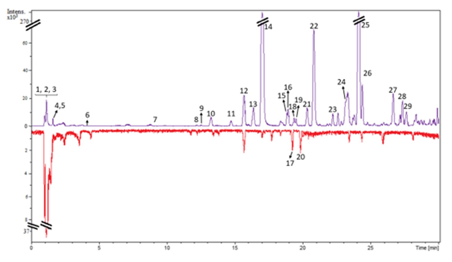
The constituent annotation of the stem bark and fruit teas (aqueous extracts) was performed based on the UFLC retention times of the peaks, accurate mass spectrometry (MS) for putative molecular formula determination, fragmentation data (MS/MS), ultraviolet (UV) spectra, and comparisons with literature data and authentic standards. Authentic standards of protocatechuic acid (6), catechin (7), and epicatechin (9) (Cordeiro et al., 2016; Costa Junior et al., 2020) were utilized to interpret the chromatograms and high-resolution mass spectra of the secondary metabolites present in the extracts. A total of 29 peaks were identified in the Guazuma ulmifolia extracts, comprising flavonoids, coumarins, and organic acids (Figure 2, Table 1).

Base Peak Chromatogram in positive ionization mode of the aqueous extracts of Guazuma ulmifolia stem bark (purple) and fruits (red).
Compounds annotated from stem bark (A) and fruit (B) ethanol extracts of Guazuma ulmifolia by LC-DAD-MS.
Peaks 2-4 did not show absorption in the UV region, revealing deprotonated ions at m/z 341.1092, 195.0504, and 191.0215, which were compatible to molecular formulas C12H22O11, C6H12O7, and C6H8O7, respectively (Table 1). Then, di-O-hexoside (2), hexonic acid (3), and citric acid (4) were putatively identified (Machado et al., 2021). Peak 6 was confirmed as being protocatechuic acid by analysis and comparison of its RT, UV, and MS data with those obtained by coinjection of an authentic sample and by the literature data (Costa Junior et al., 2020).
Flavonoids annotated in Guazuma ulmifolia extracts were flavanols (λmax 280-283; 7-9 and 11) and a flavonol (λmax 271 and 342 nm; 15), as determined by the
compatible UV spectra. The flavonols 7 and 9 were confirmed by coinjection of standards, and the data were also in accordance with the above-reported literature (Costa Junior et al., 2020; Cordeiro et al., 2016). Peaks 8 and 11 were tentatively annotated as the di- and trimeric forms of procyanidin already reported in previous studies with this plant species (Santos et al., 2018; Pereira et al., 2020). Peak 15 exhibited UV and fragmentation pathway characteristics to C-glycosylated flavonoids, showing consecutive losses of water molecules and parts of glycoside. Thus, C-hexoside was suggested by the losses of 120 and 90u, respectively, and aglycone was suggested as luteolin (Karar and Kuhnert, 2015).
Peaks 17-20 were tentatively annotated as belonging to the lignan class based on their MF and UV spectra (Mirowski et al., 2022). Coumarins were annotated for peaks 10, 12, 14, 21, 22, and 25. Generally, in the mass spectrum, coumarins display successive losses of CH3 (15u) and CO (28u) groups, which were the characteristic product ions for methoxylated coumarins. Scopoletin (12) and Scoparone (22), which differ only by the presence of a methyl group, and Fraxetin (10), which differs by the addition of oxygenation, were easily identified by their UV data and characteristic fragmentation (Herz et al., 1970; Goulart et al., 1993). Fraxidin (21) and isofraxidin
(14) isomers were tentatively differentiated by their characteristic UV, as reported by Li et al. (2011). Peak 25 was annotated as 6,7,8-trimethoxycoumarin based on its characteristic fragmentation (losses of 3xCH3, CO) and also on its UV data, which are compatible with the literature (Preat et al., 2005) (Figure 2).
3.2. Levels of phenolic compounds and antioxidant activity
The concentrations of phenolic compounds, expressed in mg of gallic acid equivalents per gram of dry extract (mg GAE.g−1), are summarized in Table 2. In
Contents of total phenolic compounds (expressed in gallic acid equivalents-GAE) of various extracts.
Guazuma ulmifolia fruit extracts, phenolic content ranged from 6.12 to 13.00 mg GAE·g−1, whereas stem bark extracts exhibited significantly higher values, ranging from
98.63 to 108.32 mg GAE·g−1. These values were comparable to those of Rosmarinus
officinalis (rosemary) leaf extract, included as a polyphenol-rich reference, with concentrations between 79.55 and 115.44 mg GAE·g−1. Ultrasound-assisted extraction using hydroethanol and hydroacetone solvents was more efficient than aqueous decoction, as evidenced by higher GAE concentrations in the resulting extracts (Table 2).
The antioxidant activity of the extracts was evaluated using the DPPH radical scavenging method, and results were expressed as IC50 values (concentration required to reduce 50% of DPPH). Lower IC50 values indicate higher antioxidant capacity. The IC50
values for Guazuma ulmifolia fruits extracts ranged from 17.89 to 28.06 μg·mL−1, while
for the stem bark extracts, they ranged from 3.97 to 7.45 μg·mL−1 (Table 3). Among extraction methods, hydroacetone extracts exhibited the highest antioxidant activity. Stem bark extracts showed superior antioxidant performance compared to rosemary, with IC50 values between 5.24 and 8.13 μg·mL−1.
Antioxidant activity (expressed in µg.mL-1 of extract) of various extracts capable of causing 50% inhibition (IC50).
3.3. Centesimal composition of the prepared diets
The centesimal composition of the diets prepared indicated a higher energy content in the high-fat diet due to the addition of 195g/kg of lard, with the aiming to induce obesity in the animals. The values for the standard diet and high-fat diet are described in Table 4.
Proximate composition of the standard and high-fat experimental diets, expressed in g.100g-1 of full sample.
3.4. Biological assay
3.4.1. Food intake and weight gain
At the end of the 10 weeks for obesity induction, it was noted that the body weight of the animals that received a high-fat diet differed statistically from the animals that received a standard diet (p<0.01), validating the diet-induced obesity model. When starting treatments with the different extracts, it was observed that among the groups that received a high-fat diet throughout the study (G2, G3 and G4), the group that received Guazuma ulmifolia stem bark extract (G4) obtained less weight gain than the other groups. Furthermore, at the end of the experimental period, G1 maintained a difference in weight gain only in relation to G2 and G3 and presented a weight variation close to that of G4, reaffirming that the stem bark extract contributed to the lower weight gain (Table 5).
Mean initial and final body weight, weight gain and daily food consumption of the groups given the standard diet (SD) and the high-fat diet (HD) during the 2nd experimental period.
Regarding feed consumption, there was no statistically significant difference between the different groups in the first experimental period since the animals in the SD group ate an average of 14.68±3.56 g/day, while the animals in the HD group ate 14.03±2.64 g/day (data not shown). During the second experimental period, the difference in weight gain between the groups can be explained by the amount of feed ingested (Table 5).
The animals that received the standard diet kept their average consumption close to the previous period (14.76 g/day per animal or 52.71 kcal/day). In contrast, G4, which received an aqueous extract of Guazuma ulmifolia stem bark, consumed the least amount of feed compared to the other animals given the high-fat diet, with an average of 13.96 g/day or 62.90 kcal/day per animal (Table 5).
3.4.2. Serum parameters
At the end of the first experimental period, significant differences were observed in the serum parameters of total cholesterol, HDL-c, and LDL-c, when comparing the group submitted with the standard diet with the groups submitted to the hyperlipid diet, as shown in Table 6.
Serum parameters of the groups subjected to standard diet (SD) and hyperlipid diet (HD) at the end of the 1nd experimental period.
The results obtained at the end of the second period are described in Table 7. The serum glucose concentration exhibited no significant difference between the groups of animals. As for the lipid profile at the end of the study, the average total cholesterol concentration of the animals in group G4 did not differ statistically from groups G1 and G2. In contrast, the G3 group presented a significantly higher value (p< 0.05) than groups G1 and G4. With regard to triglyceride levels, the lowest value was found for the G4 group, which differed statistically (p< 0.01) when compared to the G2 control group, indicating that the intervention with the stem bark extract had a positive effect on the lipid profile of the obese animals. Ther was no statistically significant difference between the groups for the mean fractions of HDL-c and LDL-c, differing only in the levels of VLDL-c.
Serum parameters of the groups subjected to standard diet (SD) and hyperlipid diet (HD) at the end of the 2nd experimental period.
4. Discussion
In Guazuma ulmifolia fruit extracts, concentrations ranged from 6.12 to 13.00 mg GAE·g−1, while stem bark extracts exhibited significantly higher levels, ranging from 98.63 to 108.32 mg GAE·g−1. These values were comparable to those of Rosmarinus officinalis (rosemary) leaf extract (79.55 to 115.44 mg GAE·g−1), used as a polyphenol-rich reference. The antioxidant activity, evaluated using the DPPH radical scavenging assay, revealed IC50 values of 17.89 to 28.06 μg·mL−1 for Guazuma ulmifolia fruit extracts and 3.97 to 7.45 μg·mL−1 for stem bark extracts, with acetone-based extractions being the most efficient for both parts of the plant. Stem bark extracts outperformed rosemary in antioxidant capacity (IC50: 5.24 to 8.13 μg·mL−1).
A positive correlation was observed between phenolic content and antioxidant activity, as extracts with higher phenolic concentrations exhibited lower IC50 values, except for the hydroethanolic fruit extract. However, this correlation is not absolute, as antioxidant efficacy is influenced by other phytochemicals and the chemical structure of active compounds, particularly the number and position of hydroxyl groups on polyphenol molecules. The solvent system also plays a critical role, with genetic, environmental, and plant maturation factors further affecting phytochemical content (Asolini et al., 2006; Melo et al., 2008). While ultrasound-assisted extraction with organic solvents was more efficient in phenolic recovery, the aqueous decoction method was chosen for anti-obesity assays due to its alignment with the traditional use of the plant, commonly consumed as a tea for this purpose (Gómez-Estrada et al., 2011).
With regard to the phytochemical constituents in this study, several compounds previously described in the literature were found in the extracts of the fruit and stem bark of Guazuma ulmifolia, including procyanidin, rutin, epicatechin, and gallic acid (Pereira et al., 2019) (see Table 1). These compounds are mainly recognized for their antioxidant activity, reducing oxidative stress through direct or indirect action, such as eliminating free radicals, chelating metal ions, improving the activity of antioxidant enzymes (Patanè et al., 2023), inhibiting oxidases (such as xanthine oxidase (XO), cyclooxygenase (COX), lipoxygenase, and phosphoinositide 3-kinase (PI3K)), and reducing α-tocopherol radicals (Shen et al., 2022).
The strong antioxidant activity of Guazuma ulmifolia stem bark may be attributed to polymeric proanthocyanidins, consisting of epicatechin and catechin units, as previously described in the literature. Beyond their antioxidant properties, these compounds, particularly procyanidin B2, offer pharmacological benefits, including anti-tumor activity, LDL-c oxidation reduction, and hair growth promotion, though their detailed mechanisms remain to be clarified.
Diseases and symptoms that are correlated with obesity, including diabetes (Aouacheri et al., 2015), inflammation (Biswas, 2016) and cardiovascular diseases (Csányi and Miller Junior, 2014), are often associated with oxidative stress, which is a condition of imbalance between the amount of reactive species and the inefficient activity of an organism's antioxidant protection system (Maurya et al., 2016). Therefore, the amount of phenolic compounds can directly influence the biological potential of natural products used for therapeutic purposes, as these compounds have antioxidant properties capable of maintaining the balance of oxidation and reduction (REDOX) and protecting cells against damage caused by excess reactive oxygen species (ROS) (Santos et al., 2018).
In this study, we exposed the rats to a high-fat diet for 10 weeks to induce obesity. After this period, the results indicated a significant increase in the weight of the animals on the high-fat diet (HD) compared to the group on the standard diet (SD). The weight gain in the group occurred without altering the consumption pattern since, in the first period, the animals in the SD group ate an average of 14.68 g (± 3.56) of feed per day, while the HD group consumed 14.03 g (± 2.64). These results are in agreement with Duarte et al. (2006), who found that even without altering the pattern of consumption, the high-lipid diet significantly increased body weight gain in rats, promoting obesity, a fact possibly due to the greater energy efficiency of the high-lipid diet. In the second period, group G4 showed the lowest average weight gain due to lower feed consumption than the other high-fat groups, with an average of 13.96 g/day, which is statistically comparable to G1, who received a standard diet. This dietary
pattern contrasts with that observed pared to those given a standard diet and is described in literature (Hariri and Thibault, 2010; Uecker et al., 2019).
In relation to the serum lipid profile, there were no significant effects of the aqueous extract of the fruit, but a decrease in the levels of total cholesterol, triglycerides, and VLDL-c was observed in the group that received the aqueous extract of the stem bark, with the result remaining similar to that for the control group fed a standard diet. Eating a high-fat diet leads to changes in particle size, numbers, and plasma levels of VLDL-c, LDL-c and HDL-c (ALNohair, 2014), resulting in conditions for the development of atherosclerosis, which is considered one of the main causes of cardiovascular disease as its development and progression is linked to abnormal lipid metabolism and inflammatory infiltration of the arterial wall (Frostegard, 2013). In this sense, Patenè and collaborators (2023) indicate that the use of flavonoids, such as catechins, increased HDL-c values, and improved cardiovascular flow by modulating endothelial nitric oxide synthesis (eNOS), which leads to increased nitric oxide production. In turn, this improves vascular relaxation by reducing LDL-c, cholesterol, and triglyceride values, which contribute to the accumulation of plaques in the arteries.
The glycemia of obese animals treated with the aqueous extract of the stem bark of Guazuma ulmifolia was lower when compared to those given the aqueous extract of the fruit of the same plant species, although there was no statistical difference. As Guazuma ulmifolia is widely used in Mexico to treat DM2, the anti-diabetic mechanisms of the aqueous stem bark extract (EAC) were studied, and it was found that it exerts its anti-diabetic properties by stimulating the influx of glucose into adipocytes, both those responsive and resistant to the effects of insulin, without affecting the development of adipose tissue. The ability of EAC to induce glucose influx in insulin-resistant adipocytes, in addition to its absence of pro-adipogenic or
antiadipogenic effects, suggests that EAC could be likely to be beneficial useful in the treatment of DM2 (Alonso-Castro and Salazar-Olivo, 2008), although our study did not find such a benefit, which may be related to the dose of the extract or the time of administration.
Flavonoids have a beneficial antidiabetic effect and help offset the negative effects caused by insulin resistance (Patanè et al., 2023). Catechins reduce plasma glucose levels by improving the signal transduction pathway that promotes glucose transport in cells via the glucose transporter-4 (GLUT4) (Snoussi et al., 2014). In particular, epigallocatechin-gallate promotes the externalization of GLUT4 via the PI3K/AKT pathway (De Los Santos et al., 2017; Xu et al., 2019). In addition, proanthocyanidins (PCAs) have been shown to contribute to the regulation of glucose homeostasis by inhibiting intestinal glucose absorption through a direct action on GLUT 1 and 2, the main glucose transporters in enterocytes (Zhong et al., 2018). Overall, all these actions contribute to improving insulin resistance and limiting the risk of hyperglycemia related to diabetic disease (Patanè et al., 2023).
5. Conclusions
The chemical study of the extracts of the fruit and stem bark of Guazuma ulmifolia revealed a total of 29 compounds, including flavonoids, coumarins, and organic acids, with recognized antioxidant and anti-inflammatory activity. In addition, the results showed high concentrations of phenolic compounds in the aqueous extract of the stem bark, justifying the greater antioxidant capacity of this extract. In our in vivo experimental design with a high-fat diet, the aqueous extract of the stem bark promoted a reduction in food intake as well as lower body weight gain in the animals and a partial improvement in serum parameters related to obesity, such as better serum levels of total cholesterol, VLDL-c, and triglycerides. However, more studies are needed to elucidate the mechanisms of action of the pharmacological activities evidenced, relating to the compounds found from the chemical study, as well as to compare them with other experimental models of obesity.
Acknowledgements
The authors acknowledge the Graduate Program in Health and Development in the Central-West Region of Brazil, Federal University of Mato Grosso do Sul-UFMS, Coordination for the Improvement of Higher Education Personnel for support. This study was financed in part by the Coordination for the Improvement of Higher Education Personnel, Brazil (CAPES)-Finance Code 001.
-
Data Availability Statement
The entire data set that supports the results of this study was published in the article itself.
References
-
AGOSTINI-COSTA, T.S., LIMA, A. and LIMA, M.V., 2003. Determination of tannin in cashew apple peduncle: vanillin method versus acid butanol method. Química Nova, vol. 26, no. 5, pp. 763-765. https://doi.org/10.1590/S0100-40422003000500022
» https://doi.org/10.1590/S0100-40422003000500022 -
AL MUQARRABUN, L.M.R. and AHMAT, N., 2015. Medicinal uses, phytochemistry and pharmacology of family Sterculiaceae: A review. European Journal of Medicinal Chemistry, vol. 92, pp. 514-530. https://doi.org/10.1016/j.ejmech.2015.01.026 PMid:25599949.
» https://doi.org/10.1016/j.ejmech.2015.01.026 -
ALNOHAIR, S., 2014. Obesity in gulf countries. International Journal of Health Sciences, vol. 8, no. 1, pp. 79-83. https://doi.org/10.12816/0006074 PMid:24899882.
» https://doi.org/10.12816/0006074 -
ALONSO-CASTRO, A.J. and SALAZAR-OLIVO, L.A., 2008. The anti-diabetic properties of Guazuma ulmifolia Lam. are mediated by the stimulation of glucose uptake in normal and diabetic adipocytes without inducing adipogenesis. Journal of Ethnopharmacology, vol. 118, no. 2, pp. 252-256. https://doi.org/10.1016/j.jep.2008.04.007 PMid:18487028.
» https://doi.org/10.1016/j.jep.2008.04.007 -
AOUACHERI, O., SAKA, S., KRIM, M., MESSAADIA, A. and MAIDI, I., 2015. The investigation of the oxidative stress-related parameters in type 2 diabetes mellitus. Canadian Journal of Diabetes, vol. 39, no. 1, pp. 44-49. https://doi.org/10.1016/j.jcjd.2014.03.002 PMid:25065473.
» https://doi.org/10.1016/j.jcjd.2014.03.002 -
ARÇARI, D.P., BARTCHEWSKY, W., DOS SANTOS, T.W., OLIVEIRA, K.A., FUNCK, A., PEDRAZZOLI, J., DE SOUZA, M.F., SAAD, M.J., BASTOS, D.H., GAMBERO, A., CARVALHO, P.O. and RIBEIRO, M.L., 2009. Antiobesity effects of yerba maté extract (Ilex paraguariensis) in high‐fat diet–induced obese mice. Obesity, vol. 17, no. 12, pp. 2127-2133. https://doi.org/10.1038/oby.2009.158 PMid:19444227.
» https://doi.org/10.1038/oby.2009.158 - ASOLINI, F.C., TEDESCO, A.M., CARPES, S.T., FERRAZ, C. and ALENCAR, S.M., 2006. Antioxidant and antibacterial activities of phenolic compounds from extracts of plants used as tea. Brazilian Journal of Food Technology, vol. 9, pp. 209-215.
- BISWAS, S.K., 2016. Does the interdependence between oxidative stress and inflammation explain the antioxidant paradox? Oxidative Medicine and Cellular Longevity, vol. 2016, pp. 5698931.
- BRASIL. Ministério da Saúde. Agência Nacional de Vigilância Sanitária – ANVISA, 2005. Physical-Chemical Methods for Food Analysis Brasília: Ministério da Saúde, 1018 p. [In portuguese].
-
BRASIL. Agência Nacional de Vigilância Sanitária – ANVISA, 2021 [viewed 18 May 2025]. Phytotherapeutic formulary of the Brazilian Pharmacopoeia [online]. Brasília: Anvisa, 2018 p. [In portuguese]. Available from: https://www.gov.br/anvisa/pt-br
» https://www.gov.br/anvisa/pt-br -
CALIXTO JÚNIOR, J.T., MORAIS, S.M., GOMEZ, C.V., MOLAS, C.C., ROLON, M., BOLIGON, A.A., ATHAYDE, M.L., OLIVEIRA, C.D.M., TINTINO, S.R. and DOUGLAS, M.C.H., 2016. Phenolic composition and antiparasitic activity of plants from the Brazilian Northeast “Cerrado”. Saudi Journal of Biological Sciences, vol. 23, no. 3, pp. 434-440. https://doi.org/10.1016/j.sjbs.2015.10.009 PMid:27081371.
» https://doi.org/10.1016/j.sjbs.2015.10.009 -
CORDEIRO, K.W., FELIPE, J.L., MALANGE, K.F., PRADO, P.R., FIGUEIREDO, P.O., GARCEZ, F.R., FREITAS, K.C., GARCEZ, W.S. and TOFFOLI-KADRI, M.C., 2016. Anti-inflammatory and antinociceptive activities of Croton urucurana Baillon bark. Journal of Ethnopharmacology, vol. 183, pp. 128-135. https://doi.org/10.1016/j.jep.2016.02.051 PMid:26944237.
» https://doi.org/10.1016/j.jep.2016.02.051 -
COSTA JUNIOR, A.G., YOSHIDA, N.C., GARCEZ, W.S., PERDOMO, R.T., MATOS, M.F.C. and GARCEZ, F.R., 2020. Metabolomics approach expands the classification of propolis samples from midwest Brazil. Journal of Natural Products, vol. 83, no. 2, pp. 333-343. https://doi.org/10.1021/acs.jnatprod.9b00783 PMid:32031802.
» https://doi.org/10.1021/acs.jnatprod.9b00783 -
CSÁNYI, G. and MILLER JUNIOR, F.J., 2014. Oxidative stress in cardiovascular disease. International Journal of Molecular Sciences, vol. 15, no. 4, pp. 6002-6008. https://doi.org/10.3390/ijms15046002 PMid:24722571.
» https://doi.org/10.3390/ijms15046002 -
DE LOS SANTOS, S., GARCÍA-PÉREZ, V., HERNÁNDEZ-RESÉNDIZ, S., PALMA-FLORES, C., GONZÁLEZ-GUTIÉRREZ, C.J., ZAZUETA, C., CANTO, P. and CORAL-VÁZQUEZ, R.M., 2017. (-)-Epicatechin induces physiological cardiac growth by activation of the PI3K/Akt pathway in mice. Molecular Nutrition & Food Research, vol. 61, no. 2, pp. 9767-9781. https://doi.org/10.1002/mnfr.201600343 PMid:27605464.
» https://doi.org/10.1002/mnfr.201600343 -
DUARTE, A.C.G.O., FONSECA, D.F., MANZONI, M.S.J., SOAVE, C.F., SENE-FIORESE, M., DÂMASO, A.R. and CHEIK, N.C., 2006. High-fat diet and secretory capacity of insulin in rats. Revista de Nutrição, vol. 19, pp. 341-348. https://doi.org/10.1590/S1415-52732006000300005
» https://doi.org/10.1590/S1415-52732006000300005 -
FROSTEGARD, J., 2013. Immunity, atherosclerosis and cardiovascular disease. BMC Medicine, vol. 11, no. 1, pp. 117. https://doi.org/10.1186/1741-7015-11-117 PMid:23635324.
» https://doi.org/10.1186/1741-7015-11-117 -
GÓMEZ-ESTRADA, H., DÍAZ-CASTILLO, F., FRANCO-OSPINA, L., MERCADO-CAMARGO, J., GUZMÁN-LEDEZMA, J., MEDINA, J.D. and GAITÁN-IBARRA, R., 2011. Folk medicine in the northern coast of Colombia: an overview. Journal of Ethnobiology and Ethnomedicine, vol. 7, no. 1, pp. 27. https://doi.org/10.1186/1746-4269-7-27 PMid:21939522.
» https://doi.org/10.1186/1746-4269-7-27 - GOULART, M.O., SANT’ANA, A.E.G., DE LIMA, R.A., CAVALCANTE, S.H., CARVALHO, M.G. and BRAZ FILHO, R., 1993. Fitoconstituintes químicos isolados de Jatrophaelliptica atribuição dos deslocamentos químicos dos átomos de carbono e hidrogênio dos diterpenos jatrofolonas A e B. Química Nova, vol. 16, no. 2, pp. 95-100.
-
HARIRI, N. and THIBAULT, L., 2010. High-fat diet-induced obesity in animal models. Nutrition Research Reviews, vol. 23, no. 2, pp. 270-299. https://doi.org/10.1017/S0954422410000168 PMid:20977819.
» https://doi.org/10.1017/S0954422410000168 -
HERZ, W., BHAT, S.V. and SANTHANAM, P.S., 1970. Coumarins of Artemisia dracunculoides and 3′,6-dimethoxy-4′,5,7-trihydroxyflavone in A. arctica. Phytochemistry, vol. 9, no. 4, pp. 891-894. https://doi.org/10.1016/S0031-9422(00)85199-7
» https://doi.org/10.1016/S0031-9422(00)85199-7 -
HÖR, M., RIMPLER, H. and HEIMRICH, M., 1996. Proanthocyanidin polymers with antisecretory activity and proanthocyanidin oligomers from Guazuma ulmifolia bark. Phytochemistry, vol. 42, no. 1, pp. 109-119. https://doi.org/10.1016/0031-9422(95)00855-1
» https://doi.org/10.1016/0031-9422(95)00855-1 - HRAPKIEWICZ, K., MEDINA, L. and HOLMES, D.D., 1997. Clinical Laboratory Animal Medicine: An Introduction 2nd ed. Ames: Iowa State University Press, 277 p.
- KARAR, M.G.E. and KUHNERT, N., 2015. UPLC-ESI-Q-TOF-MS/MS Caracterization of phenolics from Crataegusmonogyna and Cratee-guslaevigata (Hawthorn) leaves, fruits and their herbal derived drops (Crataegutt Tropfen). Journal of Chemical Biology & Therapeutics, vol. 1, pp. 102.
-
LI, X., ZHANG, Y., ZENG, X., YANG, L. and DENG, Y., 2011. Chemical profiling of bioactive constituents in Sarcandraglabra and its preparations using ultra-high-pressure liquid chromatography coupled with LTQ Orbitrap mass spectrometry. Rapid Communications in Mass Spectrometry : RCM, vol. 25, no. 17, pp. 2439-2447. https://doi.org/10.1002/rcm.5123 PMid:21818803.
» https://doi.org/10.1002/rcm.5123 -
LOPES, G.C., LONGHINI, R., DOS SANTOS, P.V.P., ARAÚJO, A.A.S., BRUSCHI, M.L. and MELLO, J.C.P., 2012. Preliminary assessment of the chemical stability of dried extracts from Guazuma ulmifolia Lam. (Sterculiaceae). International Journal of Analytical Chemistry, vol. 2012, pp. 508945. https://doi.org/10.1155/2012/508945 PMid:22291706.
» https://doi.org/10.1155/2012/508945 -
LOPES, G.C., ROCHA, J.C.B., ALMEIDA, G.C. and MELLO, J.C.P., 2009. Condensed tannins from the bark of Guazuma ulmiflia Lam. (Sterculiaceae). Journal of the Brazilian Chemical Society, vol. 20, no. 6, pp. 1103-1109. https://doi.org/10.1590/S0103-50532009000600016
» https://doi.org/10.1590/S0103-50532009000600016 -
MACHADO, C.D., KLIDER, L.M., TIRLONI, C.A.S., MARQUES, A.A.M., LORENÇONE, B.R., BATISTA, L.P., ROMÃO, P.V.M., PALOZI, R.A.C., GUARNIER, L.P., SOUZA, R.I.C., DOS SANTOS, A.C., SILVA, D.B., RAMAN, V., GASPAROTTO JÚNIOR, A. and BUDEL, J.M., 2021. Ethnopharmacological investigations of the leaves of Cecropia pachystachya Trécul (Urticaceae): a native Brazilian tree species. Journal of Ethnopharmacology, vol. 270, pp. 113740. https://doi.org/10.1016/j.jep.2020.113740 PMid:33388429.
» https://doi.org/10.1016/j.jep.2020.113740 -
MAGALHÃES, P.K.A., ARAUJO, E.N., SANTOS, A.M., VANDERLEI, M.B., SOUZA, C.C.L., CORREIA, M.S., FONSECA, S.A., PAVÃO, J.M.J.S., SOUZA, M.A., COSTA, J.G., SANTOS, A.F. and MATOS-ROCHA, T.J., 2022. Ethnobotanical and ethnopharmacological study of medicinal plants used by a traditional community in Brazil’s northeastern. Brazilian Journal of Biology = Revista Brasileira de Biologia, vol. 82, pp. e237642. https://doi.org/10.1590/1519-6984.237642 PMid:34105672.
» https://doi.org/10.1590/1519-6984.237642 -
MAGOS, G.A., MATEOS, J.C., PÁEZ, E., FERNÁNDEZ, G., LOBATO, C., MÁRQUEZ, C. and ENRÍQUEZ, R.G., 2008. Hypotensive and vasorelaxant effects of the procyanidin fraction from Guazuma ulmifolia bark in normotensive and hypertensive rats. Journal of Ethnopharmacology, vol. 117, no. 1, pp. 58-68. https://doi.org/10.1016/j.jep.2008.01.015 PMid:18314282.
» https://doi.org/10.1016/j.jep.2008.01.015 -
MAURYA, P.K., NOTO, C., RIZZO, L.B., RIOS, A.C., NUNES, S.O.V., BARBOSA, D.S., SETHI, S., ZENI, M., MANSUR, R.B., MAES, M. and BRIETZKE, E., 2016. The role of oxidative and nitrosative stress in accelerated aging and major depressive disorder. Progress in Neuro-Psychopharmacology & Biological Psychiatry, vol. 65, pp. 134-144. https://doi.org/10.1016/j.pnpbp.2015.08.016 PMid:26348786.
» https://doi.org/10.1016/j.pnpbp.2015.08.016 -
MELO, E.A., MACIEL, M.I.S., LIMA, V.L.A.G. and NASCIMENTO, R.J., 2008. Antioxidant capacity of the fruits. Revista Brasileira de Ciências Farmacêuticas, vol. 44, no. 2, pp. 193-201. https://doi.org/10.1590/S1516-93322008000200005
» https://doi.org/10.1590/S1516-93322008000200005 -
MILIAUSKAS, G., VENSKUTONIS, P.R. and VAN BEEK, T.A., 2004. Screening of radical scavenging activity of some medicinal and aromatic plant extracts. Food Chemistry, vol. 85, no. 2, pp. 231-237. https://doi.org/10.1016/j.foodchem.2003.05.007
» https://doi.org/10.1016/j.foodchem.2003.05.007 -
MIROWSKI, P.S., OJEDA, M., KOLLET, L.G., FREIRE, T.V., POTT, A., GARCEZ, W.S., PERDOMO, R.T. and GARCEZ, F.R., 2022. Selective tumor cell-growth inhibition by lignans and a seco-triterpenoid from Combretum mellifluum. Natural Product Research, vol. 36, no. 24, pp. 6224-6231. https://doi.org/10.1080/14786419.2021.2024823 PMid:35007163.
» https://doi.org/10.1080/14786419.2021.2024823 -
NAVARRO, V., VILLARREAL, M.L., ROJAS, G. and LOZOYA, X., 1996. Antimicrobial evaluation of some plants used in Mexican traditional medicine for the treatment of infectious diseases. Journal of Ethnopharmacology, vol. 53, no. 3, pp. 143-147. https://doi.org/10.1016/0378-8741(96)01429-8 PMid:8887021.
» https://doi.org/10.1016/0378-8741(96)01429-8 -
PAN, X., NIU, G. and LIU, H., 2003. Microwave-assisted extraction of tea polyphenols and tea caffeine from green tea leaves. Chemical Engineering and Processing, vol. 42, no. 2, pp. 129-133. https://doi.org/10.1016/S0255-2701(02)00037-5
» https://doi.org/10.1016/S0255-2701(02)00037-5 -
PANG, J., CHOI, T. and PARK, T., 2008. Ilex paraguariensis extract ameliorates obesity induced by high-fat diet: potential role of AMPK in the visceral adipose tissue. Archives of Biochemistry and Biophysics, vol. 476, no. 2, pp. 178-185. https://doi.org/10.1016/j.abb.2008.02.019 PMid:18314006.
» https://doi.org/10.1016/j.abb.2008.02.019 -
PATANÈ, G.T., PUTAGGIO, S., TELLONE, E., BARRECA, D., FICARRA, S., MAFFEI, C., CALDERARO, A. and LAGANÀ, G., 2023. Catechins and proanthocyanidins involvement in metabolic syndrome. International Journal of Molecular Sciences, vol. 24, no. 11, pp. 9228. https://doi.org/10.3390/ijms24119228 PMid:37298181.
» https://doi.org/10.3390/ijms24119228 -
PEREIRA, G.A., ARAÚJO, N.M.P., ARRUDA, H.S., FARIAS, D.P., MOLINA, G. and PASTORE, G.M., 2019. Phytochemicals and biological activities of mutamba (Guazuma ulmifolia Lam.): A review. Food Research International, vol. 126, pp. 108713. https://doi.org/10.1016/j.foodres.2019.108713 PMid:31732089.
» https://doi.org/10.1016/j.foodres.2019.108713 -
PEREIRA, G.A., ARRUDA, H.S., DE MORAIS, D.R., ARAUJO, N.M.P. and PASTORE, G.M., 2020. Mutamba (Guazuma ulmifolia Lam.) fruit as a novel source of dietary fibre and phenolic compounds. Food Chemistry, vol. 310, pp. 125857. https://doi.org/10.1016/j.foodchem.2019.125857 PMid:31787395.
» https://doi.org/10.1016/j.foodchem.2019.125857 -
PREAT, J., JACQUEMIN, D. and PERPÈTE, E.A., 2005. Theoretical investigations of the UV spectra of coumarin derivatives. Chemical Physics Letters, vol. 415, no. 1-3, pp. 20-24. https://doi.org/10.1016/j.cplett.2005.08.135
» https://doi.org/10.1016/j.cplett.2005.08.135 -
REEVES, P.G., NIELSEN, F.H. and FAHEY JUNIOR, G.C.J., 1993. AIN-93 purified diets for laboratory rodents: final report of the american institute of nutrition Ad Hoc writing committee on the formulation of the AIN-76A rodent diet. The Journal of Nutrition, vol. 123, no. 11, pp. 1939-1951. https://doi.org/10.1093/jn/123.11.1939 PMid:8229312.
» https://doi.org/10.1093/jn/123.11.1939 -
RODRIGUES, E.L., SANTANA, L.F., NASCIMENTO, V.A., ARAKAKI, M.A., CARDOSO, C.A.L., FILIÚ, W.F.O., GUIMARÃES, R.C.A., HIANE, P.A. and FREITAS, K.C., 2024. Use of Guazuma ulmifolia Lam. stem bark extracts to prevent high-fat diet induced metabolic disorders in mice. International Journal of Molecular Sciences, vol. 25, no. 16, pp. 8889. https://doi.org/10.3390/ijms25168889 PMid:39201576.
» https://doi.org/10.3390/ijms25168889 -
ROESLER, R., MALTA, L.G., CARRASCO, L.C., HOLANDA, R.B., SOUSA, C.A.S. and PASTORE, G.M., 2007. Antioxidant activity of cerrado fruits. Food Science and Technology, vol. 27, pp. 53-60. https://doi.org/10.1590/S0101-20612007000100010
» https://doi.org/10.1590/S0101-20612007000100010 -
SANTOS, J.M., ALFREDO, T.M., ANTUNES, K.A., CUNHA, J.S.M., COSTA, E.M.A., LIMA, E.S., SILVA, D.B., CAROLLO, C.A., SCHMITZ, W.O., BOLETI, A.P.A., SANTOS, E.L. and SOUZA, K.P., 2018. Guazuma ulmifolia Lam. decreases oxidative stress in blood cells and prevents doxorubicin-induced cardiotoxicity. Oxidative Medicine and Cellular Longevity, vol. 2018, no. 1, pp. 2935051. https://doi.org/10.1155/2018/2935051 PMid:30050650.
» https://doi.org/10.1155/2018/2935051 -
SHEN, N., WANG, T., GAN, Q., LIU, S., WANG, L. and JIN, B., 2022. Plant flavonoids: classification, distribution, biosynthesis, and antioxidant activity. Food Chemistry, vol. 383, pp. 132531. https://doi.org/10.1016/j.foodchem.2022.132531 PMid:35413752.
» https://doi.org/10.1016/j.foodchem.2022.132531 -
SNOUSSI, C., DUCROC, R., HAMDAOUI, M.H., DHAOUADI, K., ABAIDI, H., CLUZEAUD, F., NAZARET, C., GALL, M.L. and BADO, A., 2014. Green tea decoction improves glucose tolerance and reduces weight gain of rats fed normal and high-fat diet. The Journal of Nutritional Biochemistry, vol. 25, no. 5, pp. 557-564. https://doi.org/10.1016/j.jnutbio.2014.01.006 PMid:24656388.
» https://doi.org/10.1016/j.jnutbio.2014.01.006 -
TENE, V., MALAGÓN, O., FINZI, P.V., VIDARI, G., ARMIJOS, C. and ZARAGOZA, T., 2007. An ethnobotanical survey of medicinal plants used in Loja and Zamora-Chinchipe, Ecuador. Journal of Ethnopharmacology, vol. 111, no. 1, pp. 63-81. https://doi.org/10.1016/j.jep.2006.10.032 PMid:17137737.
» https://doi.org/10.1016/j.jep.2006.10.032 -
THAIPITAKWONG, T., NUMHOM, S. and ARAMWIT, P., 2018. Mulberry leaves and their potential effects against cardiometabolic risks: a review of chemical compositions, biological properties and clinical efficacy. Pharmaceutical Biology, vol. 56, no. 1, pp. 109-118. https://doi.org/10.1080/13880209.2018.1424210 PMid:29347857.
» https://doi.org/10.1080/13880209.2018.1424210 -
UECKER, J.N., SCHNEIDER, J.P., CERQUEIRA, J.H., RINCÓN, J.A.A., CAMPOS, F.T., SCHNEIDER, A., BARROS, C.C., ANDREAZZA, R., JASKULSKI, I.B. and PIENIZ, S., 2019. Ilex paraguariensis extract prevents body weight gain in rats fed a high-fat diet. Food Science and Technology, vol. 39, no. 3, pp. 620-626. https://doi.org/10.1590/fst.39817
» https://doi.org/10.1590/fst.39817 -
WORLD HEALTH ORGANIZATION – WHO, 2024 [viewed 18 Sept 2025]. Noncommunicable diseases [online]. Available from: https://www.who.int/news-room/fact-sheets/detail/noncommunicable-diseases?
» https://www.who.int/news-room/fact-sheets/detail/noncommunicable-diseases? -
XU, L., LI, W., CHEN, Z., GUO, Q., WANG, C., SANTHANAM, R.K. and CHEN, H., 2019. Inhibitory effect of epigallocatechin-3-O-gallate on α-glucosidase and its hypoglycemic effect via targeting PI3K/AKT signaling pathway in L6 skeletal muscle cells. International Journal of Biological Macromolecules, vol. 125, pp. 605-611. https://doi.org/10.1016/j.ijbiomac.2018.12.064 PMid:30529552.
» https://doi.org/10.1016/j.ijbiomac.2018.12.064 -
YAMAGUCHI, T., TAKAMURA, H., MATOBA, T. and TERAO, J., 1998. HPLC method for evaluation of the free radical-scavenging activity of foods by using 1,1 Diphenyl-2 picrylhydrazyl. Bioscience, Biotechnology, and Biochemistry, vol. 62, no. 6, pp. 1201-1204. https://doi.org/10.1271/bbb.62.1201 PMid:9692204.
» https://doi.org/10.1271/bbb.62.1201 -
ZHONG, H., XUE, Y., LU, X., SHAO, Q., CAO, Y., WU, Z. and CHEN, G., 2018. The effects of different degrees of procyanidin polymerization on the nutrient absorption and digestive enzyme activity in mice. Molecules, vol. 23, no. 11, pp. 2916. https://doi.org/10.3390/molecules23112916 PMid:30413083.
» https://doi.org/10.3390/molecules23112916
Edited by
-
Editor:
Ana Paula Peron
The entire data set that supports the results of this study was published in the article itself.