Bacterial bioactive groups for the improved and stable functionality of nanoparticles for biomedical application
Abstract
Across the world, metal nanoparticles (NPs) have received immense attention due to their vast array of applications in the biomedical field. This developing branch of nanotechnology, where the particles expand in nano size, is evolving for the pharmaceutical field in a more responsive manner. In recent years, green synthesis/nanotechnology has developed as a watchword for the fabrication and synthesis of metal NPs. In the present article, the perspective and progress achieved in recent years on the role of bacterial bioactive functional molecules during biosynthesis have been discussed. The cascades for NPs synthesis, manipulation of the number of biological metabolites as the reducing and stabilizing agents, like enzymes, proteins, redox mediators, and exopolysaccharides, have been presented. Therefore, the article elucidates the involvement of microbial metabolites to harmonize the biosynthetic NPs and the possible mechanism leading to biosynthesis of commonly studied and utilized metal NPs like silver, gold, zinc, copper, magnetite, palladium, etc., and their immense potential in biomedical applications.
Keywords:
bacteria; biosynthesis; nanoparticles; bioreduction; biomedical
Resumo
Em todo o mundo, as nanopartículas metálicas (NPs) têm recebido imensa atenção devido à sua vasta gama de aplicações na área biomédica. Este ramo em desenvolvimento da nanotecnologia, em que as partículas se expandem em nanoescala, está evoluindo para o campo farmacêutico de maneira mais responsiva. Nos últimos anos, a síntese verde/nanotecnologia se consolidou como uma palavra-chave para a fabricação e a síntese de NPs metálicas. Neste artigo, discutimos a perspectiva e o progresso alcançado nos últimos anos sobre o papel das moléculas funcionais bioativas bacterianas durante a biossíntese. Apresentamos as cascatas para a síntese de NPs e a manipulação do número de metabólitos biológicos como agentes redutores e estabilizantes, como enzimas, proteínas, mediadores redox e exopolissacarídeos. Portanto, o artigo elucida o envolvimento de metabólitos microbianos para harmonizar as NPs biossintéticas e o possível mecanismo que leva à biossíntese de NPs metálicas comumente estudadas e utilizadas, como prata, ouro, zinco, cobre, magnetita, paládio, etc., e seu imenso potencial em aplicações biomédicas.
Palavras-chave:
bactérias; biossíntese; nanopartículas; biorredução; biomédico
1. Introduction
The conventional methods, like chemical and physical, applied for the synthesis of NPs have imposed a burden on the environment, due to the usage of toxic and expensive chemicals to control the stability, aggregation, and crystal growth of NPs. (Bandeira et al., 2020; Bhattacharya and Mukherjee, 2008; Seo et al., 2006).
The need for environmentally friendly procedures for the synthesis of NPs is of paramount importance. The green synthesis of nanoparticles involves several types of plants, microbes, or their products, which are potentially safer than chemical and physical methods due to nontoxic reducers and mediators. Green synthesis is considered a bottom-up approach that involves biological systems for the conversion of atoms to clusters and later to nanostructures. The green process using microorganisms is more agreeable, being an environmentally benign method. (Gahlawat and Choudhury, 2019; Baruwati et al., 2009).
Among these microbes, bacteria are the most abundant form of microorganism, found in the biosphere. This biobased route is greatly explored by manipulating conditions like temperature, pH, duration, etc. The NPs synthesized by such processes maintain enhanced catalytic activity, increased surface area, and improve the metabolites and metal salts. (Gour and Jain, 2019; Juibari et al., 2011).
The main objective is the synthesis of NPs using cheap resources with controlled and uniform products. Using bacteria as a source for NPs synthesis offers an easy approach for increased biomass, uniformity of size, and easy handling, therefore a solution to the problems associated with the chemical synthesis, like stability and aggregation of particles. (Iravani and Varma, 2019).
Additionally, unlike other microbes, bacteria can be easily modelled and manipulated for genetic alteration, needed for biomineralization of metal ions. Bacteria are usually exposed to high concentrations of toxic metal ions in their environment. Even though they have emerged with several defense systems to overcome such stress factors, like efflux pumps, intracellular blockade, extracellular precipitation, etc. Such defense mechanisms of the bacteria could be expertly utilized to produce different kinds of NPs having application in various fields, including biomedical. (Faramarzi and Sadighi, 2013; Keat et al., 2015; Rehman et al., 2019).
Despite the fact that numerous studies have been reported for the biosynthesis of NPs by bacteria, however, the exact pathway leading to this reductive mechanism is still not fully known. Although the prevailing view is that the bacterial reduction and fabrication of NPs is carried out both intra- and extracellularly. During the extracellular process, the metal ions are reduced by the proteins, enzymes, and the cell wall components. Extracellular is considered a preferable method due to the easy extraction, higher efficiency, and cost-effective approach. Extracellular synthesis of gold NPs was first reported using Bacillus subtilis, having NPs attached to the cell wall, upon treatment with gold chloride solution. (Beveridge and Murray, 1980).
During the intracellular synthesis of silver NPs in the case of Pseudomonas stutzeri AG259, NADH reductase enzymes lead to electron supply and are oxidized to NADH ions. This conversion leads to the bioreduction, from silver ions to silver NPs ranging from 200nm. Pseudomonas aeruginosa was also found to have the ability to synthesize a wide variety of NPs like Ag, Pd, Fe, Rh, Co, Pt, Ni, etc., intracellularly, without the involvement of any external stabilizers or electron donors. (Srivastava and Constanti, 2012).
This article will briefly discuss some of the possible mechanisms and important factors involved in the process. Figure 1 represents the bacterial synthesis of NPs under controlled conditions for an intrinsic mechanism of synthesizing NPs from metallic salt.

Schematic representation of the steps involved in the biosynthesis of nanoparticles using bacteria.
1.1. Literature search strategy
A structured approach was followed to identify relevant literature. A comprehensive search was conducted in Scopus, Web of Science, and PubMed using keywords such as “bacterial biosynthesis of nanoparticles,” “biogenic metal nanoparticles,” “microbial nanotechnology,” “green synthesis nanoparticles,” and “bacteria-mediated nanoparticle fabrication.”
Articles published in English between 2010 and 2024 were considered. Both original articles and review papers were screened. Studies were included if they reported (i) bacterial strains used for nanoparticle biosynthesis, (ii) mechanisms of bioreduction or stabilization, or (iii) biomedical applications of biogenic nanoparticles. Papers focused purely on plant synthesis or physical/chemical synthesis were excluded. Additional references were collected through citation tracking. This approach ensured the inclusion of recent and relevant literature supporting the concepts discussed in this review.”
2. Proposed Mechanism of NPs Synthesis Through Bacteria
There are a huge number of studies that have explained the likely and potential mechanisms involved in the process of mediation of bacteria for the NPs synthesis. Nevertheless, the present knowledge about the mechanistic aspects of this process is yet to be fully elucidated. However, the biosynthesis process of metal NPs through bacteria can be explained through the involvement of proteins and reductase enzymes, electron shuttle quinones, and exopolysaccharides. (Singh et al., 2016).
2.1. Role of enzymes and proteins
Most of the conducted studies have proposed the role of proteins and enzymes as the essential biological moiety acting as a reducing agent during the synthesis of NPs. The formation of NPs has been mostly considered as a defense of bacteria against the environmental metal ion stress. Microorganisms seem to carry several defense mechanisms in retaliation to metal toxicity, like precipitation, reduction, formation of complexes, oxidation. (Tanzil et al., 2016; Fernández-Bertólez et al., 2024).
Previous reports have pointed out the potential involvement of NADH-dependent nitrate reductases in the reduction of metal ions. A study on Streptomyces sp. LK3 has shown the potential role of the enzyme nitrate reductase in the bioreduction of stable AgNPs (5nm), without a capping agent, hence providing a cost-effective and eco-friendly way of NPs synthesis (Rezaei et al., 2024; Karthik et al., 2014).
Another study using Alcaligenes faecalis also demonstrated the bioproduction of monodispersed AgNPs by the involvement of NADH and NADH-dependent reductases in the supernatant of the culture broth. (Divya et al., 2019).
Some scientists have studied the importance of surface proteins and conductive pili in electron transfer, leading to the reduction of metallic ions. Cologgi et al. (2011) conducted experiments on biomineralization of uranium ions to uranium NPs in extracellular reduction by Geobacter sulfurreducens, demonstrating the importance of pilin proteins, under pilin-inducing and noninducing experimental conditions. Additionally, pilin-supplemented strains were found to produce uranium NPs extracellularly, whereas the pili-deficient strains were found to form NPs inside the periplasmic space. (Kitching et al., 2016; Cologgi et al., 2011).
Therefore, pilin proteins were found to have a significant impact on the prevention of periplasmic location and enhanced the rate of bioreduction. In another study using Geobacter sulfurreducens at the molecular level, conducted by Vasylevskyi et al., it was concluded that the biomineralization of Ag+ ion to AgNPs is an endergonic reaction and its aggregation to Ag NPs clusters is an exergonic process leading to stable AgNPs synthesis. (Vasylevskyi et al., 2017).
2.2. Role of electron shuttle quinones (or redox mediators)
Many studies have reported and discussed the role of redox mediators and cytochromes in facilitating the synthesis of NPs. The direct interaction of metal ions and redox proteins like c-type cytochromes or redox mediators like NADH, ubiquinol, or oxygen/superoxide undergoes electron transfer during the nanoparticle synthesis. (Bewley et al., 2013; Du et al., 2007).
In a study, Shi et al. reported the possibility of c-type cytochrome as an electron acceptor by using the ferric oxides during anaerobic respiration by Shewanella oneidensis MR-1. S. oneidensis and its related species have emerged as metal metal-reducing pathway/machine for electron transfer through the cell surface. The proposed protein components involved in metal metal-reducing process reported so far are MtrA, MtrB, MtrC, CymA, and OmcA. CymA, belonging to quinol dehydrogenases, is an inner membrane c-type cytochrome, which is proposed to oxidize the quinol inside the membrane, for direct or indirect electron transfer to MtrA. The MtrA and MtrB are involved in the transfer of electrons to MtrC and OmcA, which are present in the outermost surface of cells and act as terminal reducing agents that may bind to the metal oxides for the transfer of electrons to these oxides. (Shi et al., 2012).
In an earlier study, Rodrigues et al studied Desulfovibrio vulgaris for the evaluation of the structure of quinol dehydrogenases, involved in the oxidation of quinol. (Rodrigues et al., 2006, 2008).
Hartshorne et al and Clarke et al, among other groups, had conducted many studies, which concluded that the electron movement across the cell membrane and cell wall via periplasm involves multiheme complexes that take part in the reduction of extracellular synthesis of NPs. (Hartshorne et al., 2009; Coursolle and Gralnick, 2010; Clarke et al., 2011; Liu et al., 2015; Ng et al., 2013).
2.3. Role of exopolysaccharides
Exopolysaccharides (EPSs) of bacteria are considered as potential agents for the biosynthesis and capping of different NPs, owing to their ability to reduce and stabilize. These biomolecules are secreted extracellularly for various functions, like surface adherence, cellular interactions, and environmental protection. (Clarke et al., 2011; Gahlawat et al., 2016).
EPS is found to contain hemiacetal and aldehyde groups, which act as reducing agents. The hemiacetal and aldehyde groups of rhamnose and pyranose, respectively, were oxidized by silver ions. Pullulan EPS has been examined as a reducing agent for the synthesis of gold NPs. The pullulan molecules get oxidized to a carboxyl group, which results in the production of gold NPs. In a similar study, using Ochrobactrum rhizosphaerae, a glycoprotein was found to be responsible for Ag NPs synthesis, where the CH2OH group of the glycoprotein molecule was oxidized to carboxyl, which resulted in the formation of silver NPs. In another study, a marine Pseudomonas aeruginosa JP-11 was found to synthesize cadmium sulphide NPs via EPS having sulfur groups, which enhance the efficiency of adsorption of cadmium metal ions. EPS produced by Arthrobacter sp. B4 was also reported for the production and stabilization of silver NPs. (Ng et al., 2013; Kang et al., 2014; Gahlawat et al., 2016; Choudhury et al., 2014; Raj, et al., 2016; Yumei et al., 2017).
In addition to carbohydrates like D-glucose, D-mannose, L-fucose, D-galactose, etc, EPS has carboxyl, phosphate, sulfate, and pyruvate substituents, conferring it with an anionic nature. The presence of these groups enhances the lipophobicity of EPS and has an impact on the interaction between other polysaccharides and cations. The various functional groups of EPS act as chelating and reducing agents on contact with metal ions. Additionally, the electrostatic interactions between metal ions and the anionic group of EPS have been reported to be involved in NPs synthesis. These functional groups of xanthan gum, dextran, and curdlan are reported to help in the bioreduction of the metal ions and form the stabilized NPs. (Freitas et al., 2011; Gutierrez et al., 2009; Sathiyanarayanan et al., 2017; Mata et al., 2009; Davidović et al., 2017).
3. Factors Affecting the Bacterial Biosynthesis of NPs
Although this conversion of metallic ions to NPs is determined by a number of factors, among them the most important is the presence of biomolecules on the cellular surfaces that activate the biomineralization. Additionally, the other contributing factors are pH, media, concentration of metallic salts, temperature, etc., affecting the size, shape, and composition of synthesized NPs (Figure 2). All such factors are of immense importance to be studied and standardized. (Klaus-Joerger et al., 2001; Hulkoti and Taranath, 2014).

Some of the important factors affecting the biosynthesis of nanoparticles using bacteria. (Holghoomi and Colagar, 2024).
Studies have demonstrated that the pH of the reaction mixture has an impact on the size and surface of the nanoparticle. This is an indication that nanoparticle characteristics can be modulated by the appropriate pH of the reaction mixture. Likewise, temperature is reported as an important requisite that influences the nanoparticles' synthesis. The chemical and physical method demands the highest temperature, ranging to (>350°C); however, the green synthesis of nanoparticles mostly requires lower temperatures or ambient temperature. Nanoparticle formation is mostly determined by the temperature of the reaction medium. For example, Morganella psychrotolerans was used for the study of growth kinetics and its effects on the synthesis of silver NPs. During the study, spherical AgNPs ranging 2-5 nm in size were obtained at 20°C on raising the temperature to 25°C, triangular and hexagonal nanoplates, along with spherical-shaped NPs were obtained. Whereas on decreasing the temperature to 15°C, a mixture of spherical and nanoplates was obtained, further reducing the temperature to 4°C resulted in the synthesis of abundant nanoplates with few spherical NPs having a large size of 70 – 100nm. (Soni and Prakash, 2011; Rai et al., 2006; Ramanathan et al., 2011).
However, optimizing green synthesis of nanoparticles by altering one parameter at a time—such as pH, temperature, inoculum size, incubation time, or metal concentration—while keeping other factors constant is impractical, expensive, time-consuming, and inefficient for exploring the interactive effects of multiple factors. (Goswami et al., 2024).
The conventional method of standardizing one factor and keeping other parameters constant at a time was found to cause experimental flaws and was more time-consuming in a study of the biosynthesis of silver nanoparticles using Bacillus cereus. This study involves the optimization via central composite design of response surface methodology. These optimized factors were, amount of metal ion AgNO3(1 mM) = 10 ml, inoculum size of Bacillus cereus = 8.7 ml, temperature = 48.5 °C, incubation time = 69 h, and pH = 9. The synthesized silver nanoparticles were small in size, 5 to 7.06 nm, with an enlarged surface area of 358.78 m2/g. In a similar study using Arthrobacter sp, it was demonstrated that metal ion concentration, along with temp and pH, can be used for the regulation of Ag NPs synthesis. Using the silver nitrate at a lower amount of 1mM and a temperature of around 70 °C and pH 7, cubic AgNPs within the range of 9 to 72 nm were obtained. However, aggregation of NPs was observed on increasing the metal ion concentration to 3mM. (Ibrahim et al., 2021).
In a similar study using Arthrobacter sp, it was demonstrated that metal ion concentration, along with temperature and pH, can be used for the regulation of AgNPs synthesis. Using the silver nitrate at a lower amount of 1mM and a temperature of around 70°C and pH 7, cubic AgNPs within the range of 9 to 72 nm were obtained. Perhaps, aggregation of NPs was observed on increasing the metal ion concentration to 3mM. These studies have clearly indicated the crucial role of various factors coupled with each other in the synthesis of NPs through microbial mediation. (Yumei et al., 2017).
4. Some Important NPs synthesized by bacteria and their biomedical applications
4.1. Silver nanoparticles (AgNPs)
AgNPs are one of the most promising and exploited particles, owing to their unique characteristics, which can be synthesized by both intra- and extracellular processes, through bacteria. Pseudomonas stutzeri and Pseudomonas aeruginosa were reported to synthesize and accumulate AgNPs intracellularly through enzyme reductase. In another report, Bacillus subtills 10833, and Bacillus amylococus 1853 were also used for the AgNPs synthesis. Using Deinococcus radiodurans, a radiation-resistant strain, Kulkarni et al. demonstrated the extracellular synthesis of AgNPs from a solution of silver chloride. (Srivastava and Constanti, 2012; Klaus-Joerger et al., 2001; Liu and Lu, 2004; Ghiuță et al., 2018).
Among the different noble metals, AgNPs exhibit unique properties in biological systems. They have received more interest as an antimicrobial agent against the ever-rising menace posed by antibiotic-resistant microbes. For cancer therapeutics, the advantages of AgNPs are beneficial and lead to an enhanced chemotherapeutic efficacy together with minimal toxicity. Additionally, several researchers have employed NPs with anticancer activity in combination with a chemotherapeutic agent. (Bankier et al., 2019; Panáček et al., 2006; Chouhan and Bajpai, 2009).
4.2. Gold (AuNPs)
AuNPs have found enormous application in biomedical sciences because of their biocompatibility, increased chemical and thermal stability. An investigation by Shen et al reported the extracellular synthesis of AuNPs ranging in size from 18.8 to 22.2nm, by using various microbes, including bacteria Labrys sp.. Bacillus subtilis obtained from Hatti Gold Mine, India, is a gold ion toxicity-resistant strain, which was used for the efficient synthesis of AuNPs. Amongst nanomaterials, the use of AuNPs is gaining popularity in nanomedicine research due to their high potential therapeutic action against cancer, through enhancers in plasmonic photo-thermal therapy (PPTT), tumor sensors, and drug delivery systems. A study conducted by Abdel-Ghany et al. indicated the role of AuNPs in inducing G2/M cell cycle arrest in breast cancer cells. The optical properties of AuNPs have been extremely appealing, since their advantageous medical and biological applications, such as radio-sensitization, surface-enhanced Raman spectroscopy (SERS), and computed tomography (CT) contrast Raman imaging. The high radiative properties for surface-enhanced Raman, including scattering, absorption, and plasmonic field, make them beneficial for molecular cancer imaging. For cancer in deep tissue, it has been important to increase the resonant absorption for near-infrared (NIR). However, recent studies have exhibited that AuNPs could be more toxic than previously thought and that toxicity effects are closely correlated with the size of NPs. Reducing the particle size associated with more tissue distribution, increased potential for deeper penetration within specific tissues, and heightened toxic response. AuNPs have high surface areas and unique physicochemical properties, which make them ideal platforms as a basis in clinical diagnosis applications. AuNP-based diagnostics can generally be classified into three approaches: (Gardea-Torresdey et al., 2002; Shen et al., 2018; Srinath et al., 2018; Lim et al., 2011; Huang et al., 2006; Jiang et al., 2018; Chen et al., 2009; Johnston et al., 2010).
-
Colorimetric detection: This approach leverages the color change of AuNPs during aggregation and is the most advanced for clinical diagnosis. AuNPs functionalized with single-stranded DNA (ssDNA) can detect specific nucleic acid sequences.
-
Nanoprobe-based detection: AuNPs serve as core seeds with versatile surface functionalities, enabling their use as nanoprobes for diagnostic applications.
-
Signal enhancement in electrochemical systems: AuNPs are employed to amplify signals in electrochemical diagnostic platforms. (Hanif et al., 2024).
4.3. Zinc Oxide NPs (ZnONPs)
Recently, the synthesis of ZnONPs mediated by bacteria has emerged rapidly with their reason of being safer, cleaner, and biocompatible. Several microbes have been reported to synthesize ZnONPs, including lactic acid bacteria, Bacillus sp, etc. These bacteria have an advantage over other microbes due to easy handling, nonpathogenic nature, and enhanced enzyme production. (Yusof et al., 2020; Rehman et al., 2019).
Owing to many features such as the fundamental role in physiological behavior, biodegradability, biocompatibility, long historical use, and high ability to functionalization, scientists work on fabricating, designing, and improving ZnO nanomaterials. Because of their unique properties, ZnONPs have gained increasing attention in recent years as a biocompatible and cost-effective nanomaterial for biomedical applications, including immunotherapy, diabetes treatment, and tissue engineering, wound healing, antibacterial, antimicrobial, antidiabetic, anticancer, anti-inflammatory, and antioxidant activity, as well as gene and drug delivery applications. Besides therapeutic applications, ZnONPs could also be employed for diagnosis purposes, using their biosensing and bioimaging properties. (Yung et al., 2017; Hamada et al., 2024; Hwa and Subramani, 2014; Xiong, 2013).
4.4. Copper NPs (Cu NPs)
Cu NPs have been synthesized extracellularly by using Phormidium cyanobacterium. This process takes place by the hydrolysis of copper cation by anionic enzymes produced by bacteria under controlled conditions like pH, temperature, etc. The properties of Cu NPs depend on the morphology, structure, size, particle distribution, aggregation, and optical characteristics. Due to these properties and other features, such as high surface area to volume rate and easy interaction with other particles, Cu NPs were used as an antimicrobial agent to improve antifungal efficiency. (Rahman et al., 2010; Kang et al., 2017; Khanna et al., 2009; Kanninen et al., 2008; Wen et al., 2009).
They have been utilized in dynamic bandages, medical attire, bedding, diabetes sensors, and wound dressing. Compared to immaculate reverse osmosis membranes, on these layers, Cu NPs prompted a 90% decrease of E. coli. The quantity of Cu reduces as the reaction of the Cu NPs increases. Enterococcus faecalis and E. coli showed high susceptibility to CuO NPs, while Klebsiella. pneumoniae was comparatively impervious to these kinds of NPs. Furthermore, Cu NPs have lower toxic effects than CuO NPs due to oxidative activity. Copper sulfide NPs exhibit absorbance in NIR-II light. This property has drawn attention in consideration of application in the biomedicine field, because the maximum light leads to only a minimal rise in temperature, which harms the typical cells. Recently, nano-copper particles have drawn attention in consideration for their low cost, high antifungal and antibacterial activity. (Govindarajan et al., 2016; Jain et al., 2015; Ahamed et al., 2014; Veerasamy et al., 2011).
4.5. Magnetic NPs (MNPs)
The Bacterial system can modify the toxic metal ions into nontoxic metal NPs through bioreduction or precipitation. This process can either take place by extracellular or intracellular reduction or precipitation using a bacterium like Magnetospirillum, Acinetobacter sp, Bacillus subtilis, and many more. Such extracellular synthesis of MNPs holds potential in industrial applications. (Kamel Madbouly and Hamdan, 2014; Rajendran, 2019).
MNPs, including pure magnetic metals, metal oxides, and magnetic nanocomposites, are undoubtedly promising in biomedical applications. By an applied magnetic field, some magnetic NPs, such as super-paramagnetic NPs, can be precisely controlled and collected in an aimed position, which is important for gene delivery, tissue engineering, targeted therapy, hyperthermia, and drug release. Recently, much interest has been concentrated on multifunctional MNPs through therapeutic (drug delivery and hyperthermia) and diagnostic (Magnetic resonance imaging, MRI). The capacity to combine the therapeutic influence created by the delivered drugs and heat release with the enhanced contrast in MRI images is very attractive, since it has an immediate control of the ability of treatment by tracking particle path and distribution by MRI, before heating the tissue. (Cardoso et al., 2018; Carregal-Romero et al., 2013; Kim et al., 2008).
Among magnetic NPs, iron oxide NPs (typically Fe2O3 or Fe3O4) are applied widely in biomedicine because of their lower toxicity. For the future design of cancer treatments, the outstanding magnetic properties and safety profiles of super-paramagnetic iron oxide NPs (SPIONs) make them a suitable candidate, due to the ability to target cancer with negligible side effects. (Li et al., 2016; Ulbrich et al., 2016).
4.6. Titanium dioxide NPs (TiO2 NPs)
TiO2 NPs are characterized as a significant category of medical nanomaterials, which were widely used in the enhancement of health care, especially cancer treatment. The photocatalytic activity of TiO2 NPs is involved in the killing of cancer cells effectively. In addition, the nanocomposites and nanosystems of TiO2, when combined, tailored or doped with several techniques, can also be applied for cell imaging, genetic engineering, drug delivery systems, and biosensors for biological assay. (Yin et al., 2013; Montero-Silva, 2018).
4.7. Platinum NPs (Pt NPs)
Nowadays, the medical application of Pt NPs is still debated, because of their unclear toxicological properties. However, Pt NPs have been steadily catching attention for many biomedical applications, such as targeted drug delivery, early detection, anticancer agents, antimicrobial, deep tissue imaging, photoablation therapy, biosensing, and hyperthermia. Although biologically prepared Pt NPs demonstrate high efficacy with low concentrations, different factors still need to be considered for clinical usage, such as the origin of raw materials, the approach of production, solubility, stability, biodistribution, controlled release, cell-specific targeting, aggregation, and toxicological case on human beings. (Pedone et al., 2017; Borowik et al., 2019; Zeng et al., 2020; Jeyaraj et al., 2019).
4.8. Palladium NPs
Besides the excellent characterizations of nanostructures of noble metals, palladium NPs (Pd NPs) are one of these materials, which own superior physicochemical properties. Due to the high optical property and thermal stability, the localized surface plasmon resonances (LSPR) properties of Pd NPs are of great significance to expand their bio-applications, such as photothermal therapy, photoacoustic imaging, and biosensors. As a novel promising imaging technique, photoacoustic imaging has been enhanced for early tumor diagnosis and management. Pd NPs show a remarkable anticancer activity through caspase-dependent apoptosis over ovarian cancer cells. It is a well-known fact that particle size plays a vital role in biomedical applications. For instance, the Pd nanosheets could be cleared out through the renal excretion route in high efficiency, only if the size is ultrasmall (<10 nm). The antimicrobial behavior of Pd NPs, as well as for several other applications, is strongly size dependent, where antimicrobial activities can be changed by a fine-scale variation in the particle size (<1 nm). Table 1 presents several other reports investigating the bacterial synthesis of NPs with their application. (Saldan et al., 2015; Phan et al., 2020; Steinberg et al., 2019; Wang et al., 2019; Tang et al., 2014; Adams et al., 2014; Liang et al., 2018).
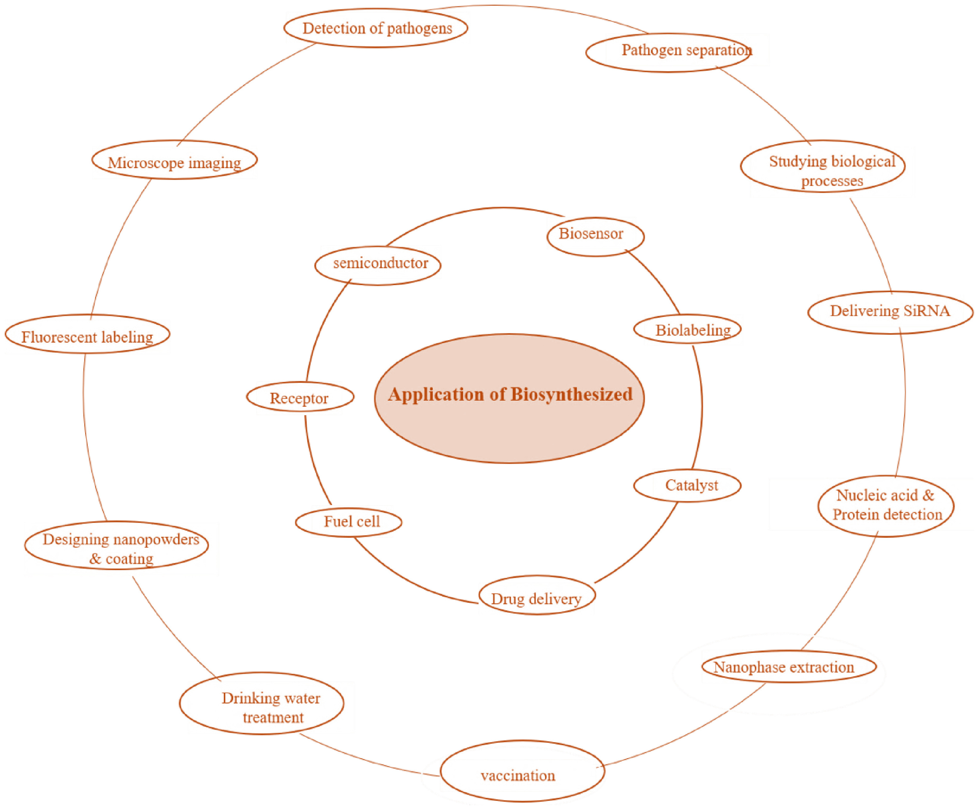
There are numerous applications of biosynthesized nanoparticles, including antimicrobial and anticancer activities, targeted drug delivery, biosensing, and vaccine development, and many others, as illustrated in Figure 3.

5. Advances and Challenges of Bacterial Synthesis of NPs
Significant advancements have been achieved in the field of nanotechnology, involving bacteria for the synthesis and application of NPs. This process has several advantages, like being an eco-friendly, biocompatible, and cost-effective method of synthesis. The biosynthesized NPs do not involve the toxic chemical constituents, which makes them desirable for biomedical applications. In contrast to physicochemical methods, the biogenic synthesis is free from an additional step of capping the NPs, in order to produce a stable and active product for pharmacological use. Additionally, biogenic synthesis requires less time, as reported by many studies, like Rehman et al, using Bacillus haynesii synthesized zinc NPs within a few minutes at room temperature. (Singh et al., 2016; Yumei et al., 2017; Rehman et al., 2019)
Despite numerous advantages displayed by the green method of nanoparticle synthesis, there are still challenges in terms of size and monodispersity of particles. The efforts are required to achieve the controlled size and morphology of NPs. Some studies have demonstrated a system for NPs synthesis using bacteria to obtain particles with monodispersity. For example, Arthrobacter sp was reported to synthesize AgNPs between pH 7.0 to 8.0, while the synthesis did not take place below a pH of 5.0 and above 8.0, owing to electrostatic repulsion between silver and EPS, under acidic and electronegativity in alkaline conditions, respectively. Moreover, the parameters of the bioprocess, like reaction time, temperature, and reactant concentrations, can alter the morphology and size, which further demands investigation on such parameters for the efficient and stabilized production of NPs. (Yumei et al., 2017; Ramanathan et al., 2011; Kalathil et al., 2011).
On understanding the mechanism of bioreduction of metals, bacteria can be genetically modified to design the process for a desirable size, shape with a high yield of NPs. One of the important and challenging issues with metal NPs is toxicity. Many studies have suggested the detrimental effects of NPs on cellular systems. The toxicity depends on various factors, like shape, size, disparity, capping, and the type of biomedical application. Physiochemical methods for the synthesis of NPs are generally more toxic than the biological process. Coating/capping of synthesized NPs with the bioagents is one of the ways to reduce toxicity. The bio coating not only makes it biocompatible, but also primarily stabilizes and prevents aggregation of NPs, making it ideal for different biomedical applications. Therefore, the NPs synthesized by biological approaches have reduced toxicity, as they mainly involve biocompatible materials for the reduction of metal ions. In a study, polymer surfactants were used to reduce the toxicity of AgNPs to obtain hemocompatible particles. In another study, heteropolysaccharides obtained from Lentinus squarrosulus were used for AgNPs capping, which resulted in a biocompatible particle with human RBCs. Such studies suggest the NPs synthesized through bio methods offer a solution to reduced cellular toxicity. (Kalathil et al., 2011; Lin et al., 2012).
5.1. Biomedical applications
Multidrug resistance of pathogenic organisms to the available antimicrobials through altering their metabolic pathways and targets has become an important challenge to tackle. Additionally, these pathogens acquire more pathogenicity due to their continuous exposure to antibiotics. Therefore, it is crucial to investigate effective, alternative, and environmentally friendly antimicrobial agents. Biogenic NPs have desired properties that help NPs to interact effectively with the pathogenic cell surfaces and penetrate to intervene during the metabolic pathways and replication. Many studies have reported the use of different NPs as antimicrobials. Banu et al. (2011) reported the effect of biofabricated AgNPs using R. stolonifer and antibiotics against Enterobacteriaceae (ESBL-strains). The antibacterial study of AgNPs synthesized usingBacillus cereus against Staphylococcus aureus,Klebsiella pneumonia,Pseudomonas aeruginosa,Escherichia coli, andSalmonella typhi showed larger inhibition zones than the commercial antibiotics. There are several such reports on the antimicrobial activities of Biogenic NPs against multiple drug-resistant disease-causing microbes. (Singh et al., 2020; Koul et al., 2021; Banu et al., 2011; Sunkar and Nachiyar, 2012).
Nowadays, cancer is the leading cause of death globally. Conventional treatment (chemotherapy, radiation, and surgery) for cancers has several side effects. Perhaps the targeted drug delivery to the required tissues or organs and its timely diagnosis of this life-threatening disease are still not possible. Hence, it is important to study alternative ways of diagnosis and treatment. Nanomedicines have been reported to be successfully utilized for the diagnosis and treatment of a tumor by targeted drug delivery. Borse et al. (2015) investigated biofabricated PtNPs synthesized fromSaccharomyces boulardii for their anticancer activity against MCF-7 and A431 cell lines. AgNPs biosynthesized fromCryptococcus laurentii showed effective anticancerous and antitumor properties against breast cancer cell lines. (Jabir et al., 2012; Sutradhar and Amin, 2014; Borse et al., 2015; Ortega et al., 2015).
The application of NPs in the food industry has a great scope in food packaging and food processing. A study presents the blended zinc oxide NPs with polymeric material that has an application as a packaging material, and this packaging material exhibited antibacterial properties. Likewise, many studies also reported that zinc oxide NPs hold promising applications for containers and covers for food packaging. AgNPs have a peculiar ability to break the bacterial biofilm, hence can be employed in the decontamination and cleaning process in the food industry. (Shukla, 2012; Espitia et al., 2012; Prasad et al., 2014; Huang et al., 2015).
NPs hold great potential in agriculture in the form of nanofertilizers, nanopesticides, and nanoinsecticides. Studied the antifungal potential of AgNPs synthesized via Pseudomonas sp. andAchromobactersp. againstFusarium oxysporum in chickpea. One of the recent studies on carbon-based nanomaterials was used as fertilizers and suggested the use of nanomaterial-based fertilizers that could minimize the use of chemical fertilizers. (Kaur et al., 2018; Bisinoti et al., 2019).
Nanoparticles as pesticides in the form of micelles, particles, nanopolymers, and metal oxides have found their applicability. In several studies, many novel materials containing NPs have been synthesized for application as pesticides. In one of the studies, investigated that baculovirusBombyx morinuclear polyhedrosis virus andSitophilus oryzaein silkworm can be managed by several NPs, like silver, zinc oxide, aluminum oxide, and titanium oxide NPs. (Goswami et al., 2010; Konishi et al., 2007; Du et al., 2007; Kaur et al., 2015).
6. Conclusion
Currently, metal NPs have been extensively studied for numerous biomedical applications owing to their huge surface area to volume ratio, giving them unique antimicrobial, antioxidant, and anticancer properties. The green route of metal NPs synthesis has emerged as an important area of nanobiotechnology, where bacteria are serving as nanofactories. Bacterial cells are rapidly growing and safer for easy and rapid synthesis of NPs with desired characteristics. However, there are several hurdles and gaps in the favorable synthesis of NPs that need to be addressed by the researchers. One of the important gaps in biogenic synthesis is the lack of full information about the metabolic cascades involved in the process. A detailed investigation of the pathway responsible for bioreduction is required for the development of ideal NPs. It would be of immense importance for biomedical purposes to understand the mechanism behind the binding of active moieties of bacteria that provide stable and biocompatible NPs. Standardization and improvement of fermentation and downstream techniques for the commercialization of particles with controlled size and shape is a major field that needs to be studied. Research on optimization of parameters like temperature, pH, etc., for better and bulk production of biocompatible NPs would offer an economically viable and sustainable solution. The involvement of genetically engineered bacteria for the modulation of NPs for desired characteristics holds potential in this arena. Such organisms are advantageous over the traditional ways of nanomaterials, for example, the rate of biosynthesis, production cost, and efficiency in energy. Besides, research on pharmaceutical issues like distribution and drug release, along with in vivo cytotoxicity, needs to be addressed. Therefore, this field of nanobiotechnology, which involves bacteria for the biosynthesis of NPs, is still in its infancy, requiring greater research efforts for many unanswered questions.
Data Availability Statement
Not applicable.
References
-
ADAMS, C.P., WALKER, K.A., OBARE, S.O. and DOCHERTY, K.M., 2014. Size-dependent antimicrobial effects of novel palladium nanoparticles. PLoS One, vol. 9, no. 1, pp. e85981. https://doi.org/10.1371/journal.pone.0085981 PMid:24465824.
» https://doi.org/10.1371/journal.pone.0085981 -
AHAMED, M., ALHADLAQ, H.A., KHAN, M., KARUPPIAH, P. and AL-DHABI, N.A., 2014. Synthesis, characterization, and antimicrobial activity of copper oxide nanoparticles. Journal of Nanomaterials, vol. 2014, no. 1, pp. 637858. https://doi.org/10.1155/2014/637858
» https://doi.org/10.1155/2014/637858 -
BANDEIRA, M., GIOVANELA, M., ROESCH-ELY, M., DEVINE, D.M. and DA SILVA CRESPO, J., 2020. Green synthesis of zinc oxide nanoparticles: a review of the synthesis methodology and mechanism of formation. Sustainable Chemistry and Pharmacy, vol. 15, pp. 100223. https://doi.org/10.1016/j.scp.2020.100223
» https://doi.org/10.1016/j.scp.2020.100223 -
BANKIER, C., MATHARU, R., CHEONG, Y.-K., REN, G., CLOUTMAN-GREEN, E. and CIRIC, L., 2019. Synergistic antibacterial effects of metallic nanoparticle combinations. Scientific Reports, vol. 9, no. 1, pp. 16074. https://doi.org/10.1038/s41598-019-52473-2 PMid:31690845.
» https://doi.org/10.1038/s41598-019-52473-2 -
BANU, A., RATHOD, V. and RANGANATH, E., 2011. Silver nanoparticle production by Rhizopus stolonifer and its antibacterial activity against extended spectrum β-lactamase producing (ESBL) strains of Enterobacteriaceae. Materials Research Bulletin, vol. 46, no. 9, pp. 1417-1423. https://doi.org/10.1016/j.materresbull.2011.05.008
» https://doi.org/10.1016/j.materresbull.2011.05.008 -
BARUWATI, B., POLSHETTIWAR, V. and VARMA, R.S., 2009. Glutathione promoted expeditious green synthesis of silver nanoparticles in water using microwaves. Green Chemistry, vol. 11, no. 7, pp. 926-930. https://doi.org/10.1039/b902184a
» https://doi.org/10.1039/b902184a -
BEVERIDGE, T.J. and MURRAY, R.G., 1980. Sites of metal deposition in the cell wall of Bacillus subtilis. Journal of Bacteriology, vol. 141, no. 2, pp. 876-887. https://doi.org/10.1128/jb.141.2.876-887.1980 PMid:6767692.
» https://doi.org/10.1128/jb.141.2.876-887.1980 -
BEWLEY, K.D., ELLIS, K.E., FIRER-SHERWOOD, M.A. and ELLIOTT, S.J., 2013. “Multi-heme proteins: nature’s electronic multi-purpose tool,” Biochimica et Biophysica Acta (BBA)-. Biochimica et Biophysica Acta. Bioenergetics, vol. 1827, no. 8-9, pp. 938-948. https://doi.org/10.1016/j.bbabio.2013.03.010
» https://doi.org/10.1016/j.bbabio.2013.03.010 -
BHATTACHARYA, R. and MUKHERJEE, P., 2008. Biological properties of “naked” metal nanoparticles. Advanced Drug Delivery Reviews, vol. 60, no. 11, pp. 1289-1306. https://doi.org/10.1016/j.addr.2008.03.013 PMid:18501989.
» https://doi.org/10.1016/j.addr.2008.03.013 -
BISINOTI, M.C., MOREIRA, A.B., MELO, C.A., FREGOLENTE, L.G., BENTO, L.R., SANTOS, J.V. and FERREIRA, O.P., 2019. Application of carbon-based nanomaterials as fertilizers in soils. In: R.F. NASCIMENTO, O.P. FERREIRA, A.J. DE PAULA and V.O. SOUSA NETO, eds. Nanomaterials applications for environmental matrices (pp. 305-333). USA: Elsevier. https://doi.org/10.1016/B978-0-12-814829-7.00008-2
» https://doi.org/10.1016/B978-0-12-814829-7.00008-2 -
BOROWIK, A., BANASIUK, R., DEREWONKO, N., RYCHLOWSKI, M., KRYCHOWIAK-MASNICKA, M., WYRZYKOWSKI, D., ZIABKA, M., WOZIWODZKA, A., KROLICKA, A. and PIOSIK, J., 2019. Interactions of newly synthesized platinum nanoparticles with ICR-191 and their potential application. Scientific Reports, vol. 9, no. 1, pp. 1-11. https://doi.org/10.1038/s41598-019-41092-6 PMid:30899037.
» https://doi.org/10.1038/s41598-019-41092-6 - BORSE, V., KALER, A. and BANERJEE, U.C., 2015. Microbial synthesis of platinum nanoparticles and evaluation of their anticancer activity. International Journal of Emerging Trends in Electrical & Electronics, vol. 11, no. 2, pp. 2320-9569.
-
CARDOSO, V.F., FRANCESKO, A., RIBEIRO, C., BAÑOBRE-LÓPEZ, M., MARTINS, P. and LANCEROS-MENDEZ, S., 2018. Advances in magnetic nanoparticles for biomedical applications. Advanced Healthcare Materials, vol. 7, no. 5, pp. 1700845. https://doi.org/10.1002/adhm.201700845 PMid:29280314.
» https://doi.org/10.1002/adhm.201700845 -
CARREGAL-ROMERO, S., CABALLERO-DÍAZ, E., BEQA, L., ABDELMONEM, A.M., OCHS, M., HÜHN, D., SUAU, B.S., VALCARCEL, M. and PARAK, W.J., 2013. Multiplexed sensing and imaging with colloidal nano- and microparticles. Annual Review of Analytical Chemistry, vol. 6, no. 1, pp. 53-81. https://doi.org/10.1146/annurev-anchem-062012-092621 PMid:23451718.
» https://doi.org/10.1146/annurev-anchem-062012-092621 -
CHEN, Y.-S., HUNG, Y.-C., LIAU, I. and HUANG, G.S., 2009. Assessment of the in vivo toxicity of gold nanoparticles. Nanoscale Research Letters, vol. 4, no. 8, pp. 858-864. https://doi.org/10.1007/s11671-009-9334-6 PMid:20596373.
» https://doi.org/10.1007/s11671-009-9334-6 -
CHOUDHURY, A.R., MALHOTRA, A., BHATTACHARJEE, P. and PRASAD, G., 2014. Facile and rapid thermo-regulated biomineralization of gold by pullulan and study of its thermodynamic parameters. Carbohydrate Polymers, vol. 106, pp. 154-159. https://doi.org/10.1016/j.carbpol.2014.01.072 PMid:24721063.
» https://doi.org/10.1016/j.carbpol.2014.01.072 -
CHOUHAN, R. and BAJPAI, A., 2009. Real time in vitro studies of doxorubicin release from PHEMA nanoparticles. Journal of Nanobiotechnology, vol. 7, no. 1, pp. 5. https://doi.org/10.1186/1477-3155-7-5 PMid:19843333.
» https://doi.org/10.1186/1477-3155-7-5 -
CLARKE, T.A., EDWARDS, M.J., GATES, A.J., HALL, A., WHITE, G.F., BRADLEY, J., REARDON, C.L., SHI, L., BELIAEV, A.S., MARSHALL, M.J., WANG, Z., WATMOUGH, N.J., FREDRICKSON, J.K., ZACHARA, J.M., BUTT, J.N. and RICHARDSON, D.J., 2011. Structure of a bacterial cell surface decaheme electron conduit. Proceedings of the National Academy of Sciences of the United States of America, vol. 108, no. 23, pp. 9384-9389. https://doi.org/10.1073/pnas.1017200108 PMid:21606337.
» https://doi.org/10.1073/pnas.1017200108 -
COLOGGI, D.L., LAMPA-PASTIRK, S., SPEERS, A.M., KELLY, S.D. and REGUERA, G., 2011. Extracellular reduction of uranium via Geobacter conductive pili as a protective cellular mechanism. Proceedings of the National Academy of Sciences of the United States of America, vol. 108, no. 37, pp. 15248-15252. https://doi.org/10.1073/pnas.1108616108 PMid:21896750.
» https://doi.org/10.1073/pnas.1108616108 -
COURSOLLE, D. and GRALNICK, J.A., 2010. Modularity of the Mtr respiratory pathway of Shewanella oneidensis strain MR-1. Molecular Microbiology, vol. 77, no. 4, pp. 995-1008. https://doi.org/10.1111/j.1365-2958.2010.07266.x PMid:20598084.
» https://doi.org/10.1111/j.1365-2958.2010.07266.x - DAS, V.L., THOMAS, R., VARGHESE, R.T., SONIYA, E., MATHEW, J., and RADHAKRISHNAN, E., 2014. Extracellular synthesis of silver nanoparticles by the Bacillus strain CS 11 isolated from industrialized area. 3 Biotech, vol. 4, no. 2, pp. 121-126.
-
DAVIDOVIĆ, S., LAZIĆ, V., VUKOJE, I., PAPAN, J., ANHRENKIEL, S.P., DIMITRIJEVIĆ, S. and NEDELJKOVIĆ, J.M., 2017. Dextran coated silver nanoparticles: chemical sensor for selective cysteine detection. Colloids and Surfaces. B, Biointerfaces, vol. 160, pp. 184-191. https://doi.org/10.1016/j.colsurfb.2017.09.031 PMid:28934661.
» https://doi.org/10.1016/j.colsurfb.2017.09.031 -
DEPLANCHE, K. and MACASKIE, L.E., 2008. Biorecovery of gold by Escherichia coli and Desulfovibrio desulfuricans. Biotechnology and Bioengineering, vol. 99, no. 5, pp. 1055-1064. https://doi.org/10.1002/bit.21688 PMid:17969152.
» https://doi.org/10.1002/bit.21688 -
DIVYA, M., KIRAN, G.S., HASSAN, S. and SELVIN, J., 2019. Biogenic synthesis and effect of silver nanoparticles (AgNPs) to combat catheter-related urinary tract infections. Biocatalysis and Agricultural Biotechnology, vol. 18, pp. 101037. https://doi.org/10.1016/j.bcab.2019.101037
» https://doi.org/10.1016/j.bcab.2019.101037 -
DU, L., JIANG, H., LIU, X. and WANG, E., 2007. Biosynthesis of gold nanoparticles assisted by Escherichia coli DH5α and its application on direct electrochemistry of hemoglobin. Electrochemistry Communications, vol. 9, no. 5, pp. 1165-1170. https://doi.org/10.1016/j.elecom.2007.01.007
» https://doi.org/10.1016/j.elecom.2007.01.007 -
ESPITIA, P., SOARES, N., COIMBRA, J., ANDRADE, N., CRUZ, R. and MEDEIROS, E., 2012. Zinc oxide nanoparticles: synthesis, antimicrobial activity and food packaging applications. Food and Bioprocess Technology, vol. 5, no. 5, pp. 1464-1477. https://doi.org/10.1007/s11947-012-0797-6
» https://doi.org/10.1007/s11947-012-0797-6 -
FARAMARZI, M.A. and SADIGHI, A., 2013. Insights into biogenic and chemical production of inorganic nanomaterials and nanostructures. Advances in Colloid and Interface Science, vol. 189–190, pp. 1-20. https://doi.org/10.1016/j.cis.2012.12.001 PMid:23332127.
» https://doi.org/10.1016/j.cis.2012.12.001 -
FERNÁNDEZ-BERTÓLEZ, N., ALBA-GONZÁLEZ, A., TOUZANI, A., RAMOS-PAN, L., MÉNDEZ, J., REIS, A.T., QUELLE-REGALDIE, A., SÁNCHEZ, L., FOLGUEIRA, M., LAFFON, B. and VALDIGLESIAS, V., 2024. Toxicity of zinc oxide nanoparticles: cellular and behavioural effects. Chemosphere, vol. 363, pp. 142993. https://doi.org/10.1016/j.chemosphere.2024.142993 PMid:39097108.
» https://doi.org/10.1016/j.chemosphere.2024.142993 -
FREITAS, F., ALVES, V.D. and REIS, M.A., 2011. Advances in bacterial exopolysaccharides: from production to biotechnological applications. Trends in Biotechnology, vol. 29, no. 8, pp. 388-398. https://doi.org/10.1016/j.tibtech.2011.03.008 PMid:21561675.
» https://doi.org/10.1016/j.tibtech.2011.03.008 -
GAHLAWAT, G. and CHOUDHURY, A.R., 2019. A review on the biosynthesis of metal and metal salt nanoparticles by microbes. RSC Advances, vol. 9, no. 23, pp. 12944-12967. https://doi.org/10.1039/C8RA10483B PMid:35520790.
» https://doi.org/10.1039/C8RA10483B -
GAHLAWAT, G., SHIKHA, S., CHADDHA, B.S., CHAUDHURI, S.R., MAYILRAJ, S. and CHOUDHURY, A.R., 2016. Microbial glycolipoprotein-capped silver nanoparticles as emerging antibacterial agents against cholera. Microbial Cell Factories, vol. 15, no. 1, pp. 25. https://doi.org/10.1186/s12934-016-0422-x PMid:26829922.
» https://doi.org/10.1186/s12934-016-0422-x -
GARDEA-TORRESDEY, J.L., PARSONS, J.G., GOMEZ, E., PERALTA-VIDEA, J., TROIANI, H.E., SANTIAGO, P. and YACAMAN, M.J., 2002. Formation and growth of Au nanoparticles inside live alfalfa plants. Nano Letters, vol. 2, no. 4, pp. 397-401. https://doi.org/10.1021/nl015673+
» https://doi.org/10.1021/nl015673+ -
GHIUȚĂ, I., CRISTEA, D., CROITORU, C., KOST, J., WENKERT, R., VYRIDES, I., ANAYIOTOS, A. and MUNTEANU, D., 2018. Characterization and antimicrobial activity of silver nanoparticles, biosynthesized using Bacillus species. Applied Surface Science, vol. 438, pp. 66-73. https://doi.org/10.1016/j.apsusc.2017.09.163
» https://doi.org/10.1016/j.apsusc.2017.09.163 -
GOSWAMI, A., ROY, I., SENGUPTA, S. and DEBNATH, N., 2010. Novel applications of solid and liquid formulations of nanoparticles against insect pests and pathogens. Thin Solid Films, vol. 519, no. 3, pp. 1252-1257. https://doi.org/10.1016/j.tsf.2010.08.079
» https://doi.org/10.1016/j.tsf.2010.08.079 -
GOSWAMI, S., BISHNOI, A., TANK, D., PATEL, P., CHAHAR, M., KHATURIA, S., MODI, N., KHALID, M., ALAM, M.W., KUMAR YADAV, V., ALRESHIDI, M.A. and YADAV, K.K., 2024. Recent trends in the synthesis, characterization and commercial applications of zinc oxide nanoparticles-a review. Inorganica Chimica Acta, vol. 573, pp. 122350. https://doi.org/10.1016/j.ica.2024.122350
» https://doi.org/10.1016/j.ica.2024.122350 -
GOUR, A. and JAIN, N.K., 2019. Advances in green synthesis of nanoparticles. Artificial Cells, Nanomedicine, and Biotechnology, vol. 47, no. 1, pp. 844-851. https://doi.org/10.1080/21691401.2019.1577878 PMid:30879351.
» https://doi.org/10.1080/21691401.2019.1577878 -
GOVINDARAJAN, M., RAJESWARY, M., VEERAKUMAR, K., MUTHUKUMARAN, U., HOTI, S.L. and BENELLI, G., 2016. Green synthesis and characterization of silver nanoparticles fabricated using Anisomeles indica: mosquitocidal potential against malaria, dengue and Japanese encephalitis vectors. Experimental Parasitology, vol. 161, pp. 40-47. https://doi.org/10.1016/j.exppara.2015.12.011 PMid:26708933.
» https://doi.org/10.1016/j.exppara.2015.12.011 -
GUTIERREZ, T., MORRIS, G. and GREEN, D.H., 2009. Yield and physicochemical properties of EPS from Halomonas sp. strain TG39 identifies a role for protein and anionic residues (sulfate and phosphate) in emulsification of n-hexadecane. Biotechnology and Bioengineering, vol. 103, no. 1, pp. 207-216. https://doi.org/10.1002/bit.22218 PMid:19160375.
» https://doi.org/10.1002/bit.22218 -
HAMADA, A.M., RADI, A.A., AL-KAHTANY, F.A. and FARGHALY, F.A., 2024. A review: zinc oxide nanoparticles: advantages and disadvantages. Journal of Plant Nutrition, vol. 47, no. 4, pp. 656-679. https://doi.org/10.1080/01904167.2023.2280127
» https://doi.org/10.1080/01904167.2023.2280127 -
HANIF, S., JAVED, R., CHEEMA, M., KIANI, M.Z., FAROOQ, S. and ZIA, M., 2024. Harnessing the potential of zinc oxide nanoparticles and their derivatives as nanofertilizers: trends and perspectives. Plant Nano Biology, vol. 10, pp. 100110. https://doi.org/10.1016/j.plana.2024.100110
» https://doi.org/10.1016/j.plana.2024.100110 -
HARTSHORNE, R.S., REARDON, C.L., ROSS, D., NUESTER, J., CLARKE, T.A., GATES, A.J., MILLS, P.C., FREDRICKSON, J.K., ZACHARA, J.M., SHI, L., BELIAEV, A.S., MARSHALL, M.J., TIEN, M., BRANTLEY, S., BUTT, J.N. and RICHARDSON, D.J., 2009. Characterization of an electron conduit between bacteria and the extracellular environment. Proceedings of the National Academy of Sciences of the United States of America, vol. 106, no. 52, pp. 22169-22174. https://doi.org/10.1073/pnas.0900086106 PMid:20018742.
» https://doi.org/10.1073/pnas.0900086106 -
HE, S., GUO, Z., ZHANG, Y., ZHANG, S., WANG, J. and GU, N., 2007. Biosynthesis of gold nanoparticles using the bacteria Rhodopseudomonas capsulata. Materials Letters, vol. 61, no. 18, pp. 3984-3987. https://doi.org/10.1016/j.matlet.2007.01.018
» https://doi.org/10.1016/j.matlet.2007.01.018 -
HOLGHOOMI, R. and COLAGAR, A.H., 2024. Applications of biocompatible nanoparticles in plant biotechnology for enhanced secondary metabolite biosynthesis. Inorganic Chemistry Communications, vol. 167, pp. 112753. https://doi.org/10.1016/j.inoche.2024.112753
» https://doi.org/10.1016/j.inoche.2024.112753 -
HUANG, J., LIN, L., SUN, D., CHEN, H., YANG, D. and LI, Q., 2015. Bio-inspired synthesis of metal nanomaterials and applications. Chemical Society Reviews, vol. 44, no. 17, pp. 6330-6374. https://doi.org/10.1039/C5CS00133A PMid:26083903.
» https://doi.org/10.1039/C5CS00133A -
HUANG, X., EL-SAYED, I.H., QIAN, W. and EL-SAYED, M.A., 2006. Cancer cell imaging and photothermal therapy in the near-infrared region by using gold nanorods. Journal of the American Chemical Society, vol. 128, no. 6, pp. 2115-2120. https://doi.org/10.1021/ja057254a PMid:16464114.
» https://doi.org/10.1021/ja057254a -
HULKOTI, N.I. and TARANATH, T.C., 2014. Biosynthesis of nanoparticles using microbes: A review. Colloids and Surfaces. B, Biointerfaces, vol. 121, pp. 474-483. https://doi.org/10.1016/j.colsurfb.2014.05.027 PMid:25001188.
» https://doi.org/10.1016/j.colsurfb.2014.05.027 -
HWA, K.-Y. and SUBRAMANI, B., 2014. Synthesis of zinc oxide nanoparticles on graphene–carbon nanotube hybrid for glucose biosensor applications. Biosensors & Bioelectronics, vol. 62, pp. 127-133. https://doi.org/10.1016/j.bios.2014.06.023 PMid:24997365.
» https://doi.org/10.1016/j.bios.2014.06.023 -
IBRAHIM, S., AHMAD, Z., MANZOOR, M.Z., MUJAHID, M., FAHEEM, Z. and ADNAN, A., 2021. Optimization for biogenic microbial synthesis of silver nanoparticles through response surface methodology, characterization, their antimicrobial, antioxidant, and catalytic potential. Scientific Reports, vol. 11, no. 1, pp. 770. https://doi.org/10.1038/s41598-020-80805-0 PMid:33436966.
» https://doi.org/10.1038/s41598-020-80805-0 -
IRAVANI, S. and VARMA, R.S., 2019. Biofactories: engineered nanoparticles via genetically engineered organisms. Green Chemistry, vol. 21, no. 17, pp. 4583-4603. https://doi.org/10.1039/C9GC01759C
» https://doi.org/10.1039/C9GC01759C - JABIR, N.R., TABREZ, S., ASHRAF, G.M., SHAKIL, S., DAMANHOURI, G.A. and KAMAL, M.A., 2012. Nanotechnology-based approaches in anticancer research. International Journal of Nanomedicine, vol. 7, pp. 4391-4408. PMid:22927757.
- JAIN, S., NAGAR, N. and DEVRA, V., 2015. Synthesis and characterization of highly efficient copper nanoparticles and their catalytic application in oxidative kinetic study. Advances in Applied Science Research, vol. 6, no. 6, pp. 171-180.
-
JAYASEELAN, C., RAHUMAN, A.A., KIRTHI, A.V., MARIMUTHU, S., SANTHOSHKUMAR, T., BAGAVAN, A., GAURAV, K., KARTHIK, L. and RAO, K.V., 2012. Novel microbial route to synthesize ZnO nanoparticles using Aeromonas hydrophila and their activity against pathogenic bacteria and fungi. Spectrochimica Acta. Part A, Molecular and Biomolecular Spectroscopy, vol. 90, pp. 78-84. https://doi.org/10.1016/j.saa.2012.01.006 PMid:22321514.
» https://doi.org/10.1016/j.saa.2012.01.006 -
JEYARAJ, M., GURUNATHAN, S., QASIM, M., KANG, M.-H. and KIM, J.-H., 2019. A comprehensive review on the synthesis, characterization, and biomedical application of platinum nanoparticles. Nanomaterials (Basel, Switzerland), vol. 9, no. 12, pp. 1719. https://doi.org/10.3390/nano9121719 PMid:31810256.
» https://doi.org/10.3390/nano9121719 -
JIANG, Y., LI, J., ZHEN, X., XIE, C. and PU, K., 2018. Dual-peak absorbing semiconducting copolymer nanoparticles for first and second near-infrared window photothermal therapy: a comparative study. Advanced Materials, vol. 30, no. 14, pp. e1705980. https://doi.org/10.1002/adma.201705980 PMid:29457284.
» https://doi.org/10.1002/adma.201705980 -
JOHNSTON, H.J., HUTCHISON, G., CHRISTENSEN, F.M., PETERS, S., HANKIN, S. and STONE, V., 2010. A review of the in vivo and in vitro toxicity of silver and gold particulates: particle attributes and biological mechanisms responsible for the observed toxicity. Critical Reviews in Toxicology, vol. 40, no. 4, pp. 328-346. https://doi.org/10.3109/10408440903453074 PMid:20128631.
» https://doi.org/10.3109/10408440903453074 -
JUIBARI, M.M., ABBASALIZADEH, S., JOUZANI, G.S. and NORUZI, M., 2011. Intensified biosynthesis of silver nanoparticles using a native extremophilic Ureibacillus thermosphaericus strain. Materials Letters, vol. 65, no. 6, pp. 1014-1017. https://doi.org/10.1016/j.matlet.2010.12.056
» https://doi.org/10.1016/j.matlet.2010.12.056 -
KALATHIL, S., LEE, J. and CHO, M.H., 2011. Electrochemically active biofilm-mediated synthesis of silver nanoparticles in water. Green Chemistry, vol. 13, no. 6, pp. 1482-1485. https://doi.org/10.1039/c1gc15309a
» https://doi.org/10.1039/c1gc15309a - KAMEL MADBOULY, A. and HAMDAN, T., 2014. Bio-synthesis of magnetite nanoparticles by bacteria. American Journal of Nano Research and Application, vol. 2, pp. 98-103.
-
KANG, F., ALVAREZ, P.J. and ZHU, D., 2014. Microbial extracellular polymeric substances reduce Ag+ to silver nanoparticles and antagonize bactericidal activity. Environmental Science & Technology, vol. 48, no. 1, pp. 316-322. https://doi.org/10.1021/es403796x PMid:24328348.
» https://doi.org/10.1021/es403796x -
KANG, Y., WU, Y.Z., HU, X., XU, X., SUN, J., GENG, R., HUANG, T., LIU, X., MA, Y., CHEN, Y., WAN, Q., QI, X., ZHANG, G., ZHAO, X. and ZENG, X., 2017. Multicolor bioimaging with biosynthetic zinc nanoparticles and their application in tumor detection. Scientific Reports, vol. 7, no. 1, pp. 45313. https://doi.org/10.1038/srep45313 PMid:28345657.
» https://doi.org/10.1038/srep45313 -
KANNINEN, P., JOHANS, C., MERTA, J. and KONTTURI, K., 2008. Influence of ligand structure on the stability and oxidation of copper nanoparticles. Journal of Colloid and Interface Science, vol. 318, no. 1, pp. 88-95. https://doi.org/10.1016/j.jcis.2007.09.069 PMid:17961585.
» https://doi.org/10.1016/j.jcis.2007.09.069 -
KARTHIK, L., KUMAR, G., KIRTHI, A.V., RAHUMAN, A. and RAO, K.B., 2014. Streptomyces sp. LK3 mediated synthesis of silver nanoparticles and its biomedical application. Bioprocess and Biosystems Engineering, vol. 37, no. 2, pp. 261-267. https://doi.org/10.1007/s00449-013-0994-3 PMid:23771163.
» https://doi.org/10.1007/s00449-013-0994-3 -
KAUR, H., DOLMA, K., KAUR, N., MALHOTRA, A., KUMAR, N., DIXIT, P., SHARMA, D., MAYILRAJ, S. and CHOUDHURY, A.R., 2015. Marine microbe as nano-factories for copper biomineralization. Biotechnology and Bioprocess Engineering; BBE, vol. 20, no. 1, pp. 51-57. https://doi.org/10.1007/s12257-014-0432-7
» https://doi.org/10.1007/s12257-014-0432-7 -
KAUR, P., THAKUR, R., DUHAN, J.S. and CHAUDHURY, A., 2018. Management of wilt disease of chickpea in vivo by silver nanoparticles biosynthesized by rhizospheric microflora of chickpea (Cicer arietinum). Journal of Chemical Technology and Biotechnology, vol. 93, no. 11, pp. 3233-3243. https://doi.org/10.1002/jctb.5680
» https://doi.org/10.1002/jctb.5680 -
KEAT, C.L., AZIZ, A., EID, A.M. and ELMARZUGI, N.A., 2015. Biosynthesis of nanoparticles and silver nanoparticles. Bioresources and Bioprocessing, vol. 2, no. 1, pp. 47. https://doi.org/10.1186/s40643-015-0076-2
» https://doi.org/10.1186/s40643-015-0076-2 -
KHANNA, P., MORE, P., JAWALKAR, J., PATIL, Y. and RAO, N.K., 2009. Synthesis of hydrophilic copper nanoparticles: effect of reaction temperature. Journal of Nanoparticle Research, vol. 11, no. 4, pp. 793-799. https://doi.org/10.1007/s11051-008-9441-9
» https://doi.org/10.1007/s11051-008-9441-9 -
KIM, D.-H., NIKLES, D.E., JOHNSON, D.T. and BRAZEL, C.S., 2008. Heat generation of aqueously dispersed CoFe2O4 nanoparticles as heating agents for magnetically activated drug delivery and hyperthermia. Journal of Magnetism and Magnetic Materials, vol. 320, no. 19, pp. 2390-2396. https://doi.org/10.1016/j.jmmm.2008.05.023
» https://doi.org/10.1016/j.jmmm.2008.05.023 -
KIMBER, R.L., LEWIS, E.A., PARMEGGIANI, F., SMITH, K., BAGSHAW, H., STARBORG, T., JOSHI, N., FIGUEROA, A.I., VAN DER LAAN, G., CIBIN, G., GIANOLIO, D., HAIGH, S.J., PATTRICK, R.A.D., TURNER, N.J. and LLOYD, J.R., 2018. Biosynthesis and characterization of copper nanoparticles using Shewanella oneidensis: application for click chemistry. Small, vol. 14, no. 10, pp. 1703145. https://doi.org/10.1002/smll.201703145 PMid:29359400.
» https://doi.org/10.1002/smll.201703145 -
KITCHING, M., CHOUDHARY, P., INGUVA, S., GUO, Y., RAMANI, M., DAS, S.K. and MARSILI, E., 2016. Fungal surface protein mediated one-pot synthesis of stable and hemocompatible gold nanoparticles. Enzyme and Microbial Technology, vol. 95, pp. 76-84. https://doi.org/10.1016/j.enzmictec.2016.08.007 PMid:27866629.
» https://doi.org/10.1016/j.enzmictec.2016.08.007 -
KLAUS-JOERGER, T., JOERGER, R., OLSSON, E. and GRANQVIST, C., 2001. Bacteria as workers in the living factory: metal-accumulating bacteria and their potential for materials science. Trends in Biotechnology, vol. 19, no. 1, pp. 15-20. https://doi.org/10.1016/S0167-7799(00)01514-6 PMid:11146098.
» https://doi.org/10.1016/S0167-7799(00)01514-6 -
KONISHI, Y., TSUKIYAMA, T., TACHIMI, T., SAITOH, N., NOMURA, T. and NAGAMINE, S., 2007. Microbial deposition of gold nanoparticles by the metal-reducing bacterium Shewanella algae. Electrochimica Acta, vol. 53, no. 1, pp. 186-192. https://doi.org/10.1016/j.electacta.2007.02.073
» https://doi.org/10.1016/j.electacta.2007.02.073 -
KOUL, B., POONIA, A.K., YADAV, D. and JIN, J.-O., 2021. Microbe-mediated biosynthesis of nanoparticles: applications and future prospects. Biomolecules, vol. 11, no. 6, pp. 886. https://doi.org/10.3390/biom11060886 PMid:34203733.
» https://doi.org/10.3390/biom11060886 -
KULKARNI, R.R., SHAIWALE, N.S., DEOBAGKAR, D.N. and DEOBAGKAR, D.D., 2015. Synthesis and extracellular accumulation of silver nanoparticles by employing radiation-resistant Deinococcus radiodurans, their characterization, and determination of bioactivity. International Journal of Nanomedicine, vol. 10, pp. 963-974. https://doi.org/10.2147/IJN.S72888 PMid:25673991.
» https://doi.org/10.2147/IJN.S72888 -
LENGKE, M.F., FLEET, M.E. and SOUTHAM, G., 2006. Morphology of gold nanoparticles synthesized by filamentous cyanobacteria from gold(I)–thiosulfate and gold(III)–chloride complexes. Langmuir : The ACS Journal of Surfaces and Colloids, vol. 22, no. 6, pp. 2780-2787. https://doi.org/10.1021/la052652c PMid:16519482.
» https://doi.org/10.1021/la052652c -
LI, X., WEI, J., AIFANTIS, K.E., FAN, Y., FENG, Q., CUI, F.Z. and WATARI, F., 2016. Current investigations into magnetic nanoparticles for biomedical applications. Journal of Biomedical Materials Research. Part A, vol. 104, no. 5, pp. 1285-1296. https://doi.org/10.1002/jbm.a.35654 PMid:26779606.
» https://doi.org/10.1002/jbm.a.35654 -
LIANG, X., ZHANG, R., LIU, C. and LIU, H., 2018. Quantitative measurement of the sustainable water resource development system in China inspired by dissipative structure theory. Sustainability (Basel), vol. 10, no. 11, pp. 3996. https://doi.org/10.3390/su10113996
» https://doi.org/10.3390/su10113996 -
LIM, Z.-Z.J., LI, J.-E.J., NG, C.-T., YUNG, L.-Y.L. and BAY, B.-H., 2011. Gold nanoparticles in cancer therapy. Acta Pharmacologica Sinica, vol. 32, no. 8, pp. 983-990. https://doi.org/10.1038/aps.2011.82 PMid:21743485.
» https://doi.org/10.1038/aps.2011.82 -
LIN, J.-J., LIN, W.-C., DONG, R.-X. and HSU, S., 2012. The cellular responses and antibacterial activities of silver nanoparticles stabilized by different polymers. Nanotechnology, vol. 23, no. 6, pp. 065102. https://doi.org/10.1088/0957-4484/23/6/065102 PMid:22248930.
» https://doi.org/10.1088/0957-4484/23/6/065102 -
LIU, J. and LU, Y., 2004. Colorimetric biosensors based on DNAzyme-assembled gold nanoparticles. Journal of Fluorescence, vol. 14, no. 4, pp. 343-354. https://doi.org/10.1023/B:JOFL.0000031816.06134.d3 PMid:15617377.
» https://doi.org/10.1023/B:JOFL.0000031816.06134.d3 -
LIU, Y., FREDRICKSON, J.K., ZACHARA, J.M. and SHI, L., 2015. Direct involvement of ombB, omaB, and omcB genes in extracellular reduction of Fe(III) by Geobacter sulfurreducens PCA. Frontiers in Microbiology, vol. 6, pp. 1075. https://doi.org/10.3389/fmicb.2015.01075 PMid:26483786.
» https://doi.org/10.3389/fmicb.2015.01075 -
LV, Q., ZHANG, B., XING, X., ZHAO, Y., CAI, R., WANG, W. and GU, Q., 2018. Biosynthesis of copper nanoparticles using Shewanella loihica PV-4 with antibacterial activity: novel approach and mechanisms investigation. Journal of Hazardous Materials, vol. 347, pp. 141-149. https://doi.org/10.1016/j.jhazmat.2017.12.070 PMid:29304452.
» https://doi.org/10.1016/j.jhazmat.2017.12.070 -
MALHOTRA, A., DOLMA, K., KAUR, N., RATHORE, Y.S., ASHISH., MAYILRAJ, S. and CHOUDHURY, A.R., 2013. Biosynthesis of gold and silver nanoparticles using a novel marine strain of Stenotrophomonas. Bioresource Technology, vol. 142, pp. 727-731. https://doi.org/10.1016/j.biortech.2013.05.109 PMid:23791020.
» https://doi.org/10.1016/j.biortech.2013.05.109 -
MATA, Y.N., TORRES, E., BLÁZQUEZ, M.L., BALLESTER, A., GONZÁLEZ, F. and MUNOZ, J.A., 2009. Gold(III) biosorption and bioreduction with the brown alga Fucus vesiculosus. Journal of Hazardous Materials, vol. 166, no. 2-3, pp. 612-618. https://doi.org/10.1016/j.jhazmat.2008.11.064 PMid:19124199.
» https://doi.org/10.1016/j.jhazmat.2008.11.064 -
MEHTA, A., SIDHU, C., PINNAKA, A.K. and ROY CHOUDHURY, A., 2014. Extracellular polysaccharide production by a novel osmotolerant marine strain of Alteromonas macleodii and its application towards biomineralization of silver. PLoS One, vol. 9, no. 6, pp. e98798. https://doi.org/10.1371/journal.pone.0098798 PMid:24932690.
» https://doi.org/10.1371/journal.pone.0098798 -
MOHD YUSOF, H., MOHAMAD, R., ZAIDAN, U.H. and ABDUL RAHMAN, N.A., 2019. Microbial synthesis of zinc oxide nanoparticles and their potential application as an antimicrobial agent and a feed supplement in animal industry: a review. Journal of Animal Science and Biotechnology, vol. 10, no. 1, pp. 57. https://doi.org/10.1186/s40104-019-0368-z PMid:31321032.
» https://doi.org/10.1186/s40104-019-0368-z -
MONTERO-SILVA, F. (2018). Synthesis of extracellular stable gold nanoparticles by Cupriavidus metallidurans CH34 cells (revision). bioRxiv, 139949. https://doi.org/10.1101/139949
» https://doi.org/10.1101/139949 -
NAIK, M.M., PRABHU, M.S., SAMANT, S.N., NAIK, P.M. and SHIRODKAR, S., 2017. Synergistic action of silver nanoparticles synthesized from silver resistant estuarine Pseudomonas aeruginosa strain SN5 with antibiotics against antibiotic resistant bacterial human pathogens. Thalassas, vol. 33, no. 1, pp. 73-80. https://doi.org/10.1007/s41208-017-0023-4
» https://doi.org/10.1007/s41208-017-0023-4 -
NG, C.K., SIVAKUMAR, K., LIU, X., MADHAIYAN, M., JI, L., YANG, L., TANG, C., SONG, H., KJELLEBERG, S. and CAO, B., 2013. Influence of outer membrane c-type cytochromes on particle size and activity of extracellular nanoparticles produced by Shewanella oneidensis. Biotechnology and Bioengineering, vol. 110, no. 7, pp. 1831-1837. https://doi.org/10.1002/bit.24856 PMid:23381725.
» https://doi.org/10.1002/bit.24856 - ORTEGA, F.G., FERNÁNDEZ-BALDO, M.A., FERNÁNDEZ, J.G., SERRANO, M.J., SANZ, M.I., DIAZ-MOCHÓN, J.J., LORENTE, J.A. and RABA, J., 2015. Study of antitumor activity in breast cell lines using silver nanoparticles produced by yeast. International Journal of Nanomedicine, vol. 10, pp. 2021-2031. PMid:25844035.
-
PANÁČEK, A., KVÍTEK, L., PRUCEK, R., KOLAR, M., VECEROVA, R., PIZÚROVA, N., SHARMA, V.K., NEVECNA, T. and ZBORIL, R., 2006. Silver colloid nanoparticles: synthesis, characterization, and their antibacterial activity. The Journal of Physical Chemistry. B, vol. 110, no. 33, pp. 16248-16253. https://doi.org/10.1021/jp063826h PMid:16913750.
» https://doi.org/10.1021/jp063826h -
PARMAR, A., KAUR, G., KAPIL, S., SHARMA, V., SHARMA, S. and CHOUDHARY, M., 2019. Novel biogenic silver nanoparticles as invigorated catalytic and antibacterial tool: a cleaner approach towards environmental remediation and combating bacterial invasion. Materials Chemistry and Physics, vol. 238, pp. 121861. https://doi.org/10.1016/j.matchemphys.2019.121861
» https://doi.org/10.1016/j.matchemphys.2019.121861 -
PEDONE, D., MOGLIANETTI, M., DE LUCA, E., BARDI, G. and POMPA, P.P., 2017. Platinum nanoparticles in nanobiomedicine. Chemical Society Reviews, vol. 46, no. 16, pp. 4951-4975. https://doi.org/10.1039/C7CS00152E PMid:28696452.
» https://doi.org/10.1039/C7CS00152E -
PHAN, T.T.V., HUYNH, T.-C., MANIVASAGAN, P., MONDAL, S. and OH, J., 2020. An up-to-date review on biomedical applications of palladium nanoparticles. Nanomaterials (Basel, Switzerland), vol. 10, no. 1, pp. 66. https://doi.org/10.3390/nano10010066 PMid:31892149.
» https://doi.org/10.3390/nano10010066 -
PHILIPSE, A.P. and MAAS, D., 2002. Magnetic colloids from magnetotactic bacteria: chain formation and colloidal stability. Langmuir : The ACS Journal of Surfaces and Colloids, vol. 18, no. 25, pp. 9977-9984. https://doi.org/10.1021/la0205811
» https://doi.org/10.1021/la0205811 -
PRASAD, R., KUMAR, V. and PRASAD, K.S., 2014. Nanotechnology in sustainable agriculture: present concerns and future aspects. African Journal of Biotechnology, vol. 13, no. 6, pp. 705-713. https://doi.org/10.5897/AJBX2013.13554
» https://doi.org/10.5897/AJBX2013.13554 -
RAHMAN, A., ISMAIL, A., JUMBIANTI, D., MAGDALENA, S. and SUDRAJAT, H., 2010. Synthesis of copper oxide nanoparticles by using Phormidium cyanobacterium. Indonesian Journal of Chemistry, vol. 9, no. 3, pp. 355-360. https://doi.org/10.22146/ijc.21498
» https://doi.org/10.22146/ijc.21498 -
RAI, A., SINGH, A., AHMAD, A. and SASTRY, M., 2006. Role of halide ions and temperature on the morphology of biologically synthesized gold nanotriangles. Langmuir : The ACS Journal of Surfaces and Colloids, vol. 22, no. 2, pp. 736-741. https://doi.org/10.1021/la052055q PMid:16401125.
» https://doi.org/10.1021/la052055q -
RAJ, R., DALEI, K., CHAKRABORTY, J. and DAS, S., 2016. Extracellular polymeric substances of a marine bacterium mediated synthesis of CdS nanoparticles for removal of cadmium from aqueous solution. Journal of Colloid and Interface Science, vol. 462, pp. 166-175. https://doi.org/10.1016/j.jcis.2015.10.004 PMid:26454375.
» https://doi.org/10.1016/j.jcis.2015.10.004 -
RAJENDRAN, K., 2019. Biological synthesis and applications of iron oxide nanoparticles. In: L. KARTHIK, A. VISHNU KIRTHI, S. RANJAN and V. MOHANA SRINIVASAN, eds. Biological synthesis of nanoparticles and their applications (pp. 85-92). Boca Raton: CRC Press. https://doi.org/10.1201/9780429265235-7
» https://doi.org/10.1201/9780429265235-7 -
RAMANATHAN, R., O’MULLANE, A.P., PARIKH, R.Y., SMOOKER, P.M., BHARGAVA, S.K. and BANSAL, V., 2011. Bacterial kinetics-controlled shape-directed biosynthesis of silver nanoplates using Morganella psychrotolerans. Langmuir : The ACS Journal of Surfaces and Colloids, vol. 27, no. 2, pp. 714-719. https://doi.org/10.1021/la1036162 PMid:21142094.
» https://doi.org/10.1021/la1036162 -
REHMAN, S., JERMY, B.R., AKHTAR, S., BORGIO, J.F., ABDUL AZEEZ, S., RAVINAYAGAM, V., AL JINDAN, R., ALSALEM, Z.H., BUHAMEID, A. and GANI, A., 2019. Isolation and characterization of a novel thermophile; Bacillus haynesii, applied for the green synthesis of ZnO nanoparticles. Artificial Cells, Nanomedicine, and Biotechnology, vol. 47, no. 1, pp. 2072-2082. https://doi.org/10.1080/21691401.2019.1620254 PMid:31126203.
» https://doi.org/10.1080/21691401.2019.1620254 -
REZAEI, H., IRANBAKHSH, A., SEPAHI, A.A., MIRZAIE, A. and LARIJANI, K., 2024. Formulation, preparation of niosome loaded zinc oxide nanoparticles and biological activities. Scientific Reports, vol. 14, no. 1, pp. 16692. https://doi.org/10.1038/s41598-024-67509-5 PMid:39030347.
» https://doi.org/10.1038/s41598-024-67509-5 -
RODRIGUES, M.L., OLIVEIRA, T.F., PEREIRA, I.A. and ARCHER, M., 2006. X-ray structure of the membrane-bound cytochrome c quinol dehydrogenase NrfH reveals novel haem coordination. The EMBO Journal, vol. 25, no. 24, pp. 5951-5960. https://doi.org/10.1038/sj.emboj.7601439 PMid:17139260.
» https://doi.org/10.1038/sj.emboj.7601439 -
RODRIGUES, M.L., SCOTT, K.A., SANSOM, M.S., PEREIRA, I.A. and ARCHER, M., 2008. Quinol oxidation by c-type cytochromes: structural characterization of the menaquinol binding site of NrfHA. Journal of Molecular Biology, vol. 381, no. 2, pp. 341-350. https://doi.org/10.1016/j.jmb.2008.05.066 PMid:18597779.
» https://doi.org/10.1016/j.jmb.2008.05.066 -
SAIFUDDIN, N.M., WONG, C.W. and YASUMIRA, A., 2009. Rapid biosynthesis of silver nanoparticles using culture supernatant of bacteria with microwave irradiation. Journal of Chemistry, vol. 6, no. 1, pp. 61-70. https://doi.org/10.1155/2009/734264
» https://doi.org/10.1155/2009/734264 -
SALDAN, I., SEMENYUK, Y., MARCHUK, I. and RESHETNYAK, O., 2015. Chemical synthesis and application of palladium nanoparticles. Journal of Materials Science, vol. 50, no. 6, pp. 2337-2354. https://doi.org/10.1007/s10853-014-8802-2
» https://doi.org/10.1007/s10853-014-8802-2 -
SARAVANAN, M., GOPINATH, V., CHAURASIA, M.K., SYED, A., AMEEN, F. and PURUSHOTHAMAN, N., 2018. Green synthesis of anisotropic zinc oxide nanoparticles with antibacterial and cytofriendly properties. Microbial Pathogenesis, vol. 115, pp. 57-63. https://doi.org/10.1016/j.micpath.2017.12.039 PMid:29248514.
» https://doi.org/10.1016/j.micpath.2017.12.039 -
SATHIYANARAYANAN, G., DINESHKUMAR, K. and YANG, Y.-H., 2017. Microbial exopolysaccharide-mediated synthesis and stabilization of metal nanoparticles. Critical Reviews in Microbiology, vol. 43, no. 6, pp. 731-752. https://doi.org/10.1080/1040841X.2017.1306689 PMid:28440091.
» https://doi.org/10.1080/1040841X.2017.1306689 -
SEO, W.S., LEE, J.H., SUN, X., SUZUKI, Y., MANN, D., LIU, Z., TERASHIMA, M., YANG, P.C., MCCONNELL, M.V., NISHIMURA, D.G. and DAI, H., 2006. FeCo/graphitic-shell nanocrystals as advanced magnetic-resonance-imaging and near-infrared agents. Nature Materials, vol. 5, no. 12, pp. 971-976. https://doi.org/10.1038/nmat1775 PMid:17115025.
» https://doi.org/10.1038/nmat1775 -
SHEN, W., QU, Y., LI, X., PEI, X., YOU, S., YIN, Q., WANG, J. and MA, Q., 2018. Comparison of gold nanoparticles biosynthesized by cell-free extracts of Labrys, Trichosporon montevideense, and Aspergillus. Environmental Science and Pollution Research International, vol. 25, no. 14, pp. 13626-13632. https://doi.org/10.1007/s11356-017-1050-7 PMid:29500590.
» https://doi.org/10.1007/s11356-017-1050-7 -
SHI, L., ROSSO, K.M., CLARKE, T.A., RICHARDSON, D.J., ZACHARA, J.M. and FREDRICKSON, J.K., 2012. Molecular underpinnings of Fe(III) oxide reduction by Shewanella oneidensis MR-1. Frontiers in Microbiology, vol. 3, pp. 50. https://doi.org/10.3389/fmicb.2012.00050 PMid:22363328.
» https://doi.org/10.3389/fmicb.2012.00050 - SHUKLA, K., 2012. Nanotechnology and emerging trends in dairy foods: the inside story to food additives and ingredients. International Journal of Nanoscience and Nanotechnology, vol. 1, no. 1, pp. 41-58.
-
SINGH, P., KIM, Y.-J., ZHANG, D. and YANG, D.-C., 2016. Biological synthesis of nanoparticles from plants and microorganisms. Trends in Biotechnology, vol. 34, no. 7, pp. 588-599. https://doi.org/10.1016/j.tibtech.2016.02.006 PMid:26944794.
» https://doi.org/10.1016/j.tibtech.2016.02.006 - SINGH, S., REHMAN, S., FATIMA, Z., and HAMEED, S., 2020. Protein kinases as potential anticandidal drug targets. Frontiers in Bioscience – Landmark, vol. 25, no. 8, pp. 1412-1432.
- SONI, N. and PRAKASH, S., 2011. Factors affecting the geometry of silver nanoparticles synthesis in Chrysosporium tropicum and Fusarium oxysporum. American Journal of Nanotechnology, vol. 2, no. 1, pp. 112-121.
-
SOUTHAM, G. and BEVERIDGE, T.J., 1994. The in vitro formation of placer gold by bacteria. Geochimica et Cosmochimica Acta, vol. 58, no. 20, pp. 4527-4530. https://doi.org/10.1016/0016-7037(94)90355-7
» https://doi.org/10.1016/0016-7037(94)90355-7 -
SPARKS, N.H.C., MANN, S., BAZYLINSKI, D.A., LOVLEY, D.R., JANNASCH, H.W. and FRANKEL, R.B., 1990. Structure and morphology of magnetite anaerobically-produced by a marine magnetotactic bacterium and a dissimilatory iron-reducing bacterium. Earth and Planetary Science Letters, vol. 98, no. 1, pp. 14-22. https://doi.org/10.1016/0012-821X(90)90084-B
» https://doi.org/10.1016/0012-821X(90)90084-B -
SRINATH, B., NAMRATHA, K. and BYRAPPA, K., 2018. Eco-friendly synthesis of gold nanoparticles by Bacillus subtilis and their environmental applications. Advanced Science Letters, vol. 24, no. 8, pp. 5942-5946. https://doi.org/10.1166/asl.2018.12224
» https://doi.org/10.1166/asl.2018.12224 -
SRIVASTAVA, S.K. and CONSTANTI, M., 2012. Room temperature biogenic synthesis of multiple nanoparticles (Ag, Pd, Fe, Rh, Ni, Ru, Pt, Co, and Li) by Pseudomonas aeruginosa SM1. Journal of Nanoparticle Research, vol. 14, no. 4, pp. 831. https://doi.org/10.1007/s11051-012-0831-7
» https://doi.org/10.1007/s11051-012-0831-7 - STEINBERG, I., HULAND, D.M., VERMESH, O., FROSTIG, H.E., TUMMERS, W.S. and GAMBHIR, S.S., 2019. Photoacoustic clinical imaging. Photoacoustics, vol. 14, pp. 77-98. PMid:31293884.
-
SUNKAR, S. and NACHIYAR, C.V., 2012. Biogenesis of antibacterial silver nanoparticles using the endophytic bacterium Bacillus cereus isolated from Garcinia xanthochymus. Asian Pacific Journal of Tropical Biomedicine, vol. 2, no. 12, pp. 953-959. https://doi.org/10.1016/S2221-1691(13)60006-4 PMid:23593575.
» https://doi.org/10.1016/S2221-1691(13)60006-4 - SUTRADHAR, K.B. and AMIN, M.L., 2014. Nanotechnology in cancer drug delivery and selective targeting. International Scholarly Research Notices, vol. 2014, no. 1, pp. 939378.
-
TANG, S., CHEN, M. and ZHENG, N., 2014. Sub-10-nm Pd nanosheets with renal clearance for efficient near-infrared photothermal cancer therapy. Small, vol. 10, no. 15, pp. 3139-3144. https://doi.org/10.1002/smll.201303631 PMid:24729448.
» https://doi.org/10.1002/smll.201303631 -
TANZIL, A.H., SULTANA, S.T., SAUNDERS, S.R., SHI, L., MARSILI, E. and BEYENAL, H., 2016. Biological synthesis of nanoparticles in biofilms. Enzyme and Microbial Technology, vol. 95, pp. 4-12. https://doi.org/10.1016/j.enzmictec.2016.07.015 PMid:27866625.
» https://doi.org/10.1016/j.enzmictec.2016.07.015 -
TARAN, M., RAD, M. and ALAVI, M., 2018. Biosynthesis of TiO2 and ZnO nanoparticles by Halomonas elongata IBRC-M 10214 in different conditions of medium. BioImpacts, vol. 8, no. 2, pp. 81-89. https://doi.org/10.15171/bi.2018.10 PMid:29977829.
» https://doi.org/10.15171/bi.2018.10 -
ULBRICH, K., HOLA, K., SUBR, V., BAKANDRITSOS, A., TUCEK, J. and ZBORIL, R., 2016. Targeted drug delivery with polymers and magnetic nanoparticles: covalent and noncovalent approaches, release control, and clinical studies. Chemical Reviews, vol. 116, no. 9, pp. 5338-5431. https://doi.org/10.1021/acs.chemrev.5b00589 PMid:27109701.
» https://doi.org/10.1021/acs.chemrev.5b00589 -
VASYLEVSKYI, S.I., KRACHT, S., CORCOSA, P., FROMM, K.M., GIESE, B. and FÜEG, M., 2017. Formation of silver nanoparticles by electron transfer in peptides and c-cytochromes. Angewandte Chemie International Edition in English, vol. 56, no. 21, pp. 5926-5930. https://doi.org/10.1002/anie.201702621 PMid:28429471.
» https://doi.org/10.1002/anie.201702621 -
VEERASAMY, R., XIN, T.Z., GUNASAGARAN, S., XIANG, T.F.W., YANG, E.F.C., JEYAKUMAR, N. and DHANARAJ, S.A., 2011. Biosynthesis of silver nanoparticles using mangosteen leaf extract and evaluation of their antimicrobial activities. Journal of Saudi Chemical Society, vol. 15, no. 2, pp. 113-120. https://doi.org/10.1016/j.jscs.2010.06.004
» https://doi.org/10.1016/j.jscs.2010.06.004 -
WANG, Y., BIBY, A., XI, Z., LIU, B., RAO, Q. and XIA, X., 2019. One-pot synthesis of single-crystal palladium nanoparticles with controllable sizes for applications in catalysis and biomedicine. ACS Applied Nano Materials, vol. 2, no. 7, pp. 4605-4612. https://doi.org/10.1021/acsanm.9b00963
» https://doi.org/10.1021/acsanm.9b00963 -
WEN, L., LIN, Z., GU, P., ZHOU, J., YAO, B., CHEN, G. and FU, J., 2009. Extracellular biosynthesis of monodispersed gold nanoparticles by a SAM capping route. Journal of Nanoparticle Research, vol. 11, no. 2, pp. 279-288. https://doi.org/10.1007/s11051-008-9378-z
» https://doi.org/10.1007/s11051-008-9378-z -
XIONG, H.M., 2013. ZnO nanoparticles applied to bioimaging and drug delivery. Advanced Materials, vol. 25, no. 37, pp. 5329-5335. https://doi.org/10.1002/adma.201301732 PMid:24089351.
» https://doi.org/10.1002/adma.201301732 -
YIN, Z.F., WU, L., YANG, H.G. and SU, Y.H., 2013. Recent progress in biomedical applications of titanium dioxide. Physical Chemistry Chemical Physics: PCCP, vol. 15, no. 14, pp. 4844-4858. https://doi.org/10.1039/c3cp43938k PMid:23450160.
» https://doi.org/10.1039/c3cp43938k -
YUMEI, L., YAMEI, L., QIANG, L. and JIE, B., 2017. Rapid biosynthesis of silver nanoparticles based on flocculation and reduction of an exopolysaccharide from Arthrobacter sp. B4: its antimicrobial activity and phytotoxicity. Journal of Nanomaterials, vol. 2017, pp. 1-8. https://doi.org/10.1155/2017/9703614
» https://doi.org/10.1155/2017/9703614 -
YUNG, M.M., FOUGÈRES, P.A., LEUNG, Y.H., LIU, F., DJURIŠIĆ, A.B., GIESY, J.P. and LEUNG, K.M.Y., 2017. Physicochemical characteristics and toxicity of surface-modified zinc oxide nanoparticles to freshwater and marine microalgae. Scientific Reports, vol. 7, no. 1, pp. 1-14. https://doi.org/10.1038/s41598-017-15988-0 PMid:29162907.
» https://doi.org/10.1038/s41598-017-15988-0 - YUSOF, H.M., MOHAMAD, R. and ZAIDAN, U.H., 2020. Sustainable microbial cell nanofactory for zinc oxide nanoparticles production by zinc-tolerant probiotic Lactobacillus plantarum strain TA4. Microbial Cell Factories, vol. 19, no. 1, pp. 1-17. PMid:31898497.
-
ZENG, X., SUN, J., LI, S., SHI, J., GAO, H., SUN LEONG, W., WU, Y., LI, M., LIU, C., LI, P., KONG, J., WU, Y.Z., NIE, G., FU, Y. and ZHANG, G., 2020. Blood-triggered generation of platinum nanoparticle functions as an anti-cancer agent. Nature Communications, vol. 11, no. 1, pp. 567. https://doi.org/10.1038/s41467-019-14131-z PMid:31992692.
» https://doi.org/10.1038/s41467-019-14131-z
Edited by
-
Editor:
Marcelo A.M. Esquisatto