Potent antioxidant and anticancer efficacy of Brassica oleracea var acephala extracts: a cytomorphological and antidiabetic assessment
Abstract
This study elucidates the antioxidant, anticancer, and antidiabetic properties of various extracts from Brassica oleracea var acephala (labeled K-29), specifically hexane (BraH), chloroform (BraC), ethyl acetate (BraE), and methanol (BraM) fractions. Antioxidant efficacy was determined through DPPH, ABTS, and FRAP assays, demonstrating a polarity-dependent increase in activity. Among the tested extracts, BraM exhibited most pronounced antioxidant activity, with IC50 values of 49.32 µg/mL, 60.10 µg/mL, and 67.73 µg/mL in DPPH, ABTS, and FRAP assays, respectively, achieving inhibition rates of 92.14%, 90.33%, and 88.35%. BraE displayed moderate antioxidant potential (71.25 µg/mL to 97.64 µg/mL), while BraH exhibited the lowest activity (170.88 µg/mL to 263.29 µg/mL). Antiproliferative activity was assessed against A549 and HeLa cell lines, with BraC showing the most potent cytotoxic effects, as reflected by GI50 values of 65.09 µg/mL and 37.00 µg/mL for A549 and HeLa cells, respectively. The antidiabetic potential of K-29 was further evaluated via α-amylase and α-glucosidase inhibition assays, where the aqueous extract demonstrated the highest inhibition rates of 85.08% and 89.55%, respectively, followed by BraM (71.52% and 81.86%), while BraE exhibited minimal inhibitory activity. These results underscore the therapeutic promise of Brassica oleracea extracts as potent natural antioxidants, anticancer agents, and antidiabetic interventions.
Keywords:
DPPH; ABTS; FRAP; apoptosis; antidiabetic; α-amylase; α-glucosidase
Resumo
Este estudo elucida as propriedades antioxidantes, anticancerígenas e antidiabéticas de vários extratos de Brassica oleracea var. acephala (denominada K-29), especificamente as frações de hexano (BraH), clorofórmio (BraC), acetato de etila (BraE) e metanol (BraM). A eficácia antioxidante foi determinada por meio de ensaios DPPH, ABTS e FRAP, demonstrando um aumento na atividade dependente da polaridade. Dentre os extratos testados, o BraM exibiu a atividade antioxidante mais pronunciada, com valores de IC50 de 49,32 µg/mL, 60,10 µg/mL e 67,73 µg/mL nos ensaios DPPH, ABTS e FRAP, respectivamente, atingindo taxas de inibição de 92,14%, 90,33% e 88,35%. O BraE apresentou potencial antioxidante moderado (71,25 µg/mL a 97,64 µg/mL), enquanto o BraH exibiu a menor atividade (170,88 µg/mL a 263,29 µg/mL). A atividade antiproliferativa foi avaliada contra as linhagens celulares A549 e HeLa, com o BraC apresentando os efeitos citotóxicos mais potentes, refletidos pelos valores de GI50 de 65,09 µg/mL e 37,00 µg/mL para as células A549 e HeLa, respectivamente. O potencial antidiabético do K-29 foi ainda avaliado por meio de ensaios de inibição da α-amilase e da α-glicosidase, onde o extrato aquoso demonstrou as maiores taxas de inibição, de 85,08% e 89,55%, respectivamente, seguido pelo BraM (71,52% e 81,86%), enquanto o BraE exibiu atividade inibitória mínima. Esses resultados ressaltam a promessa terapêutica dos extratos de Brassica oleracea como potentes antioxidantes naturais, agentes anticancerígenos e intervenções antidiabéticas.
Palavras-chave:
DPPH; ABTS; FRAP; apoptose; antidiabético; α-amilase; α-glicosidase
1. Introduction
The growing prevalence of oxidative stress-related diseases, including cancer and diabetes, has heightened interest in plant-derived bioactive compounds due to their therapeutic potential and relatively lower side effects compared to synthetic drugs (Reddy, 2023; Ahmad et al., 2024). Phytochemicals, abundantly present in fruits, vegetables, grains, and other plant sources, offer benefits beyond basic nutrition. Numerous studies have documented the anthelmintic, antifungal, anticancer, antibacterial, antiviral, antiprotozoal, and antioxidant properties of medicinal plants, primarily attributed to their phytochemical composition (Kumar et al., 2023a; Liu, 2013; Zhang et al., 2015). Among these plants, members of the Brassicaceae family, particularly Brassica oleracea var acephala, have attracted significant scientific interest due to their rich phytochemical content and extensive biological activities, including antioxidant, anticancer, and antidiabetic effects (Ramirez et al., 2020; Syed et al., 2023). The therapeutic efficacy of Brassicaceae plants is linked to their bioactive compounds, such as polyphenols, terpenoids, flavonoids, steroids, alkaloids, cardiac glycosides, tannins, saponins, volatile oils, resins, and mucilage (Ullah et al., 2020; Dias et al., 2021).
Oxidative stress, resulting from an imbalance between reactive oxygen species (ROS) and the body's antioxidant defense mechanisms, plays a central role in the pathogenesis of various chronic diseases, including cancer and diabetes (Pizzino et al., 2017; Jomova et al., 2023; Hong et al., 2024). ROS, including hydrogen peroxide (H2O2), superoxide (O2−), hydroxyl radicals (OH−), singlet oxygen (O), and alkoxyl radicals (RO), are continuously generated during cellular metabolic responses to drugs, pathogen invasion, and cytokines, as well as mitochondrial oxidative metabolism (Ray et al., 2012). These reactive species are implicated in oxidative damage to cellular components, such as DNA, lipids, and proteins, leading to pathological conditions, including DNA damage, lipid peroxidation, protein oxidation, and cellular degeneration (Akbari et al., 2022; Kumar et al., 2023b). Antioxidants play a crucial role in neutralizing ROS and protecting cells from oxidative injury (Hong et al., 2024). Plant-derived antioxidants have been extensively validated to mitigate or prevent the detrimental effects of these free radicals. Moreover, plant extracts offer a valuable source of natural antioxidants, with solvent extraction methods greatly influencing their bioactivity and potency (Xu et al., 2017; Lourenço et al., 2019; Bitwell et al., 2023). Radical scavenging assays, including DPPH, ABTS, and FRAP, are commonly employed to assess the antioxidant capacity of plant extracts, providing essential insights into their efficacy in combating oxidative stress (Bitwell et al., 2023; Ahmad et al., 2024). Dietary intake of plant-based foods, especially those rich in antioxidants like flavonoids and phenolic compounds, has been shown to promote a balance between oxidants and antioxidants in the body, effectively mitigating oxidative damage (Rudrapal et al., 2022; Sharma et al., 2022).
Beyond their antioxidant capabilities, plant-derived extracts are increasingly recognized for their antiproliferative effects on malignant cells (Lamba et al., 2024; Ahmad et al., 2024). Cancer, characterized by uncontrolled cellular proliferation and evasion of apoptosis, remains one of the foremost causes of mortality worldwide (Dandoti 2021; Rahman et al., 2021). Phytochemicals have been shown to induce apoptosis and inhibit the proliferation of cancer cells, offering a promising avenue for the development of novel anticancer therapies (Rahman et al., 2021). Extracts from Brassica oleracea have demonstrated the ability to target critical cellular pathways implicated in tumor progression, including oxidative stress reduction, mitochondrial disruption, and the activation of apoptotic pathways (Mattosinhos et al., 2022).
Furthermore, the potential of plant-based therapies in the management of type 2 diabetes, a metabolic disorder associated with impaired glucose regulation, is an area of growing interest. Enzyme inhibitors, such as α-amylase and α-glucosidase, play a crucial role in managing postprandial hyperglycemia by modulating the digestion and absorption of carbohydrates (Li et al., 2022). The discovery of natural inhibitors for these enzymes in plant extracts holds significant potential for developing safer antidiabetic agents with fewer side effects than conventional pharmacological treatments (Saeedi et al., 2019; Dirir et al., 2022). Brassica oleracea var acephala, a cruciferous vegetable, is rich in bioactive compounds such as glucosinolates, flavonoids, and phenolic compounds, which contribute to its wide-ranging biological activities (Le et al., 2020; Ortega-Hernández et al., 2021; Syed et al., 2023). However, the specific impacts of various solvent extracts of B. oleracea var acephala on antioxidant, anticancer, and antidiabetic activities remain largely unexplored. This study uniquely examines the solvent-dependant bioactivities of B. oleracea var acephala (K-29), which have not been comprehensively investigated. Antioxidant, anticancer, and antidiabetic potential of different B. oleracea var acephala (K-29) extracts obtained through hexane, chloroform, ethanol, methanol and aqueous extractions. By comparing the biological efficacy of extracts obtained from different solvents such as hexane, chloroform, ethanol, methanol, and aqueous solutions, this research seeks to fill a gap in existing knowledge. The findings provide valuable insights into the relationship between solvent polarity, antioxidant capacity, anticancer efficacy, and enzyme inhibition, offering a potential pathway for the development of effective therapeutic agents. This research is expected to pave the way for the exploitation of K-29 as a source of bioactive compounds for novel, eco-friendly treatments for chronic diseases, including cancer and diabetes.
2. Materials and Methods
2.1. Collection and preparation of plant extract
The leaves of B. oleracea var acephala (K-29), locally known as khanyari or kale, were collected from the greenhouse at SKAUST-Kashmir, India. The leaves were thoroughly washed with distilled water (dH2O) and were shade-dried. Once completely dried, the leaves were finely powdered using a mortar and pestle. A 20 g portion of the powdered leaves was sequentially macerated in solvents of increasing polarity: hexane, chloroform, ethyl acetate, and methanol, along with an aqueous extract. The mixture was left undisturbed at room temperature for 48 h, resulting in following fractions: hexane (BraH, 4.6%), chloroform (BraC, 7.25%), ethyl acetate (BraE, 3.3%), and methanol (BraM, 18.4%). This polarity-based extraction facilitated the separation of phytochemicals accordingly to their solubility. The resulting solutions were filtered through Whatman No. 1 filter paper, and the filtrates were concentrated using a rotary evaporator (Buchi Rotavapor R-210, Flawil, Switzerland) at 40 °C. The concentrated extracts were stored at 4 °C for further analysis.
2.2. Estimation of total phenolics
The quantification of total phenolics in K-29 was conducted according to a modified protocol from Yu et al. (2002) using Folin-Ciocalteu assay. Approximately 500 mg of finely dried kale leaf powder was homogenized in 10 mL of 80% ethanol, methanol, and dH2O. The resultant homogenate was subjected to centrifugation at 13,000 × g for 10 min, and the supernatant was collected. The residue was re-extracted with a five-fold volume of the solvent, and the pooled supernatants were evaporated to dryness for 24 h. The residue was reconstituted in 5 mL of dH2O. To estimate total phenolics, 0.1 mL of the kale extract was diluted with ddH2O, followed by the addition of 500 µL of Folin-Ciocalteu reagent (FCR). After a 3 min incubation, 2 mL of 20% sodium carbonate solution was added, vortexed, and subsequently heated in a water bath for 1 min. The absorbance was recorded at 650 nm against a blank. Catechol was utilized as the standard, and a calibration curve was prepared to ascertain phenolic content in the extracts. The experimental assay was performed in triplicate.
2.3. Estimation of total flavonoids
The total flavonoid content in K-29 was determined following a modified method by (Kim et al., 2003). Approximately 0.5 g of dried leaf powder was homogenized in 10 mL of 80% ethanol, methanol, and ddH2O, followed by centrifugation at 10,000 × g for 10 min. The supernatant was collected, and extraction was repeated thrice to ensure complete extraction, with the final volume adjusted to 50 mL. For the flavonoid assay, 1.5 mL of each extract was mixed with 75 µL of 5% sodium nitrate (NaNO2) solution and incubated for 6 min. Subsequently, 150 µL of 10% aluminum chloride (AlCl3) was added, followed by a 6 min incubation. Then, 500 µL of 1M NaOH was introduced, and the total volume was brought to 2.5 mL with ddH2O. Absorbance was measured at 510 nm using quercetin as the standard. A calibration curve was generated, and the total flavonoid content was expressed as milligrams of quercetin equivalents (mg QE) per 100 g of dry material.
2.4. DPPH (1,1‐diphenyl‐2‐picrylhydrazyl) assay
The free radical scavenging capacity of the extracts was evaluated by the DPPH assay, based on the method by Blois (1958) with minor modifications. Briefly, 0.3 mL of extract solution (25–800 μg/mL) from BraH, BraC, BraE, and BraM was added to 2 mL of 0.1 mM methanolic DPPH solution. The mixture was incubated in the dark at 37 °C for 30 min. Absorbance was recorded at 517 nm, with a decrease indicating enhanced radical scavenging activity. Rutin was used as the reference standard. Radical scavenging activity (RSA) was calculated using the Formula 1:
where: A is the absorbance.
2.5. ABTS (2,2-azino-bis-3-ethylbenzothiazoline-6-sulphonic acid) assay
The ABTS radical cation decolorization assay was utilized to measure antioxidant activity of the extracts (BraH, BraC, BraE, and BraM), following the method described by Re et al. (1999). ABTS radical cations were generated by reacting 7 mM ABTS with 2.45 mM potassium persulfate, followed by incubation in the dark for 12–16 h at room temperature. The ABTS•+ solution was diluted to an absorbance of 0.7 at 734 nm. A 0.1 mL aliquot of each extract (25–800 μg/mL) was mixed with 3.9 mL ABTS•+ solution, and after 6 min of incubation, the reduction in absorbance was measured at 734 nm. The percentage inhibition was determined using the Formula 2:
where: A is the absorbance.
2.6. Ferric reducing‐antioxidant power (FRAP) assay
The ferric reducing antioxidant power (FRAP) assay was employed to assess the ability of antioxidants to reduce ferric ions (Fe3+) to ferrous ions (Fe2+) under acidic conditions (pH 3.6), producing a blue-colored ferrous complex. The method followed the protocol described by Ou et al. (2002). Briefly, 200 µL of extract at various concentrations (25, 50, 100, 200, 400, and 800 µg/mL) was mixed with 3 mL of FRAP reagent, composed of 300 mM sodium acetate buffer (pH 3.6), 10 mM 2,4,6-tri(2-pyridyl)-s-triazine (TPTZ) solution, and 20 mM FeCl3·6H2O in a 10:1:1 ratio. The mixture was incubated at 37 °C for 30 min, and the increase in absorbance was recorded at 593 nm. The antioxidant activity was expressed as the percentage of inhibition, calculated by the following Formula 3:
where: A is the absorbance.
2.7. MTT assay
The cytotoxic potential of K-29 leaf extracts against A549 (lung) and HeLa (cervical) cancer cell lines was evaluated using the MTT assay (Mickisch et al., 1990). Cells were cultured in RPMI-1640 and DMEM media, supplemented with 20% fetal bovine serum (FBS), 100 U/mL penicillin, and 100 µg/mL streptomycin, and incubated under a humidified atmosphere with 5% CO2 at 37 °C. A549 and HeLa cells were seeded at a density of 8 × 10³ cells/well in 96-well plates and incubated for 24 h. Cells were then treated with various extract concentrations (31.25–1000 μg/mL) and incubated for another 24 h. Subsequently, 20 µL of MTT solution (5 mg/mL) was added and incubated for 3 h to allow formazan crystal formation. The supernatant was removed, and 100 µL of DMSO was used to dissolve the crystals. Absorbance was measured at 570 nm. The GI50, defined as the concentration required to inhibit cell growth by 50%, was calculated using the following Equation 4:
where: T24 is the number of treated cells, C+ is the number of cells in the positive control, and C− is the number of cells in the negative control after 24 h of treatment. DMSO and culture medium served as positive and negative controls, respectively.
2.8. Gas chromatography-mass spectrometry (GC-MS) analysis
GC-MS analysis of BraC extract was conducted at IIIM Jammu, India, using a Thermo Trace 1300GC system coupled with a Thermo TSQ8000 Triple Quadrupole Mass Spectrometer. The injection volume was set to 0.5 µL, and high-purity helium gas (99.999%) was employed as the carrier. The chromatographic separation was performed on a TG-5MS column (30 m × 0.25 mm, 0.25 µm). The GC oven temperature was programmed as follows: an initial temperature of 50 °C was maintained for 0.5 min, followed by a ramp of 3 °C/min to 115 °C (held for 0 min), 4 °C/min to 170 °C (held for 0 min), and a rapid increase of 35 °C/min to 200 °C, which was maintained for 5 min. The total run time was approximately 35 min. The ion source temperature was set to 280 °C, with the mass range (m/z) scanned between 40 and 600. The peak area percentages were used to generate an extracted ion chromatogram (EIC) response electronically, eliminating the need for correction factors. Data acquisition and analysis were performed using Winacds software integrated with the Aimil Chromatography Data Station. Compound identification was carried out by comparing the obtained mass spectra and MS values with those available in the National Institute of Standards and Technology (NIST) 2020 database and the Wiley library.
2.9. Morphological assessment of cancerous cells
2.9.1. Nuclear staining with DAPI
The apoptotic potential of the BraC fraction was evaluated by examining nuclear morphology in A549 and HeLa cells using DAPI (4',6-diamidino-2-phenylindole) staining (Kerrison and Steinke 2010). Cells were cultured at a density of 4 × 10⁵ cells/well in 24-well plates. After 24 h, cells were treated with the IC50 concentration of the TdRM-2 extract. Following another 24 h, the cells were washed with phosphate-buffered saline (PBS, pH 7.2) and fixed with 4% paraformaldehyde for 15-20 min in the dark. After a second PBS wash, cells were incubated with DAPI (4 μg/mL) for 30 min. After removing excess dye with PBS, coverslips were mounted using Fluoromount, and nuclear changes were visualized with a Nikon A1R Fluorescence Microscope (Nikon Eclipse Ts2, Japan).
2.9.2. Rhodamine 123 (Rh-123) staining
Mitochondrial membrane potential alterations induced by the BraC fraction were determined using Rhodamine 123 (Rh-123) staining, as described by Puja et al. (2020). A549 and HeLa cells (4 × 10⁵ cells/well) were seeded in 24 well plates and treated with the IC50 concentration of the TdRM-2 extract for 24 h. Post-treatment, 105 cells/well were stained with Rh-123 (Thermo Fisher, USA) for 1 h at 37 °C in the dark. The mitochondrial membrane potential was observed using a Nikon Eclipse Ts2 fluorescence microscope at 20X magnification.
2.10. In vitro antidiabetic activity
2.10.1. α-amylase inhibition assay
The α-amylase inhibitory activity of K-29 was assessed in vitro following the protocol described by Kunyanga et al. (2011). Briefly, the sample was combined with 100 µL of a 1% starch solution prepared in 0.02 M sodium phosphate buffer (pH 6.9) and 1 unit of α-amylase solution, which catalyzes the release of 1.9 µL of maltose from starch per minute under conditions of pH 6.9 and 25 °C. The mixture was incubated at 25 °C for 30 min. Subsequently, 1 mL of dinitrosalicylic acid (DNS) reagent was added, and the test tubes were placed in a hot water bath for 5 min. The tubes were then allowed to cool to room temperature to terminate the reaction. The reaction mixture was diluted tenfold with dH2O, and absorbance was recorded at 540 nm. The inhibitory activity of the sample extracts was compared to standard reference drugs, acarbose and voglibiose, which served as positive controls. The percentage of α-amylase inhibitory activity was calculated using the Formula 5:
here, Ac+ represents the absorbance of the reaction mixture with 100% enzyme activity (enzyme present, no sample extract), Ac− represents the absorbance of the reaction mixture without the enzyme or sample extract, As is the absorbance with enzyme and sample extract, and Ab represents the blank absorbance (no sample extract).
2.10.2. α-glucosidase inhibition assay
The α-glucosidase inhibitory activity of K-29 was determined based on the method outlined by Balan et al. (2017). A reaction mixture containing 200 µL of the leaf extract and 200 µL of 0.1 M phosphate buffer (pH 6.9) with α-glucosidase solution (1 unit/mL) was pre-incubated at 25 °C for a few minutes. After pre-incubation, 100 µL of 5 mM p-nitrophenyl-α-D-glucopyranoside solution prepared in 0.1 M phosphate buffer (pH 6.9) was added to initiate the reaction. The reaction was stopped by the immediate addition of 0.1 M sodium carbonate, followed by a tenfold dilution of the reaction mixture with dH2O. The absorbance of the sample extract was measured at 405 nm and compared to standard reference drugs, acarbose and voglibiose. The α-glucosidase inhibitory activity was calculated using the following Formula 6:
here, Ac+ represents the absorbance of the reaction mixture with 100% enzyme activity (enzyme present, no sample extract), Ac− represents the absorbance of the reaction mixture without the enzyme or sample extract, As is the absorbance with enzyme and sample extract, and Ab represents the blank absorbance (no sample extract).
3. Results
3.1. Antioxidant activity
The antioxidant capacities of various extracts (BraH, BraC, BraE, and BraM) were assessed through DPPH, ABTS, and FRAP radical scavenging assays, as illustrated in Figure 1A-C. The hexane extract (BraH) exhibited relatively low antioxidant potential, with IC50 values of 263.29 μg/mL, 170.88 μg/mL, and 258 μg/mL in the DPPH, ABTS, and FRAP assays, respectively. In contrast, BraC displayed a modest improvement in antioxidant activity, with IC50 values of 188.12 μg/mL, 108.86 μg/mL, and 147.91 μg/mL in the DPPH, ABTS, and FRAP assays, respectively. A moderate level of antioxidant activity was observed in BraE, evidenced by IC50 values of 71.25 μg/mL, 88.35 μg/mL, and 97.64 μg/mL across the DPPH, ABTS, and FRAP assays. However, BraM demonstrated significantly elevated antioxidant activity, with inhibition rates of 92.14% (IC50=49.32 μg/mL), 90.33% (IC50=60.10 μg/mL), and 88.35% (IC50=67.73 μg/mL) in the DPPH, ABTS, and FRAP assays, respectively (Figure 1A-C) The fractionation process, designed to partition phytochemicals based on polarity, revealed a positive correlation between antioxidant activity and extract polarity, with results suggesting that antioxidant efficacy is also concentration-dependent.
Antioxidant activity of Brassica oleracea (K-29) extracts BraH, BraC, BraE, and BraM. (A) DPPH radical scavenging assay (B) ABTS radical scavenging assay and (C) FRAP scavenging assay. Error bars are representative of ± SE. Different letters denote significant difference (P ≤ 0.05) between different fractions at different concentrations.
3.2. Anticancer activity
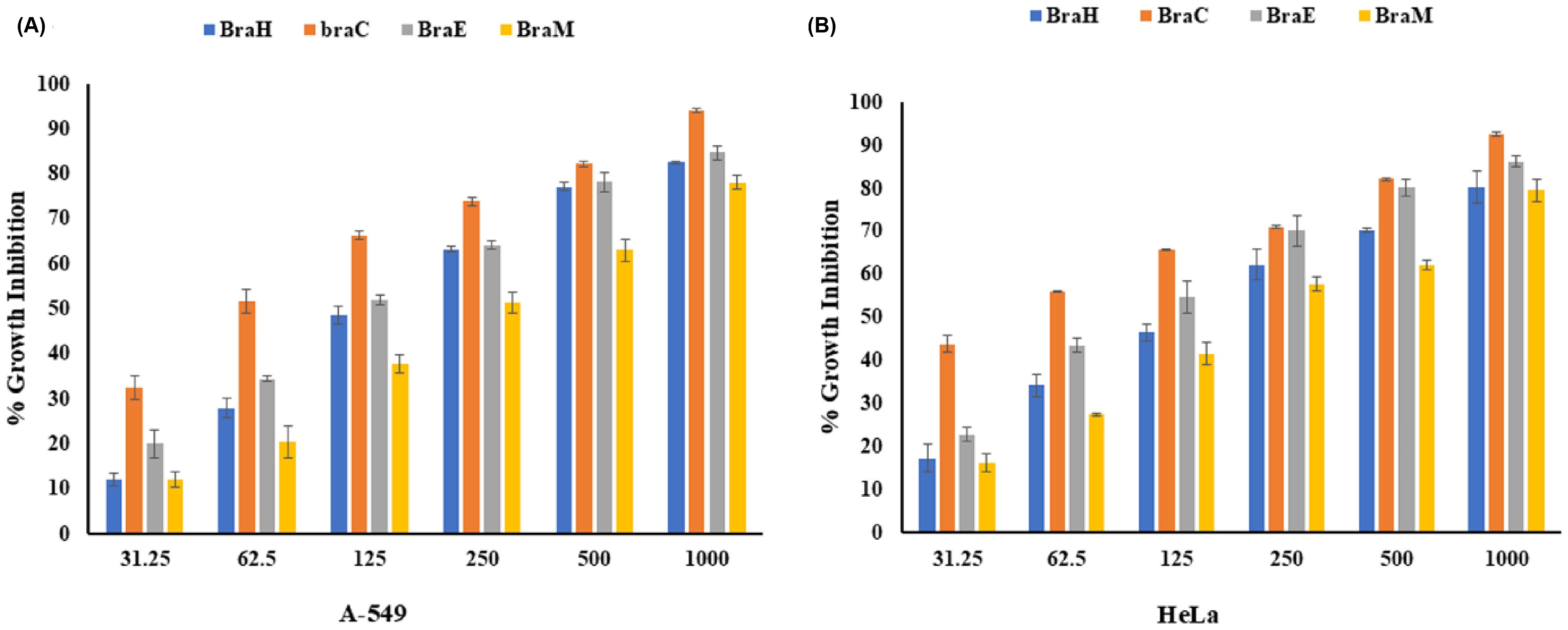
The in vitro antiproliferative potential of K-29 extracts (BraH, BraC, BraE, and BraM) was evaluated across a concentration gradient (31.25-1000 μg/mL) against A549 (human lung carcinoma) and HeLa (human cervical carcinoma) cell lines (Table 1). The antiproliferative activity was observed to increase in a concentration-dependent manner. Among the tested extracts, BraC exhibited the most pronounced antiproliferative effect, with GI50 values of 65.09 μg/mL and 37.00 μg/mL against A549 and HeLa cells, respectively, and an overall GI50 of 48.66 μg/mL, followed by BraE (132.34 μg/mL and 104.41 μg/mL) and BraH (161.50 μg/mL and 159.75 μg/mL). Conversely, BraM demonstrated the lowest antiproliferative activity, with GI50 values of 243.59 μg/mL for A549 cells (Figure 2A) and 203.73 μg/mL for HeLa cells (Figure 2B).
GI50 values of different extracts fractions of Brassica oleracea var acephala (K-29) against, HeLa and A-549 cancer cell lines.

Cytotoxic potential of BraH, BraC, BraE and BraM extracts against (A) A-549 and (B) HeLa cancer cell lines, after 24 h treatment. Values are represented in mean ± SD (p ≤ 0.05). Data labels with different letters represents significance differences among different concentrations.
3.3. Gas chromatography-mass spectrometry (GC-MS) analysis
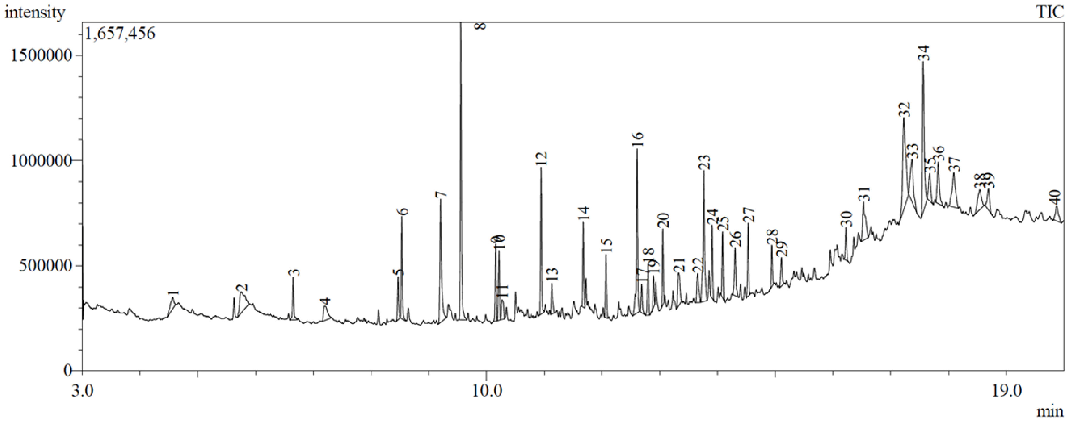
GC-MS analysis revealed the presence of various semi-polar organic compounds and volatile oil constituents, with the chromatogram depicted in Figure 3. The crude ethyl acetate extract (TrDE) exhibited the elution of 40 distinct constituents within a 20-min retention time, belonging to diverse chemical classes, including sterols, fatty acid derivatives, glycerol derivatives, phenolics, alcohols, alkanes, alkenes, esters, and sesquiterpenoids, as summarized in Table 2.
GC−MS qualitative analysis of bioactive compounds present in Brassica oleracea (K-29) BraC extract.
Notably, several compounds were present in relatively high concentrations, including 2,4-Di-tert-butylphenol (9.28%), Stigmasta-7,25-dien-3-ol, (3.β.,5.α.)- (7.25%), 13-Docosenamide, (Z)- (6.19%), Hexadecanoic acid, methyl ester (5.41%), 9-Octadecenoic acid, methyl ester, (E)- (5.33%), 1-Dodecanol (5.30%), and 2-Propenoic acid, tridecyl ester (4.18%). The concentrations of the remaining compounds ranged from 0.93% to 2.88%.
3.4. Cytomorphological changes in A-549 and HeLa cancer cells
Given the remarkable anticancer potential exhibited by the BraC fraction, an in-depth exploration of its cytomorphological effects was conducted. Treatment of A549 and HeLa cells with BraC induced significant morphological alterations, which were assessed via fluorescence microscopy. Notably, DAPI staining revealed a significantly higher occurrence of cells exhibiting intensely fluorescent, condensed, and fragmented nuclei—morphological hallmarks of apoptotic cell death (Figure 4B)— in the treated group compared with the uniformly and homogeneously stained nuclei observed in the untreated control cells (Figure 4A). This nuclear morphology is a definitive indicator of apoptosis. To further elucidate the apoptotic mechanisms, changes in mitochondrial membrane potential—a key feature of intrinsic apoptosis—were investigated using Rhodamine-123, a voltage-sensitive, mitochondria-targeting fluorophore. The analysis revealed a pronounced reduction in mitochondrial membrane potential in BraC-treated cells (Figure 4D), contrasting sharply with the uniform staining pattern maintained in the control group (Figure 4C). The loss of mitochondrial membrane integrity underscores the activation of the intrinsic apoptotic pathway. Together, these findings suggest that the BraC fraction exerts its cytotoxic effects by promoting apoptotic pathways, evidenced by nuclear condensation, fragmentation, and the collapse of mitochondrial membrane potential. Such apoptotic signatures highlight BraC's potential as a therapeutic agent targeting cancer cell viability.

BraC-induced cytomorphological alterations associated with apoptosis induction detected using fluorescence microscopy. (A) DAPI control; (B) DAPI treated with GI50 of BraC showing apoptotic nuclei; (C) Rhodamine 123 control; (D) Rhodamine 123 treated with GI50 of BraC showing disruption of mitochondrial membrane potential.
3.5. Antidiabetic potential
The evaluation of the antidiabetic activity of K-29, as determined by its inhibitory effects on α-amylase, revealed substantial variation across different solvent extractions. The aqueous extract of K-29 exhibited the most pronounced α-amylase inhibitory activity, achieving a significant inhibition rate of 85.08%. This effect was markedly higher compared to the inhibition observed with ethanolic (69.57%) and methanolic (71.52%) extracts, underscoring the enhanced efficacy of the aqueous solvent in retaining bioactive compounds responsible for antidiabetic effects (Figure 5).
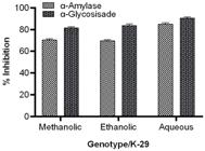
Inhibitory effects of methanolic, ethanolic, and aqueous leaf extracts of Brassica oleracea on α-amylase and α-glucosidase activities.
Similarly, the inhibitory activity against α-glucosidase, another critical enzyme in carbohydrate metabolism, showed significant variation depending on the solvent used for extraction. The aqueous extract once again demonstrated superior activity, inhibiting α-glucosidase by 89.55%, followed by the ethanolic (83.44%) and methanolic (81.86%) extracts (Figure 5). The higher potency observed in the aqueous extract suggests the presence of water-soluble phytoconstituents that are highly effective in modulating carbohydrate digestion and absorption pathways, which are crucial in managing postprandial hyperglycemia. These findings indicate that the aqueous extract of K-29 retains a more potent and bioactive phytochemical profile compared to its ethanol and methanol counterparts. The superior inhibition of both α-amylase and α-glucosidase suggests that K-29 could be a promising candidate for the development of natural antidiabetic therapies, particularly in its aqueous form.
4. Discussion
The pursuit of functional foods that provide health benefits, especially those fortified with bioactive compounds exhibiting strong antioxidant activity, is increasingly vital in disease prevention (Sorrenti et al., 2023). As consumer preferences shift toward minimally processed foods with fewer synthetic additives, the extraction of phytoconstituents rich in antioxidants from natural sources becomes even more imperative (Itam et al., 2021; López-Pedrouso et al., 2022). The antioxidant potency of compounds is often gauged by their IC50 values: compounds exhibiting IC50 values less than 50 µg/mL are classified as having very strong antioxidant activity, those between 50 and 100 µg/mL as strong, 101-150 µg/mL as moderate, and above 150 µg/mL as weak (Itam et al., 2021). Our findings reveal that K-29 demonstrates exceptional antioxidant capabilities, positioning it as a potential dietary supplement for bolstering the immune system and mitigating diseases such as diabetes, obesity, cardiovascular conditions, and certain cancers.
Research extensively supports the premise that the consumption of fruits and vegetables with elevated polyphenol levels and significant antioxidant properties correlates with a reduced risk of cancer. This is largely attributed to the bioactive constituents within these foods, which possess anticancer activities both in vitro and in vivo (Pandey and Rizvi, 2009; Ali et al., 2024). Our study identified that K-29 is abundant in phenolic compounds and exhibits noteworthy antioxidant capacity, likely due to its rich polyphenolic content. These compounds may act as cancer-preventive agents by inhibiting the onset of carcinogenesis and impeding the proliferation and progression of malignant cells. Specifically, the BraC compound was found to suppress the growth of A549 and HeLa cancer cells in a dose-dependent manner, signifying the substantial anticancer potential of K-29 in combating various malignancies.
Fluorescence microscopy has emerged as a powerful tool in detecting apoptosis through the morphological evaluation of cells. The application of fluorescent dyes that bind to specific cellular organelles allows for the visualization of critical changes associated with programmed cell death (Banfalvi, 2017). DAPI and rhodamine 123, in particular, are invaluable for identifying nuclear fragmentation, disruption of nuclear membrane integrity, and alterations in mitochondrial membrane potential (Hughes and Mehmet, 2003; Ahmad et al., 2024, 2025). These markers reveal distinctive apoptotic characteristics, enabling real-time monitoring and quantification of cell death processes, thus offering profound insights into the underlying mechanisms of apoptosis.
The study also highlights the antidiabetic potential of K-29 extracts, which exhibited marked differences in α-amylase and α-glucosidase inhibitory activities across various solvent extractions. The aqueous extract displayed the highest α-amylase inhibition (85.08%), significantly outperforming the ethanolic (69.57%) and methanolic (71.52%) extracts. Similarly, the aqueous extract demonstrated superior α-glucosidase inhibition (89.55%) compared to the ethanolic (83.44%) and methanolic (81.86%) extracts, emphasizing the solvent-specific efficacy of K-29 extracts in mitigating diabetes.
The superior antidiabetic activity observed in the aqueous extract concurs with existing literature, which posits that water-based extractions often preserve a more comprehensive range of bioactive compounds compared to organic solvents (Gopčević et al., 2019; Tran et al., 2020; Rahman et al., 2022; Maharaj et al., 2022). For example, Mohamed et al. (2022) demonstrated that aqueous extracts of Cyperus rotundus exhibited enhanced antidiabetic activity compared to ethyl acetate and ethanol extracts, likely due to the higher solubility of polar phytochemicals and phenolic compounds in water. Likewise, our findings are consistent with those of Gopčević et al. (2022), who noted that aqueous extracts of Satureja kitaibelii were enriched with flavonoids and phenolic compounds, both recognized for their inhibitory effects on α-amylase and α-glucosidase. The relatively diminished antidiabetic efficacy of ethanolic and methanolic extracts can be ascribed to the selective solubility of certain bioactive compounds in these solvents (Gopčević et al., 2022; Mohamed et al., 2022). Since water is a more effective solvent for extracting polar compounds, including phenolic acids and flavonoids, the aqueous extract exhibited a higher concentration of active compounds and thus greater inhibitory potency (Gopčević et al., 2019, 2022).
These findings suggest that the aqueous extract of K-29 may serve as a more potent therapeutic option for diabetes management, given its superior enzyme inhibitory activity. This is consistent with previous studies underscoring the efficacy of aqueous plant extracts in treating diabetes (Gopčević et al., 2022; Mohamed et al., 2022). The substantial inhibition of both α-amylase and α-glucosidase by K-29 aqueous extract emphasizes its potential as a natural antidiabetic agent, warranting further research into its specific bioactive compounds and their mechanisms of action. Given the superior antidiabetic activity observed in the aqueous extract compared to ethanolic and methanolic extracts, the selection of solvent emerges as a crucial factor in optimizing bioactive compound extraction. Future studies should aim to isolate and identify these compounds to further elucidate their role in glucose metabolism and explore their potential clinical applications in diabetes management.
Despite these promising findings, our study has certain limitations. The study was conducted in vitro, and the bioavailability and pharmacokinetics of the identified bioactive compounds in vivo remain to be explored. Additionally, potential synergistic or antagonistic interactions between the compounds were not assessed, which may influence the overall bioactivity. Future research should focus on in vivo studies and clinical trials to validate these findings and determine the safety, efficacy, and optimal dosage of K-29 extracts in disease management.
5. Conclusion
This study underscores the robust antioxidant, anticancer, and antidiabetic properties of Brassica oleracea (K-29) extracts across various solvent fractions. Among the evaluated fractions, BraM exhibited the most potent antioxidant capacity, as demonstrated by its superior radical scavenging activities in DPPH, ABTS, and FRAP assays. This suggests that the polarity of phytoconstituents is instrumental in influencing the antioxidant efficiency of K-29 extracts, with polar fractions exhibiting enhanced activity. Regarding anticancer activity, the BraC fraction displayed the most pronounced antiproliferative effects against A549 and HeLa cancer cell lines. Detailed cytomorphological analyses further revealed that BraC induces significant apoptotic events, such as nuclear condensation, DNA fragmentation, and the dissipation of mitochondrial membrane potential, establishing its potential as a pro-apoptotic agent. These findings suggest that BraC may modulate key signaling pathways involved in cancer cell proliferation and apoptosis, underscoring its potential therapeutic utility in oncological treatments.
In the context of antidiabetic potential, K-29 demonstrated notable α-amylase and α-glucosidase inhibitory activities, with the aqueous fraction exhibiting the highest efficacy. This implies that K-29 extracts may play a role in managing postprandial hyperglycemia, supporting their use as natural antidiabetic agents.
Future studies should prioritize the identification and isolation of bioactive compounds within the BraC and BraM fractions to elucidate the precise molecular mechanisms driving their antioxidant, anticancer, and antidiabetic activities. Additionally, in vivo studies are necessary to further validate the therapeutic efficacy and safety of these extracts, followed by clinical trials to evaluate their potential as adjunct therapies. Exploring the synergistic interactions between K-29 extracts and conventional chemotherapeutic or antidiabetic agents could also pave the way for novel combination therapies in the management of cancer and diabetes.
Acknowledgments
The authors of Chandigarh University, and Guru Nanak Dev University thank their universities for providing necessary lab facilities.
Data Availability Statement
All data generated or analyzed during this study are included in this published article.
References
-
AHMAD, S.S., GARG, C., KOUR, R., BHAT, A.H., RAJA, V., GANDHI, S.G., ATAYA, F.S., FOUAD, D., RADHAKRISHNAN, A. and KAUR, S., 2024. Metabolomic insights and bioactive efficacies of Tragopogon dubius root fractions: antioxidant and antiproliferative assessments. Heliyon, vol. 10, no. 16, e34746. https://doi.org/10.1016/j.heliyon.2024.e34746 PMid:39253191.
» https://doi.org/10.1016/j.heliyon.2024.e34746 -
AHMAD, S.S., GARG, C., BHAT, A.H. and KAUR, S., 2025. Phytochemicals in cancer therapy: modulating cell cycle, angiogenesis, and epithelial-mesenchymal transition. Revista Brasileira de Farmacognosia, vol. 35, no. 2, pp. 255-274. https://doi.org/10.1007/s43450-025-00620-4
» https://doi.org/10.1007/s43450-025-00620-4 -
AKBARI, B., BAGHAEI-YAZDI, N., BAHMAIE, M. and MAHDAVI ABHARI, F., 2022. The role of plant‐derived natural antioxidants in reduction of oxidative stress. BioFactors, vol. 48, no. 3, pp. 611-633. https://doi.org/10.1002/biof.1831 PMid:35229925.
» https://doi.org/10.1002/biof.1831 -
ALI, M.R., REZA, A.S.M.A., HAQUE, M.A., ISLAM, M.J., HOSSAIN, M.R., MOLLAH, M.I., ISLAM, M.B., SARKER, J., RASHID, M., SADIK, M.G., CICIA, D., CAPASSO, R., KAZI, M. and ALAM, A.K., 2024. Exploring the therapeutic potential of edible vegetables, fruits, and spices against cancer in various cell lines. Journal of Cancer, vol. 15, no. 3, pp. 577-589. https://doi.org/10.7150/jca.89539 PMid:38213720.
» https://doi.org/10.7150/jca.89539 -
BALAN, K., RATHA, P., PRAKASH, G., VISWANATHAMURTHI, P., ADISAKWATTANA, S. and PALVANNAN, T., 2017. Evaluation of invitro α-amylase and α-glucosidase inhibitory potential of N2O2 Schiff base Zn complex. Arabian Journal of Chemistry, vol. 10, no. 5, pp. 732-738. https://doi.org/10.1016/j.arabjc.2014.07.002
» https://doi.org/10.1016/j.arabjc.2014.07.002 -
BANFALVI, G., 2017. Methods to detect apoptotic cell death. Apoptosis: An International Journal on Programmed Cell Death, vol. 22, no. 2, pp. 306-323. https://doi.org/10.1007/s10495-016-1333-3 PMid:28035493.
» https://doi.org/10.1007/s10495-016-1333-3 -
BITWELL, C., INDRA, S.S., LUKE, C. and KAKOMA, M.K., 2023. A review of modern and conventional extraction techniques and their applications for extracting phytochemicals from plants. Scientific African, vol. 19, e01585. https://doi.org/10.1016/j.sciaf.2023.e01585
» https://doi.org/10.1016/j.sciaf.2023.e01585 -
BLOIS, M.S., 1958. Antioxidant determinations by the use of a stable free radical. Nature, vol. 181, no. 4617, pp. 1199-1200. https://doi.org/10.1038/1811199a0
» https://doi.org/10.1038/1811199a0 -
DANDOTI, S., 2021. Mechanisms adopted by cancer cells to escape apoptosis: a review. Biocell, vol. 45, no. 4, pp. 863-884. https://doi.org/10.32604/biocell.2021.013993
» https://doi.org/10.32604/biocell.2021.013993 -
DIAS, M.C., PINTO, D.C.G.A. and SILVA, A.M.S., 2021. Plant Flavonoids: chemical characteristics and biological activity. Molecules, vol. 26, no. 17, pp. 5377. https://doi.org/10.3390/molecules26175377 PMid:34500810.
» https://doi.org/10.3390/molecules26175377 -
DIRIR, A.M., DAOU, M., YOUSEF, A.F. and YOUSEF, L.F., 2022. A review of alpha-glucosidase inhibitors from plants as potential candidates for the treatment of type-2 diabetes. Phytochemistry Reviews, vol. 21, no. 4, pp. 1049-1079. https://doi.org/10.1007/s11101-021-09773-1 PMid:34421444.
» https://doi.org/10.1007/s11101-021-09773-1 -
GOPČEVIĆ, K., GRUJIĆ, S., ARSENIJEVIĆ, J., DŽAMIĆ, A., VELIČKOVIĆ, I., IZRAEL-ŽIVKOVIĆ, L., MEDIĆ, A., MUDRIĆ, J., SOKOVIĆ, M. and ĐURIĆ, A., 2022. Bioactivity and phenolics profile of aqueous and ethyl acetate extracts of Satureja kitaibelii Wierzb. ex Heuff. obtained by ultrasound-assisted extraction. Scientific Reports, vol. 12, no. 1, pp. 21221. https://doi.org/10.1038/s41598-022-25668-3 PMid:36481842.
» https://doi.org/10.1038/s41598-022-25668-3 -
GOPČEVIĆ, K., GRUJIĆ, S., ARSENIJEVIĆ, J., KARADŽIĆ, I., IZRAEL-ŽIVKOVIĆ, L. and MAKSIMOVIĆ, Z., 2019. Phytochemical properties of Satureja kitaibelii, potential natural antioxidants: a new insight. Plant Foods for Human Nutrition, vol. 74, no. 2, pp. 179-184. https://doi.org/10.1007/s11130-019-0716-3 PMid:30685803.
» https://doi.org/10.1007/s11130-019-0716-3 -
HONG, Y., BOITI, A., VALLONE, D. and FOULKES, N.S., 2024. Reactive oxygen species signaling and oxidative stress: transcriptional regulation and evolution. Antioxidants, vol. 13, no. 3, pp. 312. https://doi.org/10.3390/antiox13030312 PMid:38539845.
» https://doi.org/10.3390/antiox13030312 - HUGHES, D. and MEHMET, H., 2003. Cell proliferation & apoptosis. Oxford: BIOS Scientific Publishers.
-
ITAM, A., WATI, M.S., AGUSTIN, V., SABRI, N., JUMANAH, R.A. and EFDI, M., 2021. Comparative study of phytochemical, antioxidant, and cytotoxic activities and phenolic content of Syzygium aqueum (Burm. f. Alston f.) extracts growing in West Sumatera Indonesia. TheScientificWorldJournal, vol. 2021, pp. 5537597. https://doi.org/10.1155/2021/5537597 PMid:34234626.
» https://doi.org/10.1155/2021/5537597 -
JOMOVA, K., RAPTOVA, R., ALOMAR, S.Y., ALWASEL, S.H., NEPOVIMOVA, E., KUCA, K. and VALKO, M., 2023. Reactive oxygen species, toxicity, oxidative stress, and antioxidants: chronic diseases and aging. Archives of Toxicology, vol. 97, no. 10, pp. 2499-2574. https://doi.org/10.1007/s00204-023-03562-9 PMid:37597078.
» https://doi.org/10.1007/s00204-023-03562-9 - KERRISON, P. and STEINKE, M., 2010. DAPI staining protocol. Essex: Search In.
-
KIM, D.-O., JEONG, S.W. and LEE, C.Y., 2003. Antioxidant capacity of phenolic phytochemicals from various cultivars of plums. Food Chemistry, vol. 81, no. 3, pp. 321-326. https://doi.org/10.1016/S0308-8146(02)00423-5
» https://doi.org/10.1016/S0308-8146(02)00423-5 -
KUMAR, A., NIRMAL, P., KUMAR, M., JOSE, A., TOMER, V., OZ, E., PROESTOS, C., ZENG, M., ELOBEID, T., SNEHA, K. and OZ, F., 2023a. Major phytochemicals: recent advances in health benefits and extraction method. Molecules, vol. 28, no. 2, pp. 887. https://doi.org/10.3390/molecules28020887 PMid:36677944.
» https://doi.org/10.3390/molecules28020887 -
KUMAR, H., DHALARIA, R., GULERIA, S., CIMLER, R., SHARMA, R., SIDDIQUI, S.A., VALKO, M., NEPOVIMOVA, E., DHANJAL, D.S., SINGH, R., KUMAR, V., PATHERA, A.K., VERMA, N., KAUR, T., MANICKAM, S., ALOMAR, S.Y. and KUČA, K., 2023b. Anti-oxidant potential of plants and probiotic spp. in alleviating oxidative stress induced by H2O2 Biomedicine & Pharmacotherapy = Biomédecine & Pharmacothérapie, vol. 165, pp. 115022. https://doi.org/10.1016/j.biopha.2023.115022 PMid:37336149.
» https://doi.org/10.1016/j.biopha.2023.115022 -
KUNYANGA, C.N., IMUNGI, J.K., OKOTH, M., MOMANYI, C., BIESALSKI, H.K. and VADIVEL, V., 2011. Antioxidant and antidiabetic properties of condensed tannins in acetonic extract of selected raw and processed indigenous food ingredients from Kenya. Journal of Food Science, vol. 76, no. 4, pp. C560. https://doi.org/10.1111/j.1750-3841.2011.02116.x PMid:22417336.
» https://doi.org/10.1111/j.1750-3841.2011.02116.x -
LAMBA, B., MIR, M.A., RAJA, V., NEGI, N.P., BHAT, A.H., ATAYA, F.S., PAUL, N. and BATIHA, G.E., 2024. Phytochemical investigation of Tecoma capensis flower aqueous extract and in vitro biological studies: anti‐bacterial and anti‐proliferative activities of its silver nanoparticles. ChemistrySelect, vol. 9, no. 7, pp. 1. https://doi.org/10.1002/slct.202304481
» https://doi.org/10.1002/slct.202304481 -
LE, T.N., CHIU, C.H. and HSIEH, P.C., 2020. Bioactive Compounds and bioactivities of Brassica oleracea L. var. italica sprouts and microgreens: an updated overview from a nutraceutical perspective. Plants, vol. 9, no. 8, pp. 946. https://doi.org/10.3390/plants9080946 PMid:32727144.
» https://doi.org/10.3390/plants9080946 -
LI, X., BAI, Y., JIN, Z. and SVENSSON, B., 2022. Food-derived non-phenolic α-amylase and α-glucosidase inhibitors for controlling starch digestion rate and guiding diabetes-friendly recipes. Lebensmittel-Wissenschaft + Technologie, vol. 153, pp. 112455. https://doi.org/10.1016/j.lwt.2021.112455
» https://doi.org/10.1016/j.lwt.2021.112455 -
LIU, R.H., 2013. Health-promoting components of fruits and vegetables in the diet. Advances in Nutrition, vol. 4, no. 3, pp. 384S-392S. https://doi.org/10.3945/an.112.003517 PMid:23674808.
» https://doi.org/10.3945/an.112.003517 -
LÓPEZ-PEDROUSO, M., LORENZO, J.M. and FRANCO, D., 2022. Advances in natural antioxidants for food improvement. Antioxidants, vol. 11, no. 9, pp. 1825. https://doi.org/10.3390/antiox11091825 PMid:36139899.
» https://doi.org/10.3390/antiox11091825 -
LOURENÇO, S.C., MOLDÃO-MARTINS, M. and ALVES, V.D., 2019. Antioxidants of natural plant origins: from sources to food industry applications. Molecules, vol. 24, no. 22, pp. 4132. https://doi.org/10.3390/molecules24224132 PMid:31731614.
» https://doi.org/10.3390/molecules24224132 -
MAHARAJ, V., EZEOFOR, C.C., MAHARAJ, D.N., MULLER, C.J.F. and OBONYE, N.J., 2022. Identification of antidiabetic compounds from the aqueous extract of Sclerocarya birrea leaves. Molecules, vol. 27, no. 22, pp. 8095. https://doi.org/10.3390/molecules27228095 PMid:36432195.
» https://doi.org/10.3390/molecules27228095 -
MATTOSINHOS, P.D.S., SARANDY, M.M., NOVAES, R.D., ESPOSITO, D. and GONÇALVES, R.V., 2022. Anti-inflammatory, antioxidant, and skin regenerative potential of secondary metabolites from plants of the Brassicaceae family: a systematic review of in vitro and in vivo preclinical evidence (Biological activities brassicaceae skin diseases). Antioxidants, vol. 11, no. 7, pp. 1346. https://doi.org/10.3390/antiox11071346 PMid:35883837.
» https://doi.org/10.3390/antiox11071346 -
MICKISCH, G., FAJTA, S., KEILHAUER, G., SCHLICK, E., TSCHADA, R. and ALKEN, P., 1990. Chemosensitivity testing of primary human renal cell carcinoma by a tetrazolium based microculture assay (MTT). Urological Research, vol. 18, no. 2, pp. 131-136. https://doi.org/10.1007/BF00302474 PMid:1692648.
» https://doi.org/10.1007/BF00302474 -
MOHAMED, A.I., BESENI, B.K., MSOMI, N.Z., SALAU, V.F., ERUKAINURE, O.L., ALJOUNDI, A. and ISLAM, M.S., 2022. The antioxidant and antidiabetic potentials of polyphenolic-rich extracts of Cyperus rotundus (Linn.). Journal of Biomolecular Structure & Dynamics, vol. 40, no. 22, pp. 12075-12087. https://doi.org/10.1080/07391102.2021.1967197 PMid:34455935.
» https://doi.org/10.1080/07391102.2021.1967197 -
ORTEGA-HERNÁNDEZ, E., ANTUNES-RICARDO, M. and JACOBO-VELÁZQUEZ, D.A., 2021. Improving the health-benefits of kales (Brassica oleracea L. var. acephala DC) through the application of controlled abiotic stresses: a review. Plants, vol. 10, no. 12, pp. 2629. https://doi.org/10.3390/plants10122629 PMid:34961097.
» https://doi.org/10.3390/plants10122629 -
OU, B., HUANG, D., HAMPSCH-WOODILL, M., FLANAGAN, J.A. and DEEMER, E.K., 2002. Analysis of antioxidant activities of common vegetables employing oxygen radical absorbance capacity (ORAC) and ferric reducing antioxidant power (FRAP) assays: A comparative study. Journal of Agricultural and Food Chemistry, vol. 50, no. 11, pp. 3122-3128. https://doi.org/10.1021/jf0116606 PMid:12009973.
» https://doi.org/10.1021/jf0116606 -
PANDEY, K.B. and RIZVI, S.I., 2009. Plant polyphenols as dietary antioxidants in human health and disease. Oxidative Medicine and Cellular Longevity, vol. 2, no. 5, pp. 270-278. https://doi.org/10.4161/oxim.2.5.9498 PMid:20716914.
» https://doi.org/10.4161/oxim.2.5.9498 -
PIZZINO, G., IRRERA, N., CUCINOTTA, M., PALLIO, G., MANNINO, F., ARCORACI, V., SQUADRITO, F., ALTAVILLA, D. and BITTO, A., 2017. Oxidative stress: harms and benefits for human health. Oxidative Medicine and Cellular Longevity, vol. 2017, no. 1, pp. 8416763. https://doi.org/10.1155/2017/8416763 PMid:28819546.
» https://doi.org/10.1155/2017/8416763 -
PUJA, P., VINITA, N.M., DEVAN, U., VELANGANI, A.J., SRINIVASA, P., YUVALLUMAR, R., ARUL PRAKASH, P. and KUMAR, P., 2020. Fluorescence microscopy‐based analysis of apoptosis induced by platinum nanoparticles against breast cancer cells. Applied Organometallic Chemistry, vol. 34, no. 9, e5740. https://doi.org/10.1002/aoc.5740
» https://doi.org/10.1002/aoc.5740 -
RAHMAN, M.A., HANNAN, M.A., DASH, R., RAHMAN, M.H., ISLAM, R., UDDIN, M.J., SOHAG, A.A.M., RAHMAN, M.H. and RHIM, H., 2021. Phytochemicals as a complement to cancer chemotherapy: pharmacological modulation of the autophagy-apoptosis pathway. Frontiers in Pharmacology, vol. 12, pp. 639628. https://doi.org/10.3389/fphar.2021.639628 PMid:34025409.
» https://doi.org/10.3389/fphar.2021.639628 -
RAHMAN, M.M., DHAR, P.S., SUMAIA., ANIKA, F., AHMED, L., ISLAM, M.R., SULTANA, N.A., CAVALU, S., POP, O. and RAUF, A., 2022. Exploring the plant-derived bioactive substances as antidiabetic agent: an extensive review. Biomedicine & Pharmacotherapy = Biomédecine & Pharmacothérapie, vol. 152, pp. 113217. https://doi.org/10.1016/j.biopha.2022.113217 PMid:35679719.
» https://doi.org/10.1016/j.biopha.2022.113217 -
RAMIREZ, D., ABELLÁN-VICTORIO, A., BERETTA, V., CAMARGO, A. and MORENO, D.A., 2020. Functional ingredients from Brassicaceae species: overview and perspectives. International Journal of Molecular Sciences, vol. 21, no. 6, pp. 1998. https://doi.org/10.3390/ijms21061998 PMid:32183429.
» https://doi.org/10.3390/ijms21061998 -
RAY, P.D., HUANG, B.W. and TSUJI, Y., 2012. Reactive oxygen species (ROS) homeostasis and redox regulation in cellular signaling. Cellular Signalling, vol. 24, no. 5, pp. 981-990. https://doi.org/10.1016/j.cellsig.2012.01.008 PMid:22286106.
» https://doi.org/10.1016/j.cellsig.2012.01.008 -
RE, R., PELLEGRINI, N., PROTEGGENTE, A., PANNALA, A., YANG, M. and RICE-EVANS, C., 1999. Antioxidant activity applying an improved ABTS radical cation decolorization assay. Free Radical Biology & Medicine, vol. 26, no. 9-10, pp. 1231-1237. https://doi.org/10.1016/S0891-5849(98)00315-3 PMid:10381194.
» https://doi.org/10.1016/S0891-5849(98)00315-3 -
REDDY, V.P., 2023. Oxidative stress in health and disease. Biomedicines, vol. 11, no. 11, pp. 2925. https://doi.org/10.3390/biomedicines11112925 PMid:38001926.
» https://doi.org/10.3390/biomedicines11112925 -
RUDRAPAL, M., KHAIRNAR, S.J., KHAN, J., DUKHYIL, A.B., ANSARI, M.A., ALOMARY, M.N., ALSHABRMI, F.M., PALAI, S., DEB, P.K. and DEVI, R., 2022. Dietary polyphenols and their role in oxidative stress-induced human diseases: insights into protective effects, antioxidant potentials and mechanism(s) of action. Frontiers in Pharmacology, vol. 13, pp. 806470. https://doi.org/10.3389/fphar.2022.806470 PMid:35237163.
» https://doi.org/10.3389/fphar.2022.806470 -
SAEEDI, P., PETERSOHN, I., SALPEA, P., MALANDA, B., KARURANGA, S., UNWIN, N., COLAGIURI, S., GUARIGUATA, L., MOTALA, A.A., OGURTSOVA, K., SHAW, J.E., BRIGHT, D. and WILLIAMS, R., 2019. Global and regional diabetes prevalence estimates for 2019 and projections for 2030 and 2045: Results from the International Diabetes Federation Diabetes Atlas, 9th edition. Diabetes Research and Clinical Practice, vol. 157, pp. 107843. https://doi.org/10.1016/j.diabres.2019.107843
» https://doi.org/10.1016/j.diabres.2019.107843 -
SHARMA, S., PADHI, S., KUMARI, M., PATNAIK, S. and SAHOO, D., 2022. Antioxidant potential of selected wild edible leafy vegetables of sikkim himalayan region: effects of cooking methods and gastrointestinal digestion on activity. Frontiers in Nutrition, vol. 9, pp. 861347. https://doi.org/10.3389/fnut.2022.861347 PMid:35529464.
» https://doi.org/10.3389/fnut.2022.861347 -
SORRENTI, V., BURÒ, I., CONSOLI, V. and VANELLA, L., 2023. Recent advances in health benefits of bioactive compounds from food wastes and by-products: biochemical aspects. International Journal of Molecular Sciences, vol. 24, no. 3, pp. 2019. https://doi.org/10.3390/ijms24032019 PMid:36768340.
» https://doi.org/10.3390/ijms24032019 -
SYED, R.U., MONI, S.S., BREAK, M.K.B., KHOJALI, W.M.A., JAFAR, M., ALSHAMMARI, M.D., ABDELSALAM, K., TAYMOUR, S., ALRESHIDI, K.S.M., ELHASSAN TAHA, M.M. and MOHAN, S., 2023. Broccoli: a multi-faceted vegetable for health: an in-depth review of its nutritional attributes, antimicrobial abilities, and anti-inflammatory properties. Antibiotics, vol. 12, no. 7, pp. 1157. https://doi.org/10.3390/antibiotics12071157 PMid:37508253.
» https://doi.org/10.3390/antibiotics12071157 -
TRAN, N., PHAM, B. and LE, L., 2020. Bioactive compounds in anti-diabetic plants: from herbal medicine to modern drug discovery. Biology, vol. 9, no. 9, pp. 252. https://doi.org/10.3390/biology9090252 PMid:32872226.
» https://doi.org/10.3390/biology9090252 -
ULLAH, A., MUNIR, S., BADSHAH, S.L., KHAN, N., GHANI, L., POULSON, B.G., EMWAS, A.H. and JAREMKO, M., 2020. Important flavonoids and their role as a therapeutic agent. Molecules, vol. 25, no. 22, pp. 5243. https://doi.org/10.3390/molecules25225243 PMid:33187049.
» https://doi.org/10.3390/molecules25225243 -
XU, D.-P., LI, Y., MENG, X., ZHOU, T., ZHOU, Y., ZHENG, J., ZHANG, J.-J. and LI, H.-B., 2017. Natural antioxidants in foods and medicinal plants: extraction, assessment and resources. International Journal of Molecular Sciences, vol. 18, no. 1, pp. 96. https://doi.org/10.3390/ijms18010096
» https://doi.org/10.3390/ijms18010096 -
YU, L., HALEY, S., PERRET, J., HARRIS, M., WILSON, J. and QIAN, M., 2002. Free radical scavenging properties of wheat extracts. Journal of Agricultural and Food Chemistry, vol. 50, no. 6, pp. 1619-1624. https://doi.org/10.1021/jf010964p PMid:11879046.
» https://doi.org/10.1021/jf010964p -
ZHANG, Y.J., GAN, R.Y., LI, S., ZHOU, Y., LI, A.N., XU, D.P. and LI, H.B., 2015. Antioxidant phytochemicals for the prevention and treatment of chronic diseases. Molecules, vol. 20, no. 12, pp. 21138-21156. https://doi.org/10.3390/molecules201219753 PMid:26633317.
» https://doi.org/10.3390/molecules201219753
Edited by
-
Editor:
Takako Matsumura Tundisi